Submitted Successfully!
To reward your contribution, here is a gift for you: A free trial for our video production service.
Thank you for your contribution! You can also upload a video entry or images related to this topic.
Version Summary Created by Modification Content Size Created at Operation
1 + 1456 word(s) 1456 2021-12-28 07:53:11 |
2 The format is correct. -1 word(s) 1455 2022-01-11 02:23:44 |

Video Upload Options

Do you have a full video?

Confirm

Are you sure to Delete?
Cite
If you have any further questions, please contact Encyclopedia Editorial Office.
Daou, F. Current Advances in Regeneration of Degenerated Articular Cartilage. Encyclopedia. Available online: https://encyclopedia.pub/entry/17973 (accessed on 29 April 2024).
Daou F. Current Advances in Regeneration of Degenerated Articular Cartilage. Encyclopedia. Available at: https://encyclopedia.pub/entry/17973. Accessed April 29, 2024.
Daou, Farah. "Current Advances in Regeneration of Degenerated Articular Cartilage" Encyclopedia, https://encyclopedia.pub/entry/17973 (accessed April 29, 2024).
Daou, F. (2022, January 10). Current Advances in Regeneration of Degenerated Articular Cartilage. In Encyclopedia. https://encyclopedia.pub/entry/17973
Daou, Farah. "Current Advances in Regeneration of Degenerated Articular Cartilage." Encyclopedia. Web. 10 January, 2022.
Current Advances in Regeneration of Degenerated Articular Cartilage
Edit

Functional ability is the basis of healthy aging. Articular cartilage degeneration is amongst the most prevalent degenerative conditions that cause adverse impacts on the quality of life; moreover, it represents a key predisposing factor to osteoarthritis (OA). Both the poor capacity of articular cartilage for self-repair and the unsatisfactory outcomes of available clinical interventions make innovative tissue engineering a promising therapeutic strategy for articular cartilage repair. Significant progress was made in this field; however, a marked heterogeneity in the applied biomaterials, biofabrication, and assessments is nowadays evident by the huge number of research studies published to date. Accordingly, this entry assimilates the most recent advances in cell-based and cell-free tissue engineering of articular cartilage and also focuses on the assessments performed via various in vitro studies, ex vivo models, preclinical in vivo animal models, and clinical studies in order to provide a broad overview of the latest findings and clinical translation in the context of degenerated articular cartilage and OA.

cartilage regeneration tissue engineering biomaterials

1. Introduction

Cellular senescence is predominantly correlated with tissue aging and loss of function, but its physiological role must be indeed evaluated in relation to the stage of life. In early life, cellular senescence contributes to the attenuation of tissue damage, promotion of wound healing, and suppression of tumorigenesis; on the contrary, in old age, it fosters inflammation, aging, and aging-related diseases and also limits tissue regenerative potential [1]. In addition to the latter, the accumulation of senescent cells is systemic and continuous, and this results in a persistent imbalance in the homeostasis of almost all tissues. One of the organ systems that is significantly affected by cellular senescence is the musculoskeletal system, since it results in bone, muscle, and cartilage degeneration [2].
Nowadays, cartilage degeneration continues to be a major challenge for clinicians due to the limited self-healing properties originating from the low proliferation ability of chondrocytes, the sole cellular constituents of cartilage [3], as well as from the absence of blood vessels, nerves, and lymphatics [4]. The most commonly documented cartilage degeneration is that of articular cartilage covering synovial joints [3]
This high prevalence of articular cartilage degeneration represents a major clinical challenge, since, in the long-term, articular cartilage degeneration can contribute to the development of osteoarthritis (OA) (a multifactorial joint condition) [5], which is the third most common musculoskeletal disorder requiring rehabilitation after low back pain and fractures [6]
Therapeutic approaches for treating articular cartilage degeneration are diverse and can be categorized into three types: (i) symptomatic, (ii) reparative (or restorative), and (iii) regenerative. Symptomatic treatments include pain killers, anti-inflammatory drugs, and intra-articular injections of corticosteroids, hyaluronic acid (HA), or platelet-rich plasma (PRP); reparative treatments are microfracture, abrasion, drilling, osteochondral allograft, and mosaicplasty; whereas regenerative treatments entail autologous chondrocyte implantation (ACI) and matrix-induced autologous chondrocyte implantation (MACI) [7][8][9]. The reparative and regenerative treatments are reported to be effective in treating chondral and subchondral defects, but they can neither cure nor slow down articular cartilage degeneration; hence, tissue engineering strategies were introduced to overcome these limitations. Since its inception, articular cartilage tissue engineering yielded positive results and experienced major advancements, and consequently, it is now considered a promising alternative for replicating the structure and function of native articular cartilage [7][10].

2. Age-Related Changes in Articular Cartilage

Articular cartilage is classified as hyaline cartilage, and it is mainly composed of chondrocytes and a dense ECM produced by them. Its thickness ranges between 2 to 4 mm; however, it exhibits structural and compositional heterogeneity across four different zones, which are the (i) superficial (tangential), (ii) middle (transitional), (iii) deep, and (iv) calcified zones [11]. The structure of healthy articular cartilage is schematized in Figure 1 [12].
Figure 1. Schematic representation of the articular cartilage structure and biochemical composition [12].
Age-related changes in articular cartilage involve both components (chondrocytes and ECM), and the progressive nature of pathological conditions makes their classification essential, not only to detect the extent of the defects but also to guide clinical decision-making. The most commonly applied classification method is the arthroscopic grading system developed by the International Cartilage Repair Society (ICRS), which divides defects into four grades [13], as detailed below and schematized in Figure 2:
Figure 2. The International Cartilage Repair Society (ICRS) Cartilage Lesion Classification Method. Image reprinted with permission from the ICRS Cartilage Injury Evaluation Package (http://www.cartilage.org/, accessed on the 1 November 2021).

2.1. Age-Related Changes in Chondrocytes

Chondrocytes are specialized cells constituting only 2% of the total volume of articular cartilage and exhibiting variations in shape, number, and size across its zones. They play a central role in the synthesis and maintenance of a normal ECM, thus, having an adequate number of functionally competent chondrocytes is of crucial importance for articular cartilage homeostasis [11].

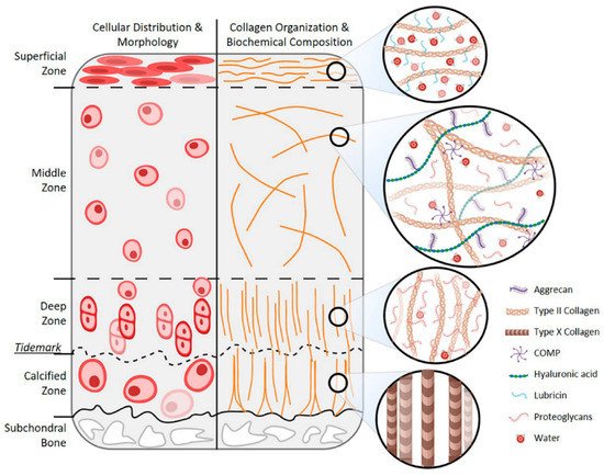
2.2. Age-Related Changes in Cartilage ECM

The ECM is responsible for the mechanical properties of articular cartilage. Its functions are mediated by two phases: the liquid phase that is composed mainly of water and inorganic ions and accounts for 65 to 80% of the wet weight, and the solid phase that is primarily made up of collagens (mainly type II collagen) and proteoglycans (including aggrecan, decorin, biglycan, and fibromodulin) and constitutes the remaining dry weight [11].

3. Tissue Engineering Strategies for Articular Cartilage Regeneration

Extensive attempts have been made to engineer cartilage tissues with structural and functional properties similar to those of native articular cartilage. These efforts have brought major advancements in the field of articular cartilage tissue engineering and, at the same time, generated a heterogeneity in the biomaterials, biofabrication, and assessments applied in this field of research [14][15]

3.1. Cell-Based Tissue Engineering Strategies

Research on cell-based articular cartilage tissue engineering has identified new cell sources, scaffolds, and bioactive molecules, as well as novel composites combining these components. 

3.1.1. Scaffold-Based Strategies

The use of scaffolds in articular cartilage tissue engineering represents a largely applied strategy, since scaffolds are intended to support cellular proliferation and differentiation, as well as to deliver pro-chondrogenic bioactive molecules, and in that pursuit, multiple scaffolds were designed for various purposes. In one of the most recent studies, kartogenin (KGN), which is a small molecule discovered by Johnson et al. (2012) to be a chondrogenic and chondroprotective agent [16], was utilized by Teng et al. (2021). 

3.1.2. Scaffold-Free Strategies

Scaffold-free tissue engineering for articular cartilage regeneration represents a promising alternative for overcoming the limitations associated with scaffold-based tissue engineering, mainly regarding the long-term safety of the devices themselves. The most commonly used technology for this purpose is the so-called cell sheet technology consisting of implantable artificial proto-tissues composed of cells in high-density and tightly interconnected to each other by a dense ECM that is harvested avoiding the use of enzymes by thermo-responsive substrates [17][18]

3.1.3. Injectables

In view of a possible clinical application, researchers are aspiring to utilize technologies that enable shifting from invasive surgical procedures to minimally invasive or non-invasive ones for the regeneration of articular cartilage. Out of the numerous developed injectables, the simplest ones are aimed at delivering cells only into the defect site. For instance, cell sheet technology was applied by Wasai et al. (2021) to fabricate injectable allogeneic polydactyly-derived chondrocyte sheets (PD) cell sheet fragments rather than large cell sheets that require invasive surgery. The results showed that there are no significant differences between the PD sheets and the PD sheets-mini in terms of cell count and viability, the number of humoral factors produced, and the histological characteristics; in addition, the injection of the PD sheets-mini did not alter cell viability [19]

3.2. Cell-Free Tissue Engineering Strategies

Despite their pivotal role in stimulating articular cartilage regeneration, cells require additional considerations when used in tissue engineering, including the surgical procedure for harvesting autologous cells and the time required for cell expansion in vitro. Moreover, it should be mentioned that chondrocytes hold a poor capacity of expansion in vitro, which often limits their use in combination with scaffolds [20]. As a result, researchers brought forward cell-free tissue engineering strategies with the ability to recruit native (endogenous) progenitor or mesenchymal cells as alternatives for cell-based ones. In this section, cell-free tissue engineering is divided into two categories: scaffold-based strategies and injectables.

3.2.1. Scaffold-Based Strategies

For cell-free, scaffold-based articular cartilage regeneration, collagen type I (Col-1)-based scaffolds are mostly applied. The latest evaluation of cell-free Col-1-based scaffolds was conducted by Szychlinska et al. (2020) in vivo using Wistar outbred rat models with knee cartilage lesions at the femoropatellar groove, and this scaffold exhibited biocompatibility and efficient recruitment of host cells for articular cartilage regeneration [21].

3.2.2. Injectables

Since scaffolds are broadly applied for cell-free articular cartilage regeneration, efforts were also exerted to move towards minimally invasive or non-invasive injectable materials and composites. To start with, the application of SF as a cell-free injectable was tested by Yuan et al. (2021) in vitro and in vivo using mice for subcutaneous injection and using New Zealand rabbit models with osteochondral defects. A novel one-step ultrasonication crosslinking method was used for this purpose, which exhibited both safety and efficacy for articular cartilage regeneration [22]

References

  1. Di Micco, R.; Krizhanovsky, V.; Baker, D.; d’Adda di Fagagna, F. Cellular Senescence in Ageing: From Mechanisms to Therapeutic Opportunities. Nat. Rev. Mol. Cell Biol. 2021, 22, 75–95.
  2. Baar, M.P.; Perdiguero, E.; Muñoz-Cánoves, P.; de Keizer, P.L. Musculoskeletal Senescence: A Moving Target Ready to Be Eliminated. Curr. Opin. Pharmacol. 2018, 40, 147–155.
  3. Lotz, M.; Loeser, R.F. Effects of Aging on Articular Cartilage Homeostasis. Bone 2012, 51, 241–248.
  4. Chang, L.-R.; Marston, G.; Martin, A. Anatomy, Cartilage; StatPearls Publishing: Treasure Island, FL, USA, 2020.
  5. Anderson, A.S.; Loeser, R.F. Why Is Osteoarthritis an Age-Related Disease? Best Pract. Res. Clin. Rheumatol. 2010, 24, 15–26.
  6. Cieza, A.; Causey, K.; Kamenov, K.; Hanson, S.W.; Chatterji, S.; Vos, T. Global Estimates of the Need for Rehabilitation Based on the Global Burden of Disease Study 2019: A Systematic Analysis for the Global Burden of Disease Study 2019. Lancet 2021, 396, 2006–2017.
  7. Medvedeva, E.V.; Grebenik, E.A.; Gornostaeva, S.N.; Telpuhov, V.I.; Lychagin, A.V.; Timashev, P.S.; Chagin, A.S. Repair of Damaged Articular Cartilage: Current Approaches and Future Directions. Int. J. Mol. Sci. 2018, 19, 2366.
  8. Pogliacomi, F.; Schiavi, P.; Paraskevopoulos, A.; Leigheb, M.; Pedrazzini, A.; Ceccarelli, F.; Vaienti, E. When Is Indicated Viscosupplementation in Hip Osteoarthritis? Acta Biomed. 2018, 90, 67–74.
  9. Leigheb, M.; Bosetti, M.; de Consoli, A.; Borrone, A.; Cannas, M.; Grassi, F. Chondral Tissue Engineering of the Reumatoid Knee with Collagen Matrix Autologous Chondrocytes Implant. Acta Biomed. 2017, 88, 107–113.
  10. Moreira-Teixeira, L.S.; Georgi, N.; Leijten, J.; Wu, L.; Karperien, M. Cartilage Tissue Engineering. Endocr. Dev. 2011, 21, 102–115.
  11. Sophia Fox, A.J.; Bedi, A.; Rodeo, S.A. The Basic Science of Articular Cartilage: Structure, Composition, and Function. Sports Health 2009, 1, 461–468.
  12. Thorp, H.; Kim, K.; Kondo, M.; Maak, T.; Grainger, D.W.; Okano, T. Trends in Articular Cartilage Tissue Engineering: 3D Mesenchymal Stem Cell Sheets as Candidates for Engineered Hyaline-like Cartilage. Cells 2021, 10, 643.
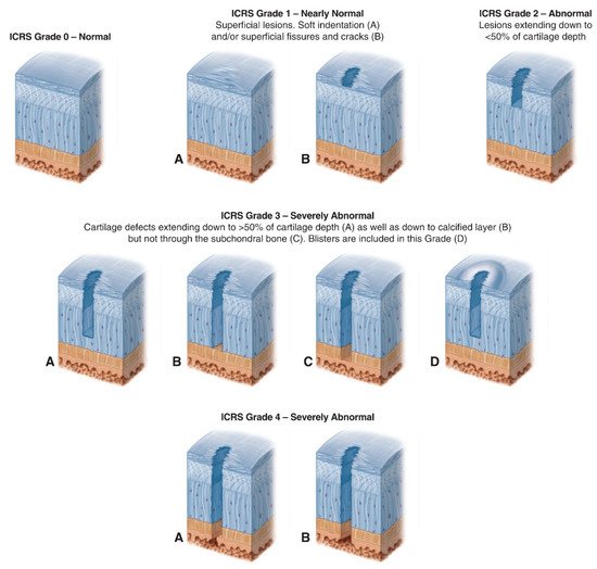
  13. International Cartilage Repair Society (ICRS) Cartilage Injury Evaluation Package. Available online: https://cartilage.org/content/uploads/2014/10/ICRS_evaluation.pdf (accessed on 18 October 2021).
  14. Kwon, H.; Brown, W.E.; Lee, C.A.; Wang, D.; Paschos, N.; Hu, J.C.; Athanasiou, K.A. Surgical and Tissue Engineering Strategies for Articular Cartilage and Meniscus Repair. Nat. Rev. Rheumatol. 2019, 15, 550–570.
  15. Zhao, X.; Hu, D.A.; Wu, D.; He, F.; Wang, H.; Huang, L.; Shi, D.; Liu, Q.; Ni, N.; Pakvasa, M.; et al. Applications of Biocompatible Scaffold Materials in Stem Cell-Based Cartilage Tissue Engineering. Front. Bioeng. Biotechnol. 2021, 9, 603444.
  16. Johnson, K.; Zhu, S.; Tremblay, M.S.; Payette, J.N.; Wang, J.; Bouchez, L.C.; Meeusen, S.; Althage, A.; Cho, C.Y.; Wu, X.; et al. A Stem Cell-Based Approach to Cartilage Repair. Science 2012, 336, 717–721.
  17. Cochis, A.; Bonetti, L.; Sorrentino, R.; Negrini, N.C.; Grassi, F.; Leigheb, M.; Rimondini, L.; Farè, S. 3D Printing of Thermo-Responsive Methylcellulose Hydrogels for Cell-Sheet Engineering. Materials 2018, 11, 579.
  18. Altomare, L.; Cochis, A.; Carletta, A.; Rimondini, L.; Farè, S. Thermo-Responsive Methylcellulose Hydrogels as Temporary Substrate for Cell Sheet Biofabrication. J. Mater. Sci. Mater. Med. 2016, 27, 95.
  19. Wasai, S.; Toyoda, E.; Takahashi, T.; Maehara, M.; Okada, E.; Uchiyama, R.; Akamatsu, T.; Watanabe, M.; Sato, M. Development of Injectable Polydactyly-Derived Chondrocyte Sheets. Int. J. Mol. Sci. 2021, 22, 3198.
  20. Cochis, A.; Grad, S.; Stoddart, M.J.; Farè, S.; Altomare, L.; Azzimonti, B.; Alini, M.; Rimondini, L. Bioreactor Mechanically Guided 3D Mesenchymal Stem Cell Chondrogenesis Using a Biocompatible Novel Thermo-Reversible Methylcellulose-Based Hydrogel. Sci. Rep. 2017, 7, 45018.
  21. Szychlinska, M.A.; Calabrese, G.; Ravalli, S.; Dolcimascolo, A.; Castrogiovanni, P.; Fabbi, C.; Puglisi, C.; Lauretta, G.; di Rosa, M.; Castorina, A.; et al. Evaluation of a Cell-Free Collagen Type I-Based Scaffold for Articular Cartilage Regeneration in an Orthotopic Rat Model. Materials 2020, 13, 2369.
  22. Yuan, T.; Li, Z.; Zhang, Y.; Shen, K.; Zhang, X.; Xie, R.; Liu, F.; Fan, W. Injectable Ultrasonication-Induced Silk Fibroin Hydrogel for Cartilage Repair and Regeneration. Tissue Eng. Part A 2021, 27, 1213–1224.
More
Information
Contributor MDPI registered users' name will be linked to their SciProfiles pages. To register with us, please refer to https://encyclopedia.pub/register :
View Times: 925
Revisions: 2 times (View History)
Update Date: 11 Jan 2022
1000/1000