Submitted Successfully!
To reward your contribution, here is a gift for you: A free trial for our video production service.
Thank you for your contribution! You can also upload a video entry or images related to this topic.
Version Summary Created by Modification Content Size Created at Operation
1 + 1344 word(s) 1344 2022-01-04 09:36:37 |
2 format correct Meta information modification 1344 2022-01-07 01:51:07 |

Video Upload Options

Do you have a full video?

Confirm

Are you sure to Delete?
Cite
If you have any further questions, please contact Encyclopedia Editorial Office.
Verhoeyen, E. CAR T Cells. Encyclopedia. Available online: https://encyclopedia.pub/entry/17828 (accessed on 02 July 2024).
Verhoeyen E. CAR T Cells. Encyclopedia. Available at: https://encyclopedia.pub/entry/17828. Accessed July 02, 2024.
Verhoeyen, Els. "CAR T Cells" Encyclopedia, https://encyclopedia.pub/entry/17828 (accessed July 02, 2024).
Verhoeyen, E. (2022, January 06). CAR T Cells. In Encyclopedia. https://encyclopedia.pub/entry/17828
Verhoeyen, Els. "CAR T Cells." Encyclopedia. Web. 06 January, 2022.
CAR T Cells
Edit

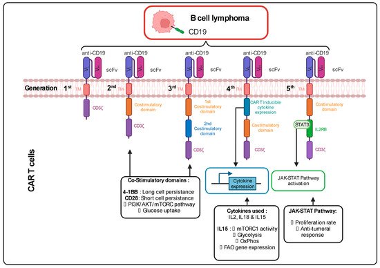
Cells are also used as a therapeutic tool. In this type of immunotherapy, the patient’s T cells are genetically engineered ex vivo to express a CAR that recognizes a specific antigen present on the surface of the tumor cells. After reinfusion of these cells in the patient’s circulation, the binding between the CAR T cells and the cancer cells induces a cytotoxic response. One example of this therapy currently being used in clinics is for the treatment of advanced B-cell lymphomas, resulting in complete remission in 30 to 40 % of the patients. Importantly, it needs to be emphasized that CAR T cells are used last in line as a therapeutic strategy to suppress tumor cells. This means that the T cells are often isolated from the patients after various treatments, including chemotherapy that can alter the metabolic phenotype of the T cells (mitochondrial damage and metabolic alterations). Therefore, metabolism is an important aspect in the conception and the anti-tumoral activity of a CAR T cell.

CAR T cell metabolism ex vivo expansion Oxphos Glycolysis costimulatory domain T cell exhaustion Memory T cell CAR NK cell

1. Continuous Improvements in the CAR Design to Stir CAR T Cell Metabolism

The CAR incorporated at the surface of T cells has a T cell receptor (TCR)-like structure. The extracellular part consists of a single variable chain fragment (scFv) of an antibody that recognizes the antigen present on the cancer cell surface. The transmembrane and intracellular parts contain co-stimulatory domains that permit the amplification of the signaling and, thus, the response of the T cell following binding to the tumor antigen. In the first-generation CAR T cell design, CD3ζ was the only signaling domain used, but in the following generations, CD28 and/or 4-1BB (CD137) co-stimulatory domains were added to this structure. The choice of these domains is also important for the metabolic status and the survival of the CAR T cells. It was observed in patients that the use of 4-1BB permitted a persistence of CAR T cells in time, without exhaustion of those CAR T cells, via the stimulation of the noncanonical nuclear factor kappa B (NF-κB) pathway [1]. In contrast, the CD28 costimulatory domain does not permit the cells to survive longer than 30 days in the patients [2][3][4][5]. This phenomenon can be explained by the fact that the 4-1BB promotes improved mitochondrial function so that T cells can rely on mitochondrial respiration as their energy source, which promotes the survival of central memory T cells by the activation of adenosine monophosphate kinase (AMPK), as demonstrated in mice [6]. When the CD28 co-stimulatory domain was included in the CAR, patient T cells were relying more on glycolysis (via activation of the PI3K/AKT/mTOR axis), which pushed their differentiation towards effector memory T cells [7][8]. Following antigen recognition by the CAR, CD28 stimulated GLUT1 via the PI3K/AKT pathway, linked to the mTOR/Myc pathways, which are also implicated in amino acid/lipid metabolism [9][10]. These data underline the importance of the choice of the co-stimulatory domain to shape CAR T cell metabolism and permit short-term or long-term anti-tumor efficacy. As for CAR T cells, TIL performance also depends strongly on the metabolic status of the malignancy and the TME.
Cytokines also play a role in the metabolic shape of the CAR T cells. Indeed, the fourth CAR T cell generation, also known as “TRUCKS” (T-cell redirected for unrestricted cytokine-mediated killing), has a transgenic cytokine expression system that improves their expansion and persistence in vivo via metabolic changes [11]. The cytokines employed in this strategy are interleukin 12 (IL12), IL15 and IL18. IL15 can, for example, lower the glycolysis level in human CAR T cells via a decrease in mTOR complex 1 (mTORC1) activity, whilst OXPHOS levels/respiratory capacity and expression of fatty acid oxidation-related genes are increased in these cells. This metabolic phenotype allows the human CAR T cells to have a stem cell memory behavior with higher cell proliferation and in vivo longevity [12]. More recent fifth-generation CAR T cells express a sub-unit of the IL2 receptor (IL2RB) between the stimulatory domains (CD3ζ and CD28/4-1BB). Since IL2RB presents a binding site for STAT3, the cells can activate the JAK-STAT pathway, following the antigen recognition, which leads to a higher proliferation rate. Moreover, these human CAR T cells seem to have a stronger anti-cancer activity against leukemic cells [13] (Figure 1).
Figure 1. Different generations of CAR T cells and their signaling pathways. TM: Transmembrane; scFv: Single-chain variable Fragment; VL: Variable Light chain; VH: Variable Heavy chain; IL2RB: interleukin 2 Receptor B; mTORC: mammalian target of rapamycin complex; FAO: Fatty Acid Oxidation; JAK-STAT: Janus Kinase-Signal Transducers and Activators of Transcription; PI3K: Phosphoinositide 3-kinase; Akt: Protein kinase; CART: Chimeric Antigen Receptor T cell. Figure generated with Biorender.

2. Ex Vivo Expansion of CAR T Cells to Manipulate Their Metabolism

As they are immune cells and similar to TILs, CAR T cells are highly sensitive to metabolic changes in their environment, which can influence their survival and their functionality. When cultured ex vivo, CAR T cells are often cultured in the presence of supra-physiological nutrient levels in order to generate enough cells for reinfusion. This also means that the cytotoxic efficiency is compromised when changing from an overdosed medium to a nutrient restrictive microenvironment. Thus, the composition of expansion media plays an important role in the process of CAR T cell generation and needs to be optimized in order to facilitate the transition between the ex vivo expansion and in vivo adaptation. For example, carnosine, an amine poorly present in sera, when added to the medium, facilitates human T cell transduction and their in vivo engraftment by switching the main metabolic state from glycolysis to OXPHOS, which allowed a better anti-tumoral response [14]. Currently, expansion media rely essentially either on fetal bovine serum (FBS) or human serum (HS), which does not take into account some blood fractions, which might highly compromise human CAR T cell fitness [15].
The effect of cytokines on T cell metabolism is only beginning to be explored in the field. Survival cytokines, IL2 and IL7, promote glycolysis in T cells [16][17], while IL-15 promotes their mitochondrial biogenesis [18]. TGFβ seems to suppress both glycolysis and mitochondrial respiration. Therefore, the choice of cytokines or growth factors in the media for CAR T cell expansion might be crucial for their in vivo adaptation.
It is therefore important to be aware that the ex vivo expansion of these CAR T cells is a tricky step in the process and can be limiting and compromise treatment efficacy. Several research teams are currently trying to improve CAR T cell therapy by circumventing the ex vivo expansion step and directly generating CAR T cells in vivo. In recent studies, lentiviral vectors specifically recognizing the CD3+ or even the CD8+ human T cells were used to generate efficiently anti-CD19 CAR T cells directly in vivo and were able to wipe out the targeted healthy and malignant B cells in humanized mouse models [19][20][21][22][23]. Although this still needs to be evaluated, one can speculate that these in vivo generated CAR T cells might expand in their proper environment and maintain their metabolic fitness in vivo.
More recently, an elegant strategy to overcome in vivo CAR T cell exhaustion has been reported with better outcomes than PD-1/PD-L1 blockade, which can improve T cell functions but do not change the epigenetic reprogramming associated with T cell exhaustion. The approach is based on inducing a transient rest in CAR signaling and, therefore, temporally blocking the sustained CAR T cell activation that leads to exhaustion [24]. This approach made use of temporal downregulation of the surface expression of the CAR by inducing its degradation by pharmacological treatments. The tunable control of CAR expression levels not only improved CAR T cell anti-tumoral functions in vivo in terms of intensity and duration but also led to non-exhausted T cells with a memory-like phenotype in xenograft mouse models of leukemia and osteosarcoma. Interestingly, the already established exhaustion of CAR T cells can even be reverted after a transient rest of the CAR signaling [24].

References

  1. Philipson, B.I.; O’Connor, R.S.; May, M.J.; June, C.H.; Albelda, S.M.; Milone, M.C. 4-1BB costimulation promotes CAR T cell survival through noncanonical NF-κB signaling. Sci. Signal. 2020, 13.
  2. Brentjens, R.J.; Davila, M.L.; Riviere, I.; Park, J.; Wang, X.; Cowell, L.G.; Bartido, S.; Stefanski, J.; Taylor, C.; Olszewska, M.; et al. CD19-Targeted T Cells Rapidly Induce Molecular Remissions in Adults with Chemotherapy-Refractory Acute Lymphoblastic Leukemia. Sci. Transl. Med. 2013, 5, 177ra38.
  3. Lee, D.W.; Kochenderfer, J.N.; Stetler-Stevenson, M.; Cui, Y.K.; Delbrook, C.; Feldman, S.A.; Fry, T.J.; Orentas, R.; Sabatino, M.; Shah, N.N.; et al. T cells expressing CD19 chimeric antigen receptors for acute lymphoblastic leukaemia in children and young adults: A phase 1 dose-escalation trial. Lancet 2015, 385, 517–528.
  4. Long, A.H.; Haso, W.M.; Shern, J.F.; Wanhainen, K.M.; Murgai, M.; Ingaramo, M.; Smith, J.P.; Walker, A.J.; Kohler, M.E.; Venkateshwara, V.R.; et al. 4-1BB costimulation ameliorates T cell exhaustion induced by tonic signaling of chimeric antigen receptors. Nat. Med. 2015, 21, 581–590.
  5. Porter, D.L.; Hwang, W.-T.; Frey, N.V.; Lacey, S.F.; Shaw, P.A.; Loren, A.W.; Bagg, A.; Marcucci, K.T.; Shen, A.; Gonzalez, V.; et al. Chimeric antigen receptor T cells persist and induce sustained remissions in relapsed refractory chronic lymphocytic leukemia. Sci. Transl. Med. 2015, 7, 303ra139.
  6. Choi, B.K.; Lee, D.Y.; Lee, D.G.; Kim, Y.H.; Kim, S.-H.; Oh, H.S.; Han, C.; Kwon, B.S. 4-1BB signaling activates glucose and fatty acid metabolism to enhance CD8+ T cell proliferation. Cell. Mol. Immunol. 2016, 14, 748–757.
  7. Kawalekar, O.U.; O’Connor, R.S.; Fraietta, J.A.; Guo, L.; Mcgettigan, S.E.; Posey, A.D.; Patel, P.R.; Guedan, S.; Scholler, J.; Keith, B.; et al. Distinct Signaling of Coreceptors Regulates Specific Metabolism Pathways and Impacts Memory Development in CAR T Cells. Immunity 2016, 44, 380–390.
  8. Zeng, H.; Cohen, S.; Guy, C.; Shrestha, S.; Neale, G.; Brown, S.A.; Cloer, C.; Kishton, R.J.; Gao, X.; Youngblood, B.; et al. mTORC1 and mTORC2 Kinase Signaling and Glucose Metabolism Drive Follicular Helper T Cell Differentiation. Immunity 2016, 45, 540–554.
  9. Frauwirth, K.A.; Riley, J.L.; Harris, M.H.; Parry, R.V.; Rathmell, J.C.; Plas, D.R.; Elstrom, R.L.; June, C.; Thompson, C.B. The CD28 Signaling Pathway Regulates Glucose Metabolism. Immunity 2002, 16, 769–777.
  10. Wieman, H.L.; Wofford, J.A.; Rathmell, J.C. Cytokine Stimulation Promotes Glucose Uptake via Phosphatidylinositol-3 Kinase/Akt Regulation of Glut1 Activity and Trafficking. Mol. Biol. Cell 2007, 18, 1437–1446.
  11. Petersen, C.T.; Krenciute, G. Next Generation CAR T Cells for the Immunotherapy of High-Grade Glioma. Front. Oncol. 2019, 9, 69.
  12. Alizadeh, D.; Wong, R.A.; Yang, X.; Wang, D.; Pecoraro, J.R.; Kuo, C.-F.; Aguilar, B.; Qi, Y.; Ann, D.K.; Starr, R.; et al. IL15 Enhances CAR-T Cell Antitumor Activity by Reducing mTORC1 Activity and Preserving Their Stem Cell Memory Phenotype. Cancer Immunol. Res. 2019, 7, 759–772.
  13. Kagoya, Y.; Tanaka, S.; Guo, T.; Anczurowski, M.; Wang, C.-H.; Saso, K.; Butler, M.O.; Minden, M.D.; Hirano, N. A novel chimeric antigen receptor containing a JAK–STAT signaling domain mediates superior antitumor effects. Nat. Med. 2018, 24, 352–359.
  14. Sukumar, M.; Liu, J.; Ji, Y.; Subramanian, M.; Crompton, J.G.; Yu, Z.; Roychoudhuri, R.; Palmer, D.C.; Muranski, P.; Karoly, E.D.; et al. Inhibiting glycolytic metabolism enhances CD8+ T cell memory and antitumor function. J. Clin. Investig. 2013, 123, 4479–4488.
  15. Ghassemi, S.; Martinez-Becerra, F.J.; Master, A.M.; Richman, S.A.; Heo, D.; Leferovich, J.; Tu, Y.; García-Cañaveras, J.C.; Ayari, A.; Lu, Y.; et al. Enhancing Chimeric Antigen Receptor T Cell Anti-tumor Function through Advanced Media Design. Mol. Ther. Methods Clin. Dev. 2020, 18, 595–606.
  16. Wofford, J.A.; Wieman, H.L.; Jacobs, S.R.; Zhao, Y.; Rathmell, J.C. IL-7 promotes Glut1 trafficking and glucose uptake via STAT5-mediated activation of Akt to support T-cell survival. Blood 2008, 111, 2101–2111.
  17. Preston, G.C.; Sinclair, L.V.; Kaskar, A.; Hukelmann, J.L.; Navarro, M.N.; Ferrero, I.; Macdonald, H.R.; Cowling, V.H.; Cantrell, D.A. Single cell tuning of Myc expression by antigen receptor signal strength and interleukin-2 in T lymphocytes. EMBO J. 2015, 34, 2008–2024.
  18. Van der Windt, G.J.W.; O’Sullivan, D.; Everts, B.; Huang, S.C.-C.; Buck, M.D.; Curtis, J.D.; Chang, C.-H.; Smith, A.M.; Ai, T.; Faubert, B.; et al. CD8 memory T cells have a bioenergetic advantage that underlies their rapid recall ability. Proc. Natl. Acad. Sci. USA 2013, 110, 14336–14341.
  19. Agarwal, S.; Weidner, T.; Thalheimer, F.B.; Buchholz, C.J. In vivo generated human CAR T cells eradicate tumor cells. OncoImmunology 2019, 8, e1671761.
  20. Frank, A.M.; Braun, A.H.; Scheib, L.; Agarwal, S.; Schneider, I.C.; Fusil, F.; Perian, S.; Sahin, U.; Thalheimer, F.B.; Verhoeyen, E.; et al. Combining T-cell-specific activation and in vivo gene delivery through CD3-targeted lentiviral vectors. Blood Adv. 2020, 4, 5702–5715.
  21. Mhaidly, R.; Verhoeyen, E. Humanized Mice Are Precious Tools for Preclinical Evaluation of CAR T and CAR NK Cell Therapies. Cancers 2020, 12, 1915.
  22. Pfeiffer, A.; Thalheimer, F.B.; Hartmann, S.; Frank, A.M.; Bender, R.R.; Danisch, S.; Costa, C.; Wels, W.S.; Modlich, U.; Stripecke, R.; et al. In vivo generation of human CD 19- CAR T cells results in B-cell depletion and signs of cytokine release syndrome. EMBO Mol. Med. 2018, 10, e9158.
  23. Weidner, T.; Agarwal, S.; Perian, S.; Fusil, F.; Braun, G.; Hartmann, J.; Verhoeyen, E.; Buchholz, C.J. Genetic in vivo engineering of human T lymphocytes in mouse models. Nat. Protoc. 2021, 16, 3210–3240.
  24. Weber, E.W.; Parker, K.R.; Sotillo, E.; Lynn, R.C.; Anbunathan, H.; Lattin, J.; Good, Z.; Belk, J.A.; Daniel, B.; Klysz, D.; et al. Transient rest restores functionality in exhausted CAR-T cells through epigenetic remodeling. Science 2021, 372.
More
Information
Contributor MDPI registered users' name will be linked to their SciProfiles pages. To register with us, please refer to https://encyclopedia.pub/register :
View Times: 422
Revisions: 2 times (View History)
Update Date: 29 Mar 2022
1000/1000
Video Production Service