Submitted Successfully!
To reward your contribution, here is a gift for you: A free trial for our video production service.
Thank you for your contribution! You can also upload a video entry or images related to this topic.
Version Summary Created by Modification Content Size Created at Operation
1 + 2513 word(s) 2513 2021-12-23 06:41:43 |
2 format correct Meta information modification 2513 2021-12-27 09:15:21 | |
3 format correct Meta information modification 2513 2021-12-29 10:11:34 |

Video Upload Options

Do you have a full video?

Confirm

Are you sure to Delete?
Cite
If you have any further questions, please contact Encyclopedia Editorial Office.
Mansorunov, D. Immune Checkpoints as Biomarkers of Gastric Cancer. Encyclopedia. Available online: https://encyclopedia.pub/entry/17568 (accessed on 27 July 2024).
Mansorunov D. Immune Checkpoints as Biomarkers of Gastric Cancer. Encyclopedia. Available at: https://encyclopedia.pub/entry/17568. Accessed July 27, 2024.
Mansorunov, Danzan. "Immune Checkpoints as Biomarkers of Gastric Cancer" Encyclopedia, https://encyclopedia.pub/entry/17568 (accessed July 27, 2024).
Mansorunov, D. (2021, December 27). Immune Checkpoints as Biomarkers of Gastric Cancer. In Encyclopedia. https://encyclopedia.pub/entry/17568
Mansorunov, Danzan. "Immune Checkpoints as Biomarkers of Gastric Cancer." Encyclopedia. Web. 27 December, 2021.
Immune Checkpoints as Biomarkers of Gastric Cancer
Edit

To increase the effectiveness of anticancer therapy based on immune checkpoint (IC) inhibition, some ICs are being investigated in addition to those used in clinic. Increased expression of the most studied ICs—PD-L1, B7-H3, and B7-H4—is associated with poor survival; their inhibition is clinically significant. Expression of IDO1, CD155, and ADAM17 is also associated with poor survival, including gastric cancer (GC). The available data indicate that CD155 and ADAM17 are promising targets for immune therapy. However, the clinical trials of anti-IDO1 antibodies have been unsatisfactory. Expression of Galectin-3 and -9, CEACAM1 and Siglec-15 demonstrates a contradictory relationship with patient survival. In conclusion, in many cases it is important to analyze the expression of other participants of the immune response besides target IC. The PD-L1, B7-H3, B7-H4, IDO1 and ADAM17 may be considered as candidates for prognosis markers for GC patient survival.

immune checkpoint expression survival therapy target immune response biomarkers

1. Introduction

Recently, therapy based on inhibition of immune checkpoints (ICs) has become the most promising approach in oncology. In practice, antibodies to PD-1/PD-L1 are mainly used as immune checkpoint inhibitors (ICIs). Despite the mostly good results of ICI therapy, a small proportion of patients responding to treatment remain a problem. In this regard, inhibition of an additional IC, such as CTLA-4, is used; other ICIs are being investigated. An increased level of PD-L1 expression in a tumor is generally recognized as the most important predictor of the response to IC inhibition [1][2]. The expression of ICs in a tumor can also be considered as associated with tumor progression, because the immune response and, accordingly, the possibility of tumor development, may depend on its level. Therefore, the level of ICs expression in a tumor can also serve as a prognostic marker. Of particular interest is the question of the possible relationship between the IC expression as a prognostic marker with the possibility of assessing the therapeutic efficacy of the studied IC inhibition. The present work is a based on the literature analysis attempt to answer these questions. Although there are some reviews on ICs and their expression [3][4][5][6], the topic has not been considered in such context.
Gastric cancer (GC) is one of the most common malignancies, ranking 5th in the world in morbidity and 2nd in mortality [7]. A big problem is the late diagnosis of GC and resistance to chemotherapy, as a result of which the 5-year survival rate is around 20% [8]. Thus, there is a need to develop more effective approaches to the GC treatment, one of which may be personalized immunotherapy based on IC inhibition and predicting its effectiveness using IC expression.
This review examines the prognostic value and association of expression with cancer clinical characteristics, as well as the response to the inhibition of a number of ICs expressed in solid tumor: B7-H1 (PD-L1, CD274), B7-H3 (CD276), B7-H4 (VTCN1), Galectin-3, Galectin-9, IDO1, CEACAM1 (CD66a), CD155 (PVR), Siglec-15 and cell surface protease ADAM17 (CD156b, TACE), which, although not an IC, is important in modulating the immune response and progression of tumors.

2. Immune Checkpoints as Biomarkers of GC

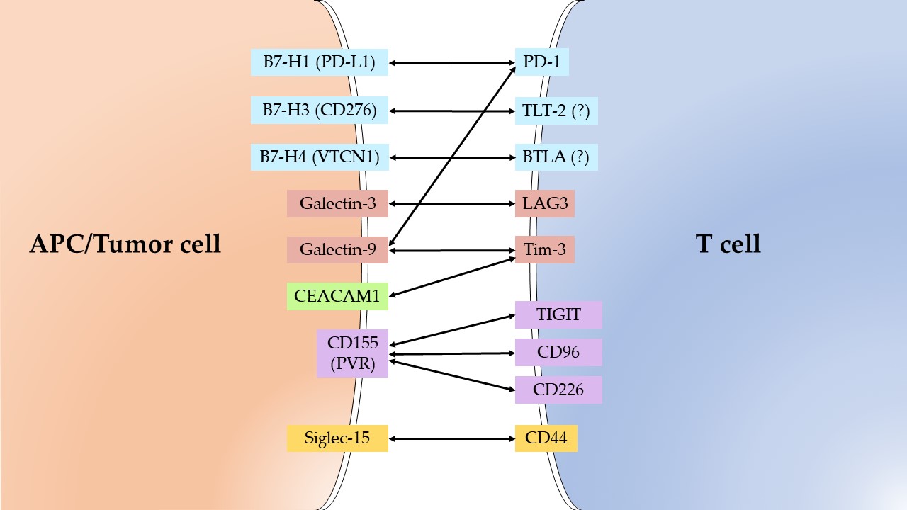
The analyzed IC ligands, expressed in tumors, and their receptors on T-cells are presented on Figure 1.

2.1. PD-L1

PD-L1 expression is used in clinical practice as a marker for predicting the response to therapy with ICI directed primarily to the PD-1/PD-L1 axis, including among patients with GC [1][2]. At the same time, the assessment of PD-L1 expression can serve as a marker for prognosis of survival during therapy with inhibitors of the IC PD-1/PD-L1 in the second and more lines: PD-L1 expression was associated with better OS (HR = 0.82, 95% CI = 0.67–0.99, p = 0.04) [9].
The results of all analyzed meta-analyses of PD-L1 expression in GC showed an association of its expression with a poor prognosis of disease development, while the HR value ranged from 1.39 to 1.74 [10][11][12][13][14].
PD-L1 expression is also associated with the clinical and pathological characteristics of GC. The most significant meta-analysis results were obtained for the association with lymph node metastasis (OR = 2.61, 95% CI = 1.78–3.84), TNM stage (OR = 2.28, 95% CI = 1.39–3.74, p = 0.006) [14], tumor size (OR = 1.87, 95% CI = 1.25–2.78, p = 0.002) [12], and venous invasion (p = 0.0003) [11].
PD-L1 expression for prognostic purposes can be measured not only in the tumor, but also in the blood serum of patients with GC. In Shigemori et al. study PD-L1 expression was measured in both tumor tissues and serum by ELISA. Multivariate analyses showed that both elevated tissue PD-L1 and serum sPD-L1 were independent prognostic factors for poor OS; the obtained HR values turned out to be close: (HR = 4.28, 95% CI = 1.43–12.8, p = 0.0094) and (HR = 11.2, 95% CI = 3.44–36.7, p = 0.0001), respectively [15]. Ito et al. reported, that PD-L1 expression measured in preoperative peripheral blood by qRT-PCR was an independent poor prognostic factor for OS (HR = 1.81, 95% CI = 1.15–2.78, p < 0.05) [16].
The results considered lead to the conclusion about PD-L1 expression as a candidate for prognostic markers of GC with moderate-risk, an important property of which is the possibility of minimally invasive use.

2.2. B7-H3

There are still few studies on B7-H3 expression in GC. The available results indicate an association of increased B7-H3 expression in tumor tissue with low OS (p = 0.003), as well as with TNM stage (p = 0.000), depth of infiltration (p = 0.001), and lymph node metastasis (p = 0.020) [17]. An increased B7-H3 expression in peripheral blood was associated with low OS in GC (HR = 1.56, 95% CI = 1.01–2.54, p = 0.046) [18].
The available data indicate that further studies of B7-H3 among patients with GC are promising for elucidating the possibility of using its expression as a prognostic marker.

2.3. B7-H4

In all publications found, increased expression of B7-H4 was associated with a poor prognosis of GC (OR = 1.63, 95% CI = 1.30–2.03), as it is reflected in the meta-analysis [19]. The most significant results on the association of B7-H4 expression in tumors with a poor prognosis of GC were obtained in studies by Jiang et al. (RR = 1.85, 95% CI = 1.15–2.96, p = 0.0087) and Arigami et al. (HR = 1.49, 95% CI = 1.03–2.17, p = 0.035) [20][21]. Similar results were obtained when measuring B7-H4 expression level in the blood of patients with GC (HR = 2.01, 95% CI = 1.08–5.03, p = 0.024) and in the serum of such patients (HR = 1.925, 95% CI = 1.033–3.857, p = 0.039) [22][23].
B7-H4 expression is also associated with certain clinical and pathological tumor characteristics. Thus, Jiang et al. study shown association with myometrial invasion (p = 0.004), lymph node metastasis (p < 0.0001), recurrence (p = 0.003) [20]. Arigami et al. reported that B7-H4 expression in blood of GC patients significantly correlated with depth of infiltration (p = 0.006), lymph node metastasis (p = 0.001), TNM stage (p < 0.001), lymphatic invasion (p < 0.001), venous invasion (p = 0.010) [22].
Therefore, B7-H4 expression is a very promising candidate in markers with moderate risk (HR = 1.5–2) for survival prognosis of GC patients.

2.4. Galectin-3

Meta-analysis of Gal-3 expression in GC showed that decreased Gal-3 expression was significantly associated with a poor prognosis (HR = 0.49, 95% CI = 0.36–0.67, p < 0.001), lymphatic vessel invasion (OR = 0.48, 95% CI = 0.26–0.89, p = 0.018), the poor TNM stage (OR = 0.47, 95% CI = 0.32–0.40, p < 0.001), greater depth of invasion (OR = 0.33, 95% CI = 0.21–0.51, p < 0.001) and the poor degree of differentiation (OR = 0.10, 95% CI = 0.04–0.25, p < 0.001) [24]. Among the individual studies, it is worth noting Okada et al. study, in which decreased Gal-3 expression was associated with a poor prognosis also (RR = 3.831, 95% CI = 1.574–9.329, p = 0.0031), the degree of differentiation (p < 0.0001), lymph node metastasis (p = 0.0495), lymphatic invasion (p = 0.0086) and TNM stage (p = 0.0433) [25]. Kim et al. reported that Gal-3 expression was associated with better OS (p = 0.006), lower depth of invasion (p < 0.001), absence of lymph node metastasis (p = 0.001), absence of distant metastasis (p = 0.004), better TNM stage (p < 0.001) and absence of lymphovascular invasion (p = 0.035) [26]. However, it should be noted that the level of Gal-3 expression in serum correlated with lymph node metastasis (p = 0.001) and distant metastasis (p < 0.001) [27].
Although most of the available studies on GC indicate the association of decreased expression of Gal-3 with a poor prognosis; however, they are few and there are conflicting results, including among data on other types of tumors. The current situation requires further research to understand the prospects of using the expression of Gal-3 as a prognostic marker.

2.5. Galectin-9

Among the studies of Gal-9 expression in GC, the most impressive results were shown by Choi et al.; in their study Gal-9 expression was associated with better OS (HR = 0.51, 95% CI = 0.35–0.76, p = 0.001), and the absence of Gal-9 expression was associated with greater depth of infiltration (p < 0.001), lymph node metastasis (p < 0.001), and TNM stage (p < 0.001) [28]. It is also worth noting the study of Jiang et al., who showed an association of increased Gal-9 expression with better OS (p = 0.002); in addition, decreased expression was associated with lymph-vascular invasion (p = 0.034), lymph node metastasis (p = 0.009), distant metastasis (p = 0.002) and TNM stage (p = 0.043) [29].
The situation with the relationship between the expression of Gal-9 and the survival of patients with GC is similar to that observed in the study of Gal-3. Some studies find better survival with increased expression of Gal-9, and there are also conflicting results in different studies (discussed in Section 1). Further studies of Gal-9 expression are needed to characterize the association with the prognosis of solid tumors, including GC.

2.6. IDO1

As evidenced by the results presented in the previous section, increased expression of IDO1 in many tumor types, including GC, is associated with worse OS and clinical characteristics indicating a poor prognosis. From the point of view of considering the expression of IDO1 as a biomarker, it should be noted that Liu et al. showed an association of IDO1 expression with a poor prognosis of GC (HR = 1.596, 95% CI = 1.156–2.204, p = 0.005), depth of infiltration (p = 0.045) and lymph node metastasis (p < 0.001) [30]. Similar results on the association of IDO1 expression with a poor prognosis of GC were obtained by Nishi et al. (HR = 2.75, 95% CI = 1.01–7.58, p < 0.05) [31].
So far, few studies have been devoted to the study of IDO1 expression in connection with the prognosis of GC. Nevertheless, in combination with the results for other types of cancer, the data obtained indicate that further work is promising to establish the parameters of the relationship between IDO1 expression and the prognosis of GC development. The available data indicate a moderate risk of poor prognosis with overexpression of IDO1.

2.7. CEACAM1,CD155 and Siglec-15

Data on the expression of CEACAM1 and CD155 in relation to survival, including among patients with GC, are contradictory. Therefore, at the moment, it is premature to consider their expression as candidates for biomarkers. There are still little data on Siglec-15. Further research is needed.

2.8. ADAM17

In all analyzed studies with a significant association of ADAM17 expression with survival, ADAM17 expression was associated with a poor prognosis of GC. So, in a meta-analysis by Ni et al. ADAM17 expression was associated with poor OS (HR = 2.04, 95% CI = 1.66–2.50), TNM stage (OR = 4.09, 95% CI = 1.85–9.04) and lymph node metastasis (OR = 3.08, 95% CI = 1.13–8.36) [32]. Significant HR values were obtained by Zhang et al.: in their study ADAM17 expression was associated with poor OS (HR = 5.87, 95% CI = 1.59–20.52, p = 0.008), degree of tumor differentiation (p = 0.006), depth of invasion (p < 0.0001), lymph node metastasis (p = 0.02), distant metastasis (p = 0.02) and TNM stage (p = 0.03) [33]. In Shou et al., HR values for the association of increased ADAM17 expression with poor OS were lower (HR = 2.067, 95% CI = 1.475–2.883, p = 0.000); however, high levels of significance of the relationship between expression and other clinical parameters of poor prognosis were observed: tumor size (p = 0.000), depth of invasion (p = 0.000), TNM stage (p = 0.000), lymph node metastasis (p = 0.000) and distant metastasis (p = 0.000) [34]. Li et al. obtained similar results: ADAM17 expression was associated with poor OS (HR = 2.239, 95% CI = 1.516–3.305, p < 0.001) and lymph node metastasis (OR = 2.161, 95% CI = 1.115–4.190, p = 0.022) [35]. Hence, an association of increased ADAM17 expression with reduced survival in GC patients has been identified, possibly with a higher than moderate risk. At the same time, the parameters of association with other clinical indicators of poor prognosis, such as metastasis, remain to be elucidated.
So, the considered members of the B7 family—B7-H1 (PD-L1), B7-H3, B7-H4, as well as IDO1 and ADAM17, can be recognized as candidates for prognosis markers for the survival of patients with GC. The risk of poor survival associated with their increased expression is moderate, except for ADAM17, which may be associated with a higher risk. The expression of the remaining analyzed ICs—Gal-3 and -9, CEACAM1, CD155 and Siglec-15—requires further in-depth study.

3. Conclusions

The published data on the relationship between the expression of several ICs—PD-L1, B7-H3, B7-H4, IDO1, Gal-3 and -9, CEACAM1, CD155, Siglec-15, and the ADAM17 immune response modulator—with the prognosis of cancer tumors, including GC, are analyzed. These data are compared with the results of clinical studies on their inhibition. Expression of PD-L1, B7-H3, B7-H4, IDO1, CD155, and ADAM17 is associated with the prognosis of tumors, including GC. Increased expression of the most studied ICs—PD-L1, B7-H3, and B7-H4—is associated with poor patient survival and other unfavorable clinical parameters, and their inhibition leads to clinically significant therapeutic results. The available data indicate that CD155 inhibition is promising as a target for immune therapy. The unsatisfactory results of clinical studies of several anti-IDO1 drugs turned out to be unexpected, in light of the confident relationship between IDO1 expression and clinical parameters, which indicates an insufficient understanding of the role of IDO1 in immune suppression and the need for further research in this direction. Expression of Gal-3 and -9, as well as CEACAM1 and Siglec-15, demonstrates a contradictory relationship with the development of solid tumors in different studies. The lack of satisfactory results from clinical studies of inhibitors of these ICs additionally indicates the complex nature of their functioning. Thus, according to the data of Yang et al., the resulting effect of Gal-9 may depend on the ratios of the three immune components, and in order to achieve a positive effect, the accumulation of Treg cells in the tumor space due to Gal-9 inhibition should be eliminated [36]. Accordingly, when studying the relationship between the expression of Gal-9 and the survival rate of patients with malignant tumors, one should, apparently, analyze the expression of other participants in the immune response to the tumor. This also applies to other ICs that do not show a reproducible relationship with clinical indicators. In general, on the combination of an IC expression association with clinical features and a response to their therapeutic inhibition, three IC groups can be distinguished. In the first group, the association with clinical features corresponds to the clinically significant therapeutic results (PD-L1, B7-H3, B7-H4). In the second group, the controversial association of expression with the development of tumors corresponds to unsatisfactory results of clinical studies of inhibitors of these ICs (Gal-3 and -9, CEACAM1). The third unexpected combination is a confident association of IDO1 expression with clinical features and unsatisfactory results of clinical studies. This case is the most mysterious for interpretation. It can be assumed that, in the IDO1 inhibition, a side reaction occurs similar to the accumulation of Treg cells in the TME under the inhibition of Gal-9, but that there are no multicomponent interactions, apparently inherent for the second group. In the first group, if such complications occur, it is not often.
Based on the available data, the considered members of the B7 family—B7-H1 (PD-L1), B7-H3, B7-H4, as well as IDO1 and ADAM17—can be recognized as candidates for prognosis markers for the survival of patients with GC.

References

  1. Doroshow, D.B.; Bhalla, S.; Beasley, M.B.; Sholl, L.M.; Kerr, K.M.; Gnjatic, S.; Wistuba, I.I.; Rimm, D.L.; Tsao, M.S.; Hirsch, F.R. PD-L1 as a biomarker of response to immune-checkpoint inhibitors. Nat. Rev. Clin. Oncol. 2021, 18, 345–362.
  2. Patel, S.P.; Kurzrock, R. PD-L1 Expression as a Predictive Biomarker in Cancer Immunotherapy. Mol. Cancer Ther. 2015, 14, 847–856.
  3. Bolandi, N.; Derakhshani, A.; Hemmat, N.; Baghbanzadeh, A.; Asadzadeh, Z.; Afrashteh Nour, M.; Brunetti, O.; Bernardini, R.; Silvestris, N.; Baradaran, B. The Positive and Negative Immunoregulatory Role of B7 Family: Promising Novel Targets in Gastric Cancer Treatment. Int. J. Mol. Sci. 2021, 22, 10719.
  4. Cao, Y.; Wang, X.; Jin, T.; Tian, Y.; Dai, C.; Widarma, C.; Song, R.; Xu, F. Immune checkpoint molecules in natural killer cells as potential targets for cancer immunotherapy. Signal Transduct. Target. Ther. 2020, 5, 250.
  5. Zhang, Y.; Zheng, J. Functions of Immune Checkpoint Molecules Beyond Immune Evasion. Adv. Exp. Med. Biol. 2020, 1248, 201–226.
  6. Qin, S.; Xu, L.; Yi, M.; Yu, S.; Wu, K.; Luo, S. Novel immune checkpoint targets: Moving beyond PD-1 and CTLA-4. Mol. Cancer 2019, 18, 155.
  7. Sung, H.; Ferlay, J.; Siegel, R.L.; Laversanne, M.; Soerjomataram, I.; Jemal, A.; Bray, F. Global Cancer Statistics 2020: GLOBOCAN Estimates of Incidence and Mortality Worldwide for 36 Cancers in 185 Countries. CA. Cancer J. Clin. 2021, 71, 209–249.
  8. Etemadi, A.; Safiri, S.; Sepanlou, S.G.; Ikuta, K.; Bisignano, C.; Shakeri, R.; Amani, M.; Fitzmaurice, C.; Nixon, M.; Abbasi, N.; et al. The global, regional, and national burden of stomach cancer in 195 countries, 1990–2017: A systematic analysis for the Global Burden of Disease study 2017. Lancet Gastroenterol. Hepatol. 2020, 5, 42–54.
  9. Roviello, G.; Corona, S.P.; D’Angelo, A.; Rosellini, P.; Nobili, S.; Mini, E. Immune Checkpoint Inhibitors in Pre-Treated Gastric Cancer Patients: Results from a Literature-Based Meta-Analysis. Int. J. Mol. Sci. 2020, 21, 448.
  10. Qiu, Z.; Du, Y. Clinicopathological and prognostic significance of programmed death ligant-1 expression in gastric cancer: A meta-analysis. J. Gastrointest. Oncol. 2021, 12, 112–120.
  11. Gu, L.; Chen, M.; Guo, D.; Zhu, H.; Zhang, W.; Pan, J.; Zhong, X.; Li, X.; Qian, H.; Wang, X. PD-L1 and gastric cancer prognosis: A systematic review and meta-analysis. PLoS ONE 2017, 12, e0182692.
  12. Zhang, M.; Dong, Y.; Liu, H.; Wang, Y.; Zhao, S.; Xuan, Q.; Wang, Y.; Zhang, Q. The clinicopathological and prognostic significance of PD-L1 expression in gastric cancer: A meta-analysis of 10 studies with 1,901 patients. Sci. Rep. 2016, 6, 37933.
  13. Feng, X.-S.; Wang, X.-S.; Wang, Y.-F.; Hu, X.-C.; Yan, J.-Q.; Wang, W.; Yang, R.-J.; Feng, Y.-Y.; Gao, S.-G.; Liu, Y.-X.; et al. Prognostic significance of PD-L1 expression in patients with gastric cancer in East Asia: A meta-analysis. Onco. Targets. Ther. 2016, 9, 2649.
  14. Xu, F.; Feng, G.; Zhao, H.; Liu, F.; Xu, L.; Wang, Q.; An, G. Clinicopathologic Significance and Prognostic Value of B7 Homolog 1 in Gastric Cancer: A Systematic Review and Meta-Analysis. Medicine 2015, 94, e1911.
  15. Shigemori, T.; Toiyama, Y.; Okugawa, Y.; Yamamoto, A.; Yin, C.; Narumi, A.; Ichikawa, T.; Ide, S.; Shimura, T.; Fujikawa, H.; et al. Soluble PD-L1 Expression in Circulation as a Predictive Marker for Recurrence and Prognosis in Gastric Cancer: Direct Comparison of the Clinical Burden Between Tissue and Serum PD-L1 Expression. Ann. Surg. Oncol. 2019, 26, 876–883.
  16. Ito, S.; Fukagawa, T.; Noda, M.; Hu, Q.; Nambara, S.; Shimizu, D.; Kuroda, Y.; Eguchi, H.; Masuda, T.; Sato, T.; et al. Prognostic Impact of Immune-Related Gene Expression in Preoperative Peripheral Blood from Gastric Cancer Patients. Ann. Surg. Oncol. 2018, 25, 3755–3763.
  17. Zhan, S.; Liu, Z.; Zhang, M.; Guo, T.; Quan, Q.; Huang, L.; Guo, L.; Cao, L.; Zhang, X. Overexpression of B7-H3 in α-SMA-Positive Fibroblasts Is Associated with Cancer Progression and Survival in Gastric Adenocarcinomas. Front. Oncol. 2020, 9, 1466.
  18. Arigami, T.; Uenosono, Y.; Hirata, M.; Yanagita, S.; Ishigami, S.; Natsugoe, S. B7-H3 expression in gastric cancer: A novel molecular blood marker for detecting circulating tumor cells. Cancer Sci. 2011, 102, 1019–1024.
  19. Cui, Y.; Li, Z. B7-H4 is Predictive of Poor Prognosis in Patients with Gastric Cancer. Med. Sci. Monit. 2016, 22, 4233–4237.
  20. Jiang, J.; Zhu, Y.; Wu, C.; Shen, Y.; Wei, W.; Chen, L.; Zheng, X.; Sun, J.; Lu, B.; Zhang, X. Tumor expression of B7-H4 predicts poor survival of patients suffering from gastric cancer. Cancer Immunol. Immunother. 2010, 59, 1707–1714.
  21. Arigami, T.; Uenosono, Y.; Ishigami, S.; Hagihara, T.; Haraguchi, N.; Natsugoe, S. Clinical significance of the B7-H4 coregulatory molecule as a novel prognostic marker in gastric cancer. World J. Surg. 2011, 35, 2051–2057.
  22. Arigami, T.; Uenosono, Y.; Hirata, M.; Hagihara, T.; Yanagita, S.; Ishigami, S.; Natsugoe, S. Expression of B7-H4 in blood of patients with gastric cancer predicts tumor progression and prognosis. J. Surg. Oncol. 2010, 102, 748–752.
  23. Shi, H.; Ji, M.; Wu, J.; Zhou, Q.; Li, X.; Li, Z.; Zheng, X.; Xu, B.; Zhao, W.; Wu, C.; et al. Serum B7-H4 expression is a significant prognostic indicator for patients with gastric cancer. World J. Surg. Oncol. 2014, 12, 188.
  24. Long, B.; Yu, Z.; Zhou, H.; Ma, Z.; Ren, Y.; Zhan, H.; Li, L.; Cao, H.; Jiao, Z. Clinical characteristics and prognostic significance of galectins for patients with gastric cancer: A meta-analysis. Int. J. Surg. 2018, 56, 242–249.
  25. Okada, K.; Shimura, T.; Suehiro, T.; Mochiki, E.; Kuwano, H. Reduced galectin-3 expression is an indicator of unfavorable prognosis in gastric cancer. Anticancer Res. 2006, 26, 1369–1376.
  26. Kim, S.J.; Kim, D.C.; Kim, M.C.; Jung, G.J.; Kim, K.H.; Jang, J.S.; Kwon, H.C.; Kim, Y.M.; Jeong, J.S. Fascin expression is related to poor survival in gastric cancer. Pathol. Int. 2012, 62, 777–784.
  27. Li, Y. Serum Galectin-3 as a Potential Marker for Gastric Cancer. Med. Sci. Monit. 2015, 21, 755–760.
  28. Choi, S.I.; Seo, K.W.; Kook, M.C.; Kim, C.G.; Kim, Y.W.; Cho, S.J. Prognostic value of tumoral expression of galectin-9 in gastric cancer. Turkish J. Gastroenterol. 2017, 28, 166–170.
  29. Jiang, J.; Jin, M.-S.; Kong, F.; Cao, D.; Ma, H.-X.; Jia, Z.; Wang, Y.-P.; Suo, J.; Cao, X. Decreased galectin-9 and increased Tim-3 expression are related to poor prognosis in gastric cancer. PLoS ONE 2013, 8, e81799.
  30. Liu, H.; Shen, Z.; Wang, Z.; Wang, X.; Zhang, H.; Qin, J.; Qin, X.; Xu, J.; Sun, Y. Increased expression of IDO associates with poor postoperative clinical outcome of patients with gastric adenocarcinoma. Sci. Rep. 2016, 6, 21319.
  31. Nishi, M.; Yoshikawa, K.; Higashijima, J.; Tokunaga, T.; Kashihara, H.; Takasu, C.; Ishikawa, D.; Wada, Y.; Shimada, M. The Impact of Indoleamine 2,3-dioxygenase (IDO) Expression on Stage III Gastric Cancer. Anticancer Res. 2018, 38, 3387–3392.
  32. Ni, P.; Yu, M.; Zhang, R.; He, M.; Wang, H.; Chen, S.; Duan, G. Prognostic Significance of ADAM17 for Gastric Cancer Survival: A Meta-Analysis. Medicina 2020, 56, 322.
  33. Zhang, T.; Zhu, W.; Huang, M.; Fan, R.; Chen, X. Prognostic value of ADAM17 in human gastric cancer. Med. Oncol. 2012, 29, 2684–2690.
  34. Shou, Z.-X.; Jin, X.; Zhao, Z.-S. Upregulated Expression of ADAM17 Is a Prognostic Marker for Patients with Gastric Cancer. Ann. Surg. 2012, 256, 1014–1022.
  35. Li, W.; Wang, D.; Sun, X.; Zhang, Y.; Wang, L.; Suo, J. ADAM17 promotes lymph node metastasis in gastric cancer via activation of the Notch and Wnt signaling pathways. Int. J. Mol. Med. 2019, 43, 914–926.
  36. Yang, R.; Sun, L.; Li, C.-F.; Wang, Y.-H.; Yao, J.; Li, H.; Yan, M.; Chang, W.-C.; Hsu, J.-M.; Cha, J.-H.; et al. Galectin-9 interacts with PD-1 and TIM-3 to regulate T cell death and is a target for cancer immunotherapy. Nat. Commun. 2021, 12, 832.
More
Information
Subjects: Oncology
Contributor MDPI registered users' name will be linked to their SciProfiles pages. To register with us, please refer to https://encyclopedia.pub/register :
View Times: 431
Revisions: 3 times (View History)
Update Date: 29 Mar 2022
1000/1000
Video Production Service