Submitted Successfully!
To reward your contribution, here is a gift for you: A free trial for our video production service.
Thank you for your contribution! You can also upload a video entry or images related to this topic.
Version Summary Created by Modification Content Size Created at Operation
1 + 3384 word(s) 3384 2021-12-22 07:46:17 |
2 format correct Meta information modification 3384 2021-12-23 02:32:46 |

Video Upload Options

Do you have a full video?

Confirm

Are you sure to Delete?
Cite
If you have any further questions, please contact Encyclopedia Editorial Office.
Zhang, W. Gliotoxins. Encyclopedia. Available online: https://encyclopedia.pub/entry/17440 (accessed on 27 July 2024).
Zhang W. Gliotoxins. Encyclopedia. Available at: https://encyclopedia.pub/entry/17440. Accessed July 27, 2024.
Zhang, Weimin. "Gliotoxins" Encyclopedia, https://encyclopedia.pub/entry/17440 (accessed July 27, 2024).
Zhang, W. (2021, December 22). Gliotoxins. In Encyclopedia. https://encyclopedia.pub/entry/17440
Zhang, Weimin. "Gliotoxins." Encyclopedia. Web. 22 December, 2021.
Gliotoxins
Edit

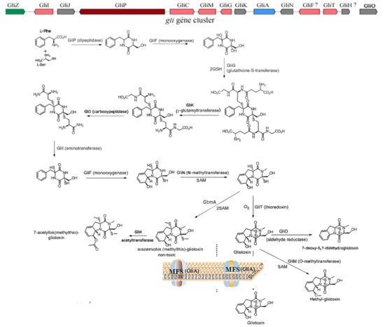
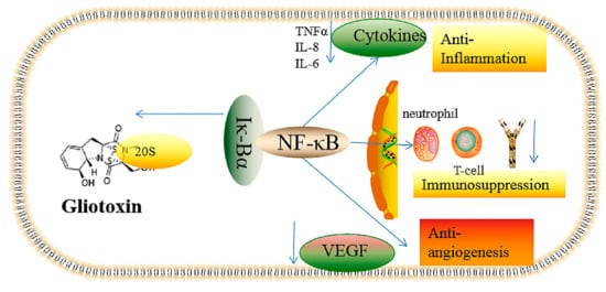
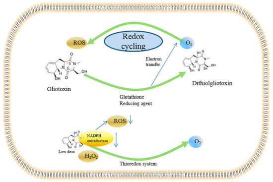
Gliotoxin is a kind of epipolythiodioxopiperazine derived from different fungi that is characterized by a disulfide bridge. Gliotoxins can be biosynthesized by a gli gene cluster and regulated by a positive GliZ regulator. Gliotoxins show cytotoxic effects via the suppression the function of macrophage immune function, inflammation, antiangiogenesis, DNA damage by ROS production, peroxide damage by the inhibition of various enzymes, and apoptosis through different signal pathways. In the other hand, gliotoxins can also be beneficial with different doses. Low doses of gliotoxin can be used as an antioxidant, in the diagnosis and treatment of HIV, and as an anti-tumor agent in the future. Gliotoxins have also been used in the control of plant pathogens, including Pythium ultimum and Sclerotinia sclerotiorum.

gliotoxin toxic mechanism NF-κB detoxification biosynthetic strategies

1. The Biosynthesis of Gliotoxin

Gliotoxin is the first isolated ETP with the most simple structure; more than 100 kinds of gliotoxin-like compounds have been excavated, such as chaetocin, chetomin, and sporidesmin. Gliotoxin was firstly isolated in Trichoderma virens [1] and then was found in other fungi, including A. fumigatus [2]Aspergillus nigerAspergillus terreusEurotium chevalieri, and other Penicillium and Acremonium species [3]. The biosynthetic mechanism of gliotoxin mediated by the gli gene cluster in A. fumigatus has been elucidated [4][5]. The non-ribosomal peptide synthetase encoded by the gliP gene catalyzes the diketopiperazine scaffold formed by the non-ribosomal peptide synthetase (GliP); then, the hydroxyl group is added to the amino acid by cytochrome P450 monooxygenase (GliC), followed by the sulfurization catalyzed by glutathione S-transferase (GliG). The sulfur bond is important for the toxicity of gliotoxins. Next, the -glutamyl moieties are cut off by the catalyzation of GliK and GliJ, and a notorious epidithiol moiety is then formed, intermediated by gliI-encoding amino transferase, the N-methyltransferase (GliN) encoded by gliN and/or O-methyltransferase GliM encoded by gliM, which play the role of freestanding amide to accelerate amide methylation and confer stability on the ETP. Additionally, gliotoxin is generated by the formation of a disulfide bond catalyzed by oxidoreductase (GliT). GtmA, which is not in the gliotoxin biosynthesis gene cluster, can intermediate the cleavage of the disulfide bond in the gliotoxin to form bisdethiobis(methylthio)-gliotoxin; moreover, GliA is responsible for pumping out the gliotoxin to the outer membrane; therefore, both GtmA and GliA can attenuate the cytotoxicity of gliotoxin to the host, thus proving an effective strategy to alleviate the hazard and cytotoxicity of gliotoxin to different hosts [5]. The biosynthetic pathway for the biosynthesis of gliotoxin and its derivatives is proposed in Figure 1. Moreover, GliZ has been demonstrated as a positive regulator for the biosynthesis of gliotoxins; the disruption of gliZ would abolish the production and gliotoxin [6]. Moreover, LaeA [7] and VeA [8] global regulators have also been demonstrated to play an important role in the generation of gliotoxins in A. fumigutas. In addition, the in vitro function of genes related to gliotoxin biosynthesis, including gliZgliGgliI, and gliO in deep-sea fungus Dichotomyces cejpii FS110 [9] and gliotoxin biosynthesis-related genes gliKgliM, and gliT in deep-sea fungus Geosmithia pallida FS140 [10], have been characterized by their biochemical reaction. Interestingly, GliK was reported to exert the function of gliotoxin acetylation, thus facilitating the generation of gliotoxin derivates in G. pallida FS140. The function of GliF (monooxygenase) and GliH in the gli gene cluster has remained obscure until now, which needs further investigation.
Figure 1. The proposed biosynthetic pathway of gliotoxin and its derivatives [5]. The gli gene cluster depicts the biosynhetic pathway of gliotoxin in A. fumigutas, the coding protein for each gene was annoated in the biosynthetic pathway of gliotoxin and thereof derivatives. The SAM refers to S-adenosylmethionine, MFS refers to major facilitator superfamily. GtmA refers to S-methyltransferase.
Gliotoxin and its derivatives have been excavated from deep-sea fungus D. cejpii FS110 [9] and G. pallida FS140 [10] in our group. Additionally, epipolythiodioxopiperazine derivatives of gliotoxin with the dithiol group showed weaker cytotoxicity compared with gliotoxin, and gliotoxin derivates of the methylthio group showed much weaker cytotoxicity than that of dithiol gliotoxin. Furthermore, the depletion of the methythio group in gliotoxin derivative leads to the loss of cytotoxicity, suggesting the important role of the disulfide bridge and thiol group in the biological activity of gliotoxin and its derivatives.

2. The Underlying Toxic Mechanism of Gliotoxin

Gliotoxin can induce cytotoxicities via different mechanisms, including the inhibition of angiogenesis, immunosuppressive activity, the inducement of cell apoptosis, the suppression of the activity of protease, the production of ROS and the inhibition of the enzymatic activity of peroxidases, the imbalance of oxireductase reaction, the induction of DNA damage, and the repression of specific genes’ expression, including NF-κB factor [11].

2.1. The Inhibition of Angiogenesis

Previous studies have demonstrated that gliotoxin can inhibit angiogenesis in humans with the symptom of invasive aspergillosis, which was intermediated by the gliotoxin produced by A. fumigatus [12]. Meanwhile, invasive aspergillosis is the most common infected fungal disease that causes high mortality, especially for immune deficiency patients, because of the deficiency in the approaches for diagnosis and treatment [13]. Additionally, the incidence rate of invasive aspergillus is increasing year by year due to the wider application of organ transplants and antibiotics [13]. As a consequence, it is important to elucidate the mechanism of aspergillosis mediated by gliotoxin. Gliotoxin can repress angiogenesis in the lung of mice and patients suffering from aspergillosis; furthermore, A. fumigatus could impede capillary tube formation in a murine model of cutaneous infection via the generation of secondary metabolites, especially gliotoxins [14]. Additionally, pure gliotoxin can also suppress angiogenesis and endothelial cell migration in vitro [12][15]. The deletion of gliP in the biosynthetic gene cluster and LaeA global transcriptional regulator can significantly attenuate the angiogenic effect caused by A. fumigatus [12][16], thus further supporting the function of angiogenesis caused by gliotoxin [17]. In addition, the immune response can also be inhibited via the antiangiogenic effect mediated by gliotoxin, thus further deteriorating the symptom of invasive aspergillus.
The proangiogenic and antianogentic effects caused by A. fumigatus has been investigated. The antiangiogenesis effect of gliotoxin produced by A. fumigatus was exerted via the reduction of the generation of ROS and, thereby, the suppression of NF-κB [18]. Furthermore, gliotoxin can also repress the proliferation of polymorphonuclear leucocytes [19]. Moreover, gliotoxin can suppress the generation of NF-κB, thus blocking the production of cytokines and inflammatory factors to reduce the angiogenic effect [20]. To compensate the antigiogenic effect, thus protecting the host, the infection with A. fumigatus triggers proangiogenic effects, including the production of proinflammatory factors such as tumor necrosis factor alpha (TNFα), hypoxia-inducible factor 1-alpha (HIF1α), and interleukin (IL-8), thus recruiting the polymorphonuclear leucocytes (PMNLs) and leading to the burst of ROS, including H2O2. The gene ratio of ROS can induce the upregulation of NF-κB, thus elevating the expression of vascular endothelial growth factor and other proangiogenic factors to compensate the antanginogenic effect caused by A. fumigatus [12][21]. Moreover, gliotoxin can abrogate the angiogensis induced by H2O2 in low concentrations, and gliotoxin can completely inhibit the angiogenic effect in immunosuppressive mice treated with cyclophosphamide via the abolishment of the production of ROS, which is mediated by gliotoxin-catalyzed reduction by the assistance of NAPDH [22]. Therefore, these studies also provide strategies for the prevention of angiogenic effects of invasive aspergillus via the upregulation of proangiogenic mediators, including NF-κB, cytokines, and vascular endothelial growth factor (VGEFs). Different concentrations of gliotoxins can induce different proangiogenic and antiangiogenic effects; thus, the angiogenic effect can be balanced via the control of gliotoxin concentrations.

2.2. Immunosuppressive Activity

Gliotoxin has been reported to induce immunosuppressive activity via cell apoptosis mediated by the repression of phagocytosis and macrophages [23] as well as the suppression of the proliferation of giant cells and T-cells [23]. Moreover, gliotoxin can further deteriorate the diseases caused by fungi infection via the inhibition of the activities of immune-related cells [23][24]. The amounts of immune cells, including leucocyte, lymphocyte, and Langerhans cells, significantly decreased after the treatment with gliotoxins in adult camels for 7 days. Furthermore, gliotoxin from A. fumigatus abolish the generation of oleukotriene B4 through the suppression of leukotriene A4 hydrolase, thus exerting the function of immunosuppression [25]. In addition, the knockout of gliP and gliZ genes attenuated the toxicity of gliotoxin towards nonneutrophil cells in mice [26]; 50 nM of gliotoxin can exert immunosuppressive activity via the suppression of the phagocytosis of astrocytes [14][27], which led us to exploit low-dose gliotoxins as immunosuppressive agents during an organ-transplant operation, thus reducing immunological rejection in organ transplant patients.
The immunosuppressive mechanism of gliotoxin has been widely investigated. It is well known that gliotoxin can inhibit the activation of NF-κB mediated by the repression of IκBα degradation caused by the chymotrypsin-like activity of the 20S proteasome [28]. The disulfide of glitoxin can target the protease, which can be reversed by reducing agent dithiothreitol, which can be reduced from gliotoxin to dithiol gliotoxin. Low doses of gliotoxin, with a concentration of 1 μM, exhibited 63% inhibition of the LLVY-amc peptide-hydrolyzing activity mediated by the 20S proteasome, suggesting the high immunosuppressive efficiency of gliotoxin [28].
NF-κB is a kind of inducible transcriptional factor; some examples are NF-κB1 (p50), NF-κB2 (p52), RelA, RelB, and c-Rel. A typical active NF-κB is the P50-P65 dimer that can bind DNA; thus, activating NF-κB will modulate inflammation and innate and adaptive immune system reactions. The IκBα protein can inhibit the activity of NF-κB in the cytoplasm via binding with NF-κB, which can be activated by the degradation of the IκBα protein, mediated by the 20S protease, and by the binding of the DNA in the nucleus [29][30][31]. Glitoxin can stabilize the expression level of IκBα via the inhibition of the 20S protease, blocking the activation of NF-κB and thus exert the function of immunosuppression; the disulfide bond in gliotoxin is essential for its immunorepression activity [28]. In consequence, the disruption of gliP can abolish the immunosuppression function of A. fumigatus via the suppression of gliotoxin production [32]. Furthermore, gliotoxin can assist A. fumigatus in achieving immune escape from the phagocytosis of macrophages by targeting the metabolism of phosphatidylinositol 3,4,5-trisphosphate [PtdIns(3,4,5)P3] [33]; the gliotoxin can promote the immune escape of A. fumigatus by decreasing the content of intracellular lipid acyl inositol 3,4,5-trisphosphate [33], thus leading to the function deficiency of the integrin and actin backbone and preventing cell membrane protrusion extension [34], further impairing the function of macrophages. Furthermore, it was reported that gliotoxin contributed to the invasion of A. fumigatus spores into cells via the activation of phospholipase D in lung epithelial cells and by inducing actin backbone rearrangement [35][36], but the specific regulatory mechanism remains obscure.

2.3. Inflammatory and Anti-Inflammatory Effects

Gliotoxin can also initiate the production of inflammatory factors, thus eliciting further cytotoxicity. Gliotoxin incubation with A459 cells can induce the secretion of proinflammatory cytokines, including IL-4, IL-8, and IL-10, whereas the upregulation of IL-6 and IL-8 cytokines was observed in gliotoxin-treated L132 cells, with only IL-8 upregulation observed in A459 and L132 cells treated with fumagillin [24][37]. In addition, the expression of a few cytokines, such as IL-1β, IL-17, interferon-gamma (IFN-γ), TNF-α, and granulocyte macrophage-colony stimulating factor (GM-CSF), was downregulated in the two cells treated with gliotoxin and fumagillin, suggesting neutrophil-mediated tissue damage after A. fumigutas infection [37]. The results also suggest a stronger inflammation-eliciting effect by gliotoxin compared with fumagillin. Gliotoxin was administrated to female C57BL/6 mice immunized with myelin oligodendrocyte glycoprotein with a dose of 1 mg/kg, which can aggravate the symptom of neuroinflammation via the upregulation of inflammatory genes, including T-box transcription factor (TBX21), inducible nitric oxide synthase (iNOS), arginase 1 (ARG1), and cytokines such as IFN-γ, IL-17, and IL-2 [38].
In contrast, in vivo anti-inflammatory activity was also investigated. Herfarth et al. reported that gliotoxin can suppress intestinal inflammation in dextran sulfate sodium (DSS)-induced colitis mediated by NF-κB activation [39]. The expression of cytokines, including TNF-α, was downregulated in RAW-264.7 mouse macrophage-like cells; meanwhile, TNF-α and IL-1α mRNA expressions were also significantly suppressed in DSS-induced colitis mice by gliotoxin treatment for 8 days, thus leading to the alleviation of the symptom of colonic inflammation in mice [39]. Furthermore, the gliotoxin treatment also reduced the DNA-binding activity of NF-κB, thus impeding the inflammation mediated by NF-κB activation in the colon [20]. In addition, gliotoxin can ameliorate trinitrobenzene sulfonic acid (TNBS)-induced mouse colitis via the suppression of the expression levels of TNF-, IL-1, and intercellular adhesion molecule-1 (ICAM-1) proteins as well as the upregulation of heme oxygenase-1, thus indicating the potential application of gliotoxins in the clinical treatment of Crohn’s disease [40] via the induction of hemooxygenase-1, which can oxidize low-density lipoproteins with the potential to form chemo-attractants, thus alleviating the inflammation [41].
In summary, gliotoxin’s toxic effects, including the antiangiogenesis and inflammatory-related effects mediated by NF-κB activity and redox cycling, are summarized in Figure 1. Gliotoxin can target the 20S proteasome, which is responsible for the degradation of the IK-Bα protein that specifically binds to NF-κB; thus, gliotoxin can suppress NF-κB activity. NF-κB is closely associated with the function of angiogenesis, immune response, and cell proliferation; thus, gliotoxin treatment can induce the effects of immunosuppression, antiangiogenesis, and anti-inflammation (Figure 2).
Figure 2. The cytotoxic effects, including immunosuppression, antiangiogenesis, and anti-inflammation, of gliotoxin mediated by NF-κB. The arrow to the left indicates that gliotoxin can detach 20S protease from Iκ-Bα, thus inhibiting the activation of NF-κB. The downward arrow indicates the down-regulation caused by the inactivation of NF-κB mediated by gliotoxin.

2.4. Inducing the Production of ROS and the Inhibition of Peroxidase

Gliotoxin can induce the production of reactive oxygen species (ROS) via intracellular redox cycling [42], thus resulting in damage from the peroxidation of macromolecules and biofilms, mediated by oxidative stress and the abrogation of antioxidation. In human neutrophils, reduced gliotoxin acts as a generator of superoxide by inhibiting NADPH oxidase [43]. Alternatively, gliotoxin can also served as an antioxidant. Reduced nicotinamide adenine dinucleotid phosphate (NADPH) peroxidase can only produce low contents of ROS to scavenge pathogenic bacteria and fungi, thus protecting the hosts. Meanwhile, Tsunawaki et al. reported that gliotoxin can inhibit the enzymatic activity of NADPH peroxidase with a concentration of higher than 1 μg/mL, thus causing damage to the host, whereas the NADPH peroxidase in neutrophils was not inhibited by the treatment with this dose of gliotoxin. Thus, different doses of gliotoxin induce different oxidative damage and different physiological effects; 30–100 ng/mL gliotoxin can inhibit the phagocytosis of zymosan without affecting ROS production. Gliotoxin can also lead to the reshaping of the actin cytoskeleton [44], thus contributing to cell shrinkage and filopodia disappearance, which can be reversed by Cyclic Adenosine monophosphate (cAMP) antagonist Rp-cAMP; however, gliotoxin-induced phagocytosis can not be inversed by cAMP [44].
The detailed mechanism of the gliotoxin-induced mechanism mediated by ROS has been investigated. Bernardo et al. described a redox-uptake system mediated by gliotoxin [45]: intracellular gliotoxin can be reduced to dithiol gliotoxin by a cellular reducing agent, thus releasing electrons to cellular O2 to form O2-mediated gliotoxin, which can yield ROS. Furthermore, dithiol gliotoxin can also be oxidized into gliotoxin by cellular O2 in turn, thereby resulting in the accumulation of ROS via the redox-uptake system mediated by gliotoxin. Thus, gliotoxin can cause cell peroxide damage mediated by the redox-uptake system in a dose-dependent manner.
On the other hand, gliotoxin can also be explored as an antioxidant. Gliotoxins exhibited the ability to reduce H2O2 to H2O in a thioredoxin redox system composed of thioredoxin, thioredoxin reductase, and NADPH [46][47], whereas gliotoxin did not show H2O2-reducing activity in a glutathione (GSH) / glutathione disulfide (GSSG) reductase system. The H2O2-reducing activity in the yeast Trx system depends on the concentrations of gliotoxin and H2O2, which was applicable to the rat Trx system [47], suggesting gliotoxin as a potent antioxidant for mammalian cells. The in vitro H2O2-reducing activity of gliotoxin in Hela cells was also investigated; the results indicated that the H2O2 level was significantly decreased by the addition of gliotoxin, with concentrations ranging from 40–160 nmol, in a dose-dependent manner in Hela cells, which was demonstrated by 2′,7′-dichlorofluorescein diacetate (DCFH-DA) probes [47]. In addition, gliotoxin can target NADPH oxireductase to prevent the onset of O2- generation in human neutrophils [48] and lead to the release of cytochrome c through a reaction with intracellular reductants in an oxygen-dependent manner; the prevention of redox cycling facilitated the damage of gliotoxin to NADPH oxireductase with IC50 of 9 nM [49]. Furthermore, the detailed targeting of gliotoxin to the NADPH oxidase system was investigated by a cell-free activation assay with NADPH oxidase components. The ROS generation ability of the membrane was reduced by 40% after gliotoxin treatment [19][49][50], and the in vitro addition of gliotoxin to flavocytochrome b558 and cytosolic components also exhibited an inhibition rate of 50% with the concentration of 3.3 μM [50], suggesting that flavocytochrome b558 is the target of gliotoxin. Moreover, the cell surface of alkaline phosphatase activity, which is a marker enzyme of oxidant-producing intracellular compartments (OPICs), is also suppressed by gliotoxin in human neutrophils stimulated with phorbol myristate acetate (PMA) as well as the repression of NADPH oxidase [51], thus contributing to the inhibition of superoxide production in PMA-treated human neutrophils.
Moreover, the block of oxidative stress by gliotoxin would impede the proliferation of human umbilical vein endothelial cells in a dose-dependent manner with the IC50 of gliotoxin of 250 nM [46][52], which was attributed to the fact that oxidative stress can trigger angiogenesis. Nevertheless, gliotoxin exhibited cytotoxicity to human umbilical vein endothelial cells (HUVECs) with concentrations higher than 500 nM [46][52], whereas there was no obvious cytotoxicity with concentrations lower than 300 nM, which is consistent with a previous study that found that gliotoxin induces apoptosis in activated human hepatic stellate cells with concentrations ranged from 0.3 to 7.5 μM [53]. To further elucidate the role of H2O2 and gliotoxin in angiogenesis, 100 nM H2O2 was added to induce the sprout of endothelial cells and the formation of thick tubes, which were inhibited by the addition of gliotoxin with concentrations of 125 to 250 nM [52]. In addition, gliotoxin potently inhibited the H2O2-stimulated invasion of HUVECs to the control level at 125 nM [52]. Thus, gliotoxin can exert the function of antiangiogenesis via the regulation of the intracellular redox state. The thioredoxin-SH group can reduce the gliotoxin to dithiol gliotoxin while leading to the reduction of H2O2 to H2O [46][54][55], thereby alleviating the angiogenic and invasive effects mediated by H2O2. Therefore, low doses of gliotoxin show great potential to be developed as an antioxidant to encounter the peroxide damage caused by H2O2.
Taken together, the ROS-inducing effects mediated by the redox cycling system caused by gliotoxin are summarized in Figure 3. Gliotoxin can be reduced to dithiol gliotoxin by in vivo reducing agents, including dithiothreitol (DTT), glutathione, and reducing enzymes, thus providing electrons to oxygen atoms and, thereby, producing intracellular ROS. Excessive ROS can cause peroxide damage and single- and double-strand breaks, causing great damage to the human body. In contrast, gliotoxin can be served as an antioxidant in the presence of the thioredoxin redox system through the inhibition of NADPH oxidase. Moreover, both gliotoxin and ditholgliotoxin are cytotoxic; however, gliotoxin is more toxic, probably due to the existence of the disulfide bridge, which can bind to many physiological-related proteins.
Figure 3. Gliotoxin-mediated redox cycling system and the antioxidant effect. The downward arrow indicates the ROS content was decreased by low dose of gliotoxin via the inhibition of NADPH oxireducatase activity in the presence of thioredox system.

2.5. Genotoxicity of Gliotoxin

Gliotoxin can also cause genotoxicity mediated by DNA damage in vitro and in vivo. Ditholgliotoxin can cause single- and double-stranded breaks in the presence of DNA and Fe3+, which was determined by neutral agarose gel electrophoresis [56][57]. Gliotoxin did not induce single- and double-stranded breaks except by the addition of reducing agents, including glutathione, DTT, and pyridine reducing enzymes [57], suggesting the genotoxicity was probably mediated by ditholgliotoxin and this DNA damage effect can be abrogated by metal chelator and catalase. This supports that the DNA damage effect was mediated by the ROS produced during the redox cycling process to form hydroxylated and other altered DNA products, which was determined by 32P DNA radiolabelling and two-dimensional thin-layer chromatography [58].
DNA damage was observed in mouse RAW264.7 macrophages after gliotoxin treatment for 2 h in plain medium in a single cell electrophoresis assay [57][59][60][61]; however, a clear, dose-related increase in sister-chromatid exchange was not observed in Chinese hamster ovary (CHO) cells [62]. Gliotoxin can also cause mitochondrial damage, thus producing ROS to induce DNA damage and causing toxicity to lung epithelial cells A459 and L132; this is stronger than another toxin fumagillin produced by A. fumigatus [37]. It is also speculated that gliotoxin can induce lung epithelial cells damage mediated by endoplasmic reticulum (ER) stress due to the observation of the accumulation of unfolded proteins in the lumen of the ER. Additionally, the cytotoxicity of gliotoxin towards different organ-derived cells is as follows: renal epithelial cells > type II epithelial cells > hepatocytes > normal lung epithelial cells [37], indicating that gliotoxin has selective cytotoxicity towards organ-derived cells to some extent.

References

  1. Singh, S.; Dureja, P.; Tanwar, R.; Singh, A. Production and antifungal activity of secondary metabolites of Trichoderma virens. Pestic. Res. J. 2005, 17, 26–29.
  2. Kupfahl, C.; Michalka, A.; Lass-Flörl, C.; Fischer, G.; Haase, G.; Ruppert, T.; Geginat, G.; Hof, H. Gliotoxin production by clinical and environmental Aspergillus fumigatus strains. Int. J. Med. Microbiol. Suppl. 2008, 298, 319–327.
  3. Lewis, R.E.; Wiederhold, N.P.; Lionakis, M.S.; Prince, R.A.; Kontoyiannis, D.P. Frequency and Species Distribution of Gliotoxin-Producing Aspergillus Isolates Recovered from Patients at a Tertiary-Care Cancer Center. J. Clin. Microbiol. 2005, 43, 6120–6122.
  4. Scharf, D.H.; Heinekamp, T.; Remme, N.; Hortschansky, P.; Brakhage, A.A.; Hertweck, C. Biosynthesis and function of gliotoxin in Aspergillus fumigatus. Appl. Microbiol. Biotechnol. 2011, 93, 467–472.
  5. Dolan, S.K.; O’Keeffe, G.; Jones, G.W.; Doyle, S. Resistance is not futile: Gliotoxin biosynthesis, functionality and utility. Trends Microbiol. 2015, 23, 419–428.
  6. Bok, J.W.; Chung, D.; Balajee, S.A.; Marr, K.A.; Andes, D.; Nielsen, K.F.; Frisvad, J.C.; Kirby, K.A.; Keller, N.P. GliZ, a tran-scriptional regulator of gliotoxin biosynthesis, contributes to Aspergillus fumigatus virulence. Infect. Immun. 2006, 74, 6761–6768.
  7. Bok, J.W.; Keller, N.P. LaeA, a Regulator of Secondary Metabolism in Aspergillus spp. Eukaryot. Cell 2004, 3, 527–535.
  8. Dhingra, S.; Andes, D.; Calvo, A.M. VeA Regulates Conidiation, Gliotoxin Production, and Protease Activity in the Opportunistic Human Pathogen Aspergillus fumigatus. Eukaryot. Cell 2012, 11, 1531–1543.
  9. Ye, W.; Zhang, W.; Liu, T.; Huang, Z.; Zhu, M.; Chen, Y.; Li, H.; Li, S. De Novo Transcriptome Sequencing of the Deep-Sea-Derived Fungus Dichotomomyces cejpii and Analysis of Gliotoxin Biosynthesis Genes. Int. J. Mol. Sci. 2018, 19, 1910.
  10. Ye, W.; Li, S.; Liu, S.; Kong, Y.; Zhang, W.; Liu, S.; Liu, T.; Zhang, W. Characterization of novel gliotoxin biosynthesis-related genes from deep-sea-derived fungus Geosmithia pallida FS140. Biochimie 2021, 191, 1–10.
  11. Scharf, D.H.; Brakhage, A.A.; Mukherjee, P.K. Gliotoxin–bane or boon? Environ. Microbiol. 2016, 18, 1096–1109.
  12. Ben-Ami, R.; Lewis, R.E.; Leventakos, K.; Kontoyiannis, D.P. Aspergillus fumigatus inhibits angiogenesis through the production of gliotoxin and other secondary metabolites. Blood 2009, 114, 5393–5399.
  13. Erjavec, Z.; Kluin-Nelemans, H.; Verweij, P. Trends in invasive fungal infections, with emphasis on invasive aspergillosis. Clin. Microbiol. Infect. 2009, 15, 625–633.
  14. Ben-Ami, R.; Lewis, R.E.; Kontoyiannis, D.P. Enemy of the (immunosuppressed) state: An update on the pathogenesis of Aspergillus fumigatus infection. Br. J. Haematol. 2010, 150, 406–417.
  15. Lee, H.J.; Lee, J.H.; Hwang, B.Y.; Kim, H.S.; Lee, J.J. Anti-angiogenic activities of gliotoxin and its methylthio-derivative, fungal metabolites. Arch. Pharm. Res. 2001, 24, 397–401.
  16. Ben-Ami, R. Angiogenesis at the mold–host interface: A potential key to understanding and treating invasive aspergillosis. Future Microbiol. 2013, 8, 1453–1462.
  17. Dikmen, M.; Cantürk, Z.; Engur, S.; Kaya Tilki, E. Inhibitory effects of secondary metabolites of halotolerant Pythium ultimum terreus on angiogenesis. Biomed. Res. 2017, 28, 8.
  18. Maulik, N. Redox signaling of angiogenesis. Antioxid. Redox Signal. 2002, 4, 805–815.
  19. Orciuolo, E.; Stanzani, M.; Canestraro, M.; Galimberti, S.; Carulli, G.; Lewis, R.; Petrini, M.; Komanduri, K.V. Effects of As-pergillus fumigatus gliotoxin and methylprednisolone on human neutrophils: Implications for the pathogenesis of invasive as-pergillosis. J. Leukoc. Biol. 2007, 82, 839–848.
  20. Gupta, S.C.; Sundaram, C.; Reuter, S.; Aggarwal, B.B. Inhibiting NF-κB activation by small molecules as a therapeutic strategy. Biochim. Biophys. Acta Gene Regul. Mech. 2010, 1799, 775–787.
  21. Lignelli, E.; Palumbo, F.; Myti, D.; Morty, R.E. Recent advances in our understanding of the mechanisms of lung alveolarization and bronchopulmonary dysplasia. Am. J. Physiol. Cell. Mol. Physiol. 2019, 317, L832–L887.
  22. Ben-Ami, R.; Albert, N.D.; Lewis, R.E.; Kontoyiannis, D.P. Proangiogenic Growth Factors Potentiate In Situ Angiogenesis and Enhance Antifungal Drug Activity in Murine Invasive Aspergillosis. J. Infect. Dis. 2013, 207, 1066–1074.
  23. Bondy, G.S.; Pestka, J.J. Immunomodulation by fungal toxins. J. Toxicol. Environ. Health Part B Crit. Rev. 2000, 3, 109–143.
  24. Higurashi, H.; Arai, M.; Watanabe, A.; Igari, H.; Seki, N.; Kamei, K.; Kuriyama, T. Gene expression profiling of polymorphonuclear leukocytes treated with the culture filtrate of Aspergillus fumigatus and gliotoxin. Microbiol. Immunol. 2007, 51, 407–419.
  25. König, S.; Pace, S.; Pein, H.; Heinekamp, T.; Kramer, J.; Romp, E.; Straßburger, M.; Troisi, F.; Proschak, A.; Dworschak, J.; et al. Gliotoxin from Aspergillus fumigatus Abrogates Leukotriene B4 Formation through Inhibition of Leukotriene A4 Hydrolase. Cell Chem. Biol. 2019, 26, 524–534.
  26. Coméra, C.; André, K.; Laffitte, J.; Collet, X.; Galtier, P.; Maridonneau-Parini, I. Gliotoxin from Aspergillus fumigatus affects phagocytosis and the organization of the actin cytoskeleton by distinct signaling pathways in human neutrophils. Microbes Infect. 2007, 9, 47–54.
  27. Speth, C.; Kupfahl, C.; Pfaller, K.; Hagleitner, M.; Deutinger, M.; Würzner, R.; Mohsenipour, I.; Lass-Flörl, C.; Rambach, G. Gliotoxin as putative virulence factor and immunotherapeutic target in a cell culture model of cerebral aspergillosis. Mol. Immunol. 2011, 48, 2122–2129.
  28. Kroll, M.; Arenzana-Seisdedos, F.; Bachelerie, F.; Thomas, D.; Friguet, B.; Conconi, M. The secondary fungal metabolite gliotoxin targets proteolytic activities of the proteasome. Chem. Biol. 1999, 6, 689–698.
  29. Yamamoto, Y.; Gaynor, R.B. Role of the NF-κB pathway in the pathogenesis of human disease states. Curr. Mol. Med. 2001, 1, 287–296.
  30. Dev, A.; Iyer, S.; Razani, B.; Cheng, G. NF-κB and innate immunity. Curr. Top. Microbiol. Immunol. 2011, 349, 115–143.
  31. Bacher, S.; Schmitz, M.L. The NF-κB pathway as a potential target for autoimmune disease therapy. Curr. Pharm. Des. 2004, 10, 2827–2837.
  32. Kupfahl, C.; Heinekamp, T.; Geginat, G.; Ruppert, T.; Härtl, A.; Hof, H.; Brakhage, A.A. Deletion of the gliP gene of Aspergillus fumigatus results in loss of gliotoxin production but has no effect on virulence of the fungus in a low-dose mouse infection model. Mol. Microbiol. 2006, 62, 292–302.
  33. Schlam, D.; Canton, J.; Carreño, M.; Kopinski, H.; Freeman, S.A.; Grinstein, S.; Fairn, G.D. Gliotoxin suppresses macrophage immune function by subverting phosphatidylinositol 3, 4, 5-trisphosphate homeostasis. MBio 2016, 7, e02242–15.
  34. Haun, F.; Neumann, S.; Peintner, L.; Wieland, K.; Habicht, J.; Schwan, C.; Østevold, K.; Koczorowska, M.M.; Biniossek, M.; Kist, M.; et al. Identification of a novel anoikis signalling pathway using the fungal virulence factor gliotoxin. Nat. Commun. 2018, 9, 3524.
  35. Jia, X.; Chen, F.; Pan, W.; Yu, R.; Tian, S.; Han, G.; Fang, H.; Wang, S.; Zhao, J.; Li, X.; et al. Gliotoxin promotes Aspergillus fumigatus internalization into type II human pneumocyte A549 cells by inducing host phospholipase D activation. Microbes Infect. 2014, 16, 491–501.
  36. Li, X.; Gao, M.; Han, X.; Tao, S.; Zheng, D.; Cheng, Y.; Yu, R.; Han, G.; Schmidt, M.; Han, L. Disruption of the Phospholipase D Gene Attenuates the Virulence of Aspergillus fumigatus. Infect. Immun. 2011, 80, 429–440.
  37. Gayathri, L.; AkbκBarsha, M.A.; Ruckmani, K. In vitro study on aspects of molecular mechanisms underlying invasive asper-gillosis caused by gliotoxin and fumagillin, alone and in combination. Sci. Rep. 2020, 10, 14473.
  38. Fraga-Silva, T.F.D.C.; Mimura, L.A.N.; Leite, L.D.C.T.; Borim, P.A.; Ishikawa, L.L.W.; Venturini, J.; De Arruda, M.S.P.; Sartori, A. Gliotoxin Aggravates Experimental Autoimmune Encephalomyelitis by Triggering Neuroinflammation. Toxins 2019, 11, 443.
  39. Herfarth, H.; Brand, K.; Rath, H.; Rogler, G.; Schölmerich, J.; Falk, W. Nuclear factor-κB activity and intestinal inflammation in dextran sulphate sodium (DSS)-induced colitis in mice is suppressed by gliotoxin. Clin. Exp. Immunol. 2000, 120, 59–65.
  40. Oh, J.; Hur, J.; Kim, Y.; Kwon, Y.-M.; Kim, K.; Chung, Y.; Choi, M. Gliotoxin Protects Trinitrobenzene Sulfonic Acid-Induced Colonic Damage through Induction of Heme Oxygenase-1. Toxicol. Res. 2004, 20, 293–298.
  41. Araujo, J.A.; Zhang, M.; Yin, F. Heme oxygenase-1, oxidation, inflammation, and atherosclerosis. Front. Pharmacol. 2012, 3, 119.
  42. Waring, P.; Sjaarda, A.; Lin, Q.H. Gliotoxin inactivates alcohol dehydrogenase by either covalent modification or free radical damage mediated by redox cycling. Biochem. Pharmacol. 1995, 49, 1195–1201.
  43. Yoshida, L.S.; Abe, S.; Tsunawaki, S. Fungal Gliotoxin Targets the Onset of Superoxide-Generating NADPH Oxidase of Human Neutrophils. Biochem. Biophys. Res. Commun. 2000, 268, 716–723.
  44. Konstantinovas, C.; de Oliveira Mendes, T.A.; Vannier-Santos, M.A.; Lima-Santos, J. Modulation of human immune response by fungal biocontrol agents. Front Microbiol. 2017, 8, 39.
  45. Bernardo, P.H.; Brasch, N.; Chai, C.L.; Waring, P. A Novel Redox Mechanism for the Glutathione-dependent Reversible Uptake of a Fungal Toxin in Cells. J. Biol. Chem. 2003, 278, 46549–46555.
  46. Choi, H.S.; Shim, J.S.; Kim, J.-A.; Kang, S.W.; Kwon, H.J. Discovery of gliotoxin as a new small molecule targeting thioredoxin redox system. Biochem. Biophys. Res. Commun. 2007, 359, 523–528.
  47. Piaz, F.; Braca, A.; Belisario, M.; De Tommasi, N. Thioredoxin System Modulation by Plant and Fungal Secondary Metabolites. Curr. Med. Chem. 2010, 17, 479–494.
  48. Tsunawaki, S.; Yoshida, L.S.; Nishida, S.; Kobayashi, T.; Shimoyama, T. Fungal Metabolite Gliotoxin Inhibits Assembly of the Human Respiratory Burst NADPH Oxidase. Infect. Immun. 2004, 72, 3373–3382.
  49. Warris, A.; Ballou, E.R. Oxidative responses and fungal infection biology. Semin. Cell Dev. Biol. 2019, 89, 34–46.
  50. Nishida, S.; Yoshida, L.S.; Shimoyama, T.; Nunoi, H.; Kobayashi, T.; Tsunawaki, S. Fungal Metabolite Gliotoxin Targets Flavocytochrome b558 in the Activation of the Human Neutrophil NADPH Oxidase. Infect. Immun. 2005, 73, 235–244.
  51. Li, G.; Kobayashi, T.; Tsunawaki, S.; Ogawa, Y.; Seguchi, H. Gliotoxin Inhibits Superoxide Production and Exocytosis of the Oxidant-producing Intracellular Compartments in Human Neutrophils Stimulated with Phorbol Myristate Acetate. Acta Histochem. Cytochem. 2004, 37, 183–189.
  52. Zhang, B.; Zhang, J.; Peng, S.; Liu, R.; Li, X.; Hou, Y.; Fang, J. Thioredoxin reductase inhibitors: A patent review. Expert Opin. Ther. Pat. 2017, 27, 547–556.
  53. Kweon, Y.-O.; Paik, Y.-H.; Schnabl, B.; Qian, T.; Lemasters, J.J.; Brenner, D.A. Gliotoxin-mediated apoptosis of activated human hepatic stellate cells. J. Hepatol. 2003, 39, 38–46.
  54. Miralles, L.M. Investigating and Exploiting Fungal Natural Products in Aspergillus Spp.; National University of Ireland: Maynooth, Ireland, 2015.
  55. Chai, C.; Waring, P. Redox sensitive epidithiodioxopiperazines in biological mechanisms of toxicity. Redox Rep. 2000, 5, 257–264.
  56. Nouri, M.A.; Al-Halbosiy, M.M.; Dheeb, B.I.; Hashim, A.J. Cytotoxicity and genotoxicity of gliotoxin on human lymphocytes in vitro. J. King Saud Univ. Sci. 2015, 27, 193–197.
  57. Nieminen, S.M.; Mäki-Paakkanen, J.; Hirvonen, M.R.; Roponen, M.; von Wright, A. Genotoxicity of gliotoxin, a secondary metabolite of Aspergillus fumigatus, in a battery of short-term test systems. Mutat. Res. Genet. Toxicol. Environ. Mutagen. 2002, 520, 161–170.
  58. Golden, M.C.; Hahm, S.J.; Elessar, R.; Saksonov, S.; Steinberg, J. DNA damage by gliotoxin from Aspergillus fumigatus. An occupational and environmental propagule: Adduct detection as measured by 32P DNA radiolabelling and two-dimensional thin-layer chromatography: DNA-Schädigung durch Gliotoxin von Aspergillus fumigatus. Eine arbeits-und umwelt-medizinische Studie: Addukt-Nachweis mittels 32P-Markierung und zweidimensionaler Dünnschichtchromatographie. Mycoses 1998, 41, 97–104.
  59. Suen, Y.K.; Fung, K.P.; Lee, C.Y.; Kong, S.K. Gliotoxin induces apoptosis in cultured macrophages via production of reactive oxygen species and cytochrome c release without mitochondrial depolarization. Free Radic. Res. 2001, 35, 1–10.
  60. Eichner, R.D.; Waring, P.; Geue, A.M.; Braithwaite, A.W.; Müllbacher, A. Gliotoxin causes oxidative damage to plasmid and cellular DNA. J. Biol. Chem. 1988, 263, 3772–3777.
  61. Lin, H.; Wang, Q.; Niu, Y.; Gu, L.; Hu, L.; Li, C.; Zhao, G. Antifungal and anti-inflammatory effect of punicalagin on murine Aspergillus fumigatus keratitis. Curr. Eye Res. 2021.
  62. Ramalingam, S.; Bahuguna, A.; Kim, M. The effects of mycotoxin patulin on cells and cellular components. Trends Food Sci. Technol. 2018, 83, 99–113.
More
Information
Contributor MDPI registered users' name will be linked to their SciProfiles pages. To register with us, please refer to https://encyclopedia.pub/register :
View Times: 628
Revisions: 2 times (View History)
Update Date: 23 Dec 2021
1000/1000
Video Production Service