Submitted Successfully!
To reward your contribution, here is a gift for you: A free trial for our video production service.
Thank you for your contribution! You can also upload a video entry or images related to this topic.
Version Summary Created by Modification Content Size Created at Operation
1 + 1471 word(s) 1471 2021-11-24 07:35:34 |
2 format correct Meta information modification 1471 2021-12-13 01:55:22 |

Video Upload Options

Do you have a full video?

Confirm

Are you sure to Delete?
Cite
If you have any further questions, please contact Encyclopedia Editorial Office.
Astruc, C. Antibiotic Therapies and Calves’ Gastrointestinal Microbiota. Encyclopedia. Available online: https://encyclopedia.pub/entry/16924 (accessed on 27 July 2024).
Astruc C. Antibiotic Therapies and Calves’ Gastrointestinal Microbiota. Encyclopedia. Available at: https://encyclopedia.pub/entry/16924. Accessed July 27, 2024.
Astruc, Chloé. "Antibiotic Therapies and Calves’ Gastrointestinal Microbiota" Encyclopedia, https://encyclopedia.pub/entry/16924 (accessed July 27, 2024).
Astruc, C. (2021, December 09). Antibiotic Therapies and Calves’ Gastrointestinal Microbiota. In Encyclopedia. https://encyclopedia.pub/entry/16924
Astruc, Chloé. "Antibiotic Therapies and Calves’ Gastrointestinal Microbiota." Encyclopedia. Web. 09 December, 2021.
Antibiotic Therapies and Calves’ Gastrointestinal Microbiota
Edit

The main components of calves’ GIM are Ruminococcaceae and Lachnospiraceae (40%) (Firmicutes phylum), and Bacteroidaceae (15%) (Bacteroides phylum), followed by Enterobacterales (25%) (Proteobacteria phylum), which decreases during GIM maturation (5%), whereas Prevotellaceae increases (20%) (Bacteroidetes phylum). The composition of feedcolostrum and GIM in neonate calves is similar, and GIM’s evolution occurs rapidly during the first 10 weeks of life. Amounts of ARGs were found higher in calves than in adult animals reared in the same environment. Living conditions, such as wet soil and the number of cattle residing in the farm (>500), were risk factors for colonization with cefotaxime (third generation cephalosporin, GC) resistant bacteria. A decrease of Enterobacterales during the first weeks of life has been associated with a general decrease in ARGs abundance in calves, with breed influencing the abundance of certain ARGs and ampC gene (copy number).

intestinal microbiota beta-lactams bacitracin tetracycline

1. Effect of Waste Milk Feeding on Calves’ GIM

Exposure of calves GIM to antibiotics is frequent through feeding waste milk containing antibiotics residual. Penati et al. [1] observed that calves fed with milk containing residual cefalexin (first GC) differed for GIM composition, with a final higher abundance of Chlamydiae phylum compared to an untreated group until 6 weeks after cefalexin residual fed withdrawal. Dupouy et al. [2] investigated the selective power of cefquinome (fourth GC) administered to calves colonized by different amounts of Extended-Spectrum-Beta-Lactamase (ESBL) producing Ecoli. The administration mimicked residual concentration found in waste milk (2 mg/L) and that of udder milk of treated cows (20 mg/L). Cefquinome had a selective effect, irrespective of the administrated concentration, on all calves colonized by ESBL-producing Ecoli prior to antibiotic exposure. Maynou et al. [3] compared the GIM of Holstein calves receiving raw milk and a formula with waste milk containing residues of beta-lactams and probably lincosamides at unknown concentrations. No difference between the two groups was observed in terms of GIM composition. Effects of low-concentration antibiotics (penicillin, ampicillin, and oxytetracycline) were associated with a decrease of certain microbial functions, such as stress response, regulation of the cell signaling, and nitrogen metabolism, in neonatal GIM of treated calves, potentially affecting the adaptation of GIM to environmental challenges [4]. In another study, a very low concentration of a cocktail of antibiotics in waste milk did not alter calves’ GIM composition at the phylum level. Besides, a significant decrease of the Veillonella genus was observed in calves exposed to antibiotic residues compared to an unexposed control group [5].

2. Therapeutic Concentration of Antibiotics in Calves

2.1. Beta-Lactams

In 6-months-old Norwegian Red calves treated by intramuscular injection (IMI) of benzyl-penicillin, Grønvold et al. [6] observed the emergence in Ecoli of resistance to benzyl-penicillin and to other classes of antibiotics, whereas no resistance was observed in non-treated calves. Considering that Ecoli has low permeability to benzyl-penicillin, the mechanisms underlining the emergence of such resistance remain unclear.
Antibiotic therapy based on trimethoprim-sulfamethoxazole, a folic acid synthesis inhibitor, followed by ceftiofur (3rd GC), delayed diversification in species composition of calves GIM, whereas inter-individual variability, which usually decreases with maturation, remained overall elevated, suggesting that antibiotics delayed maturation of the GIM [7].

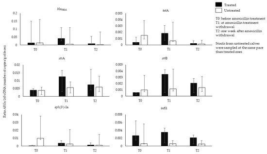
2.2. Original Data on the Analysis of Amoxicillin Effects on Calves’ GIM

History of amoxicillin therapy has been associated to the rise of resistance in Ecoli isolates from calves’ feces [8]. To our knowledge, longitudinal studies analyzing the effects of amoxicillin on calves’ GIM were lacking. We thus prospectively collected feces from calves (n = 16) aged from 5 to 26 days, belonging to five breeds (Charolais/Montbeliard, Montbeliard, Prim’ Holstein, Charolais/Prim’ Holstein, Limousin/Montbeliard), and resident in different farms (n = 7) in the Rhône-Alpes region (France), during the period October 2018–March 2020. Eleven out of 16 calves were suffering from omphalitis (umbilical cord infection) and were treated with IMI of amoxicillin (Longamox®, 15 mg/kg) for a duration varying between 4 and 16 days. The remaining five calves did not receive antibiotic treatment, and their feces were sampled at the same pace of the treated ones. The abundance of 41 ARGs, intI1/2/3, and of 16S rDNA was analyzed by qPCR [9]. Seventeen out of 41 investigated ARGs were found in the feces of all calves before amoxicillin treatment. The blaTEM gene and bacterial abundance were comparable between the treated and untreated group before treatment (ratio blaTEM/16S rDNA: 0.013 in both groups). At the end of amoxicillin treatment (T1), the amount of blaTEM increased in treated calves (blaTEM/16S rDNA ratio: 0.040) along with other ARGs (tetA, strA and strB), and intI1, index of class 1 integrons. These data suggest co-selection by amoxicillin treatment of ARGs related to other antibiotic classes and potential multidrug development (Figure 1). A decrease of all ARGs was observed 1 week after amoxicillin withdrawal (T2) (blaTEM/16S rDNA ratio: 0.008). The amount of blaTEM constantly decreased in the untreated group (blaTEM/16S rDNA ratio: T1, 0.005; T2: 0.002). The difference observed in the amount of ARGs at pretreatment and post-treatment, or between treated and untreated group, was not statistically significant (Wilcoxon Mann Whitney or Wilcoxon signed-rank test, p > 0.05). Several factors could confound the effect of the amoxicillin treatment on the blaTEM amount, for instance variation of the blaTEM gene amount among individuals, probably due to variation of calves’ age, which ranged from 5 to 26 days. Age is a determinant for GIM composition and ARGs amount at early life. In addition, calves were distributed in seven commercial farms probably contributing to the difference in ARGs content as well, because of different farm management. Environmental exposure of all calves to ARGs cannot be excluded, as calves of the untreated and treated group lived in the same farm, thus probably influencing the level of difference of blaTEM amount between the two groups. For a better understanding of antibiotics action on the GIM and selection of ARGs, experiments in environmentally controlled set-up would be a benefit for avoiding confounding factors influencing GIM composition and ARGs variation further than antibiotic action. However, studies in commercial farms are necessary to model antibiotic therapies effects in a real-life environment and evaluate ARGs propagation to other hosts or in the farm environment.
Figure 1. Representation of ARGs/16S rDNA ratio (number of copies/g of feces) evaluated by qPCR in feces of treated and untreated calves at amoxicillin pre-treatment (T0), at amoxicillin withdrawal (T1), and 1 week after amoxicillin withdrawal (T2).

2.3. Macrolides

Prophylactic subcutaneous injection (SCI) of tulathromycin caused a decrease of Bifidobacterium genus (Actinobacteria) in treated calves [10]. On the contrary, comparison of metaphylactic therapy based on enrofloxacin, a fluoroquinolone, or tulathromycin in Holstein calves did not evidence major changes in the GIM neither at the phylum level nor for gene function. Besides, Desulfovibrionales (Proteobacteria), which include species of potential pathogens for humans [11], had a higher relative abundance in the enrofloxacin-treated group 56 days post-withdrawal [12].
Metaphylactic SCI of tildipirosin did not alter the GIM of Holstein calves, at least at the phylum level [13]. Several antibiotic therapies caused a decrease of GIM diversity and E. coli amount during the treatment and until 15 days after withdrawal in Holstein calves observed in three different commercial farms [14].

2.4. Tetracyclines

Keijser et al. [15] analyzed the effects of a low and high dose of oxytetracycline in treated calves. The high-dose was administered for 5 days, whereas the low-dose was administered for 7 weeks. Major changes compared to a group of untreated animals were observed over time. Both oxytetracycline doses correlated with a decrease of RuminococcusCoprobacillus, and Lachnospiraceae, all belonging to the Firmicutes phylum, along with an increase of Prevotella (phylum Bacteroidetes), Faecalibacterium, and Blautia (phylum Firmicutes), compared to an untreated group. The selection of tetM gene and other ARGs, such as mel and floR, occurred only in high-dose treated calves and lasted for all the study period (42 days). Oultram et al. [16] analyzed the effects of oxytetracycline (IMI), tulathromycin (SCI), and florfenicol (SCI) used to treat pneumonia and otitis occurring in 7-week-old Holstein calves, hosted in a commercial farm. Considering the five most abundant detected genera, LactobacillusFaecalibacteriumBacteroidesParabacteroides, and Sharpea, a statistically significant decrease in the oxytetracycline-treated calves was observed for Lactobacillus genus compared to the control group. Overall, antibiotic treatment slightly decreased species richness in the calves’ microbiota 1 week after withdrawal. However, no statistical significance was observed compared with control group. Thames et al. [17] studied by qPCR the effects of neomycin and tetracycline orally administered on the abundance of selected ARGs (tetC/G/O/W/X, ermB/F, sul1/2; intI1), and found that only tetO was significantly more abundant in the treated group.

2.5. Other Antibiotics

Lhermie et al. [18] analyzed the effect of fluoroquinolones at low (2 mg/L) and high (10 mg/L) doses administered by IMI in young bulls (7–10-months-old) and calves (2–5-weeks-old). The therapy moderately selected for resistant Enterobacterales compared to the untreated group, and with less detectable effects in young bulls, probably because of a more mature GIM compared to calves that was expected to contain less Enterobacterales and more species difficult to cultivate (the study was conducted by cultivation). However, calves were colonized by fluoroquinolone-resistant bacteria before the treatment. Dobrzanska et al. [19] analyzed the effect of thiamphenicol. At 7 days from thiamphenicol administration, Proteobacteria increased because of Ecoli expansion, along with the emergence of mcr-2, a less prevalent gene than mcr-1 responsible for colistin resistance, and oqxB gene, encoding for antibiotic efflux pump. In the treated group, a rise of methanogenic Archaea and Prevotellaceae, typically associated to weight gain, was also observed.

References

  1. Penati, M.; Sala, G.; Biscarini, F.; Boccardo, A.; Bronzo, V.; Castiglioni, B.; Cremonesi, P.; Moroni, P.; Pravettoni, D.; Addis, M.F. Feeding pre-weaned calves with waste milk containing antibiotic residues is related to a higher incidence of diarrhea and alterations in the fecal microbiota. Front. Vet. Sci. 2021, 8, 650150.
  2. Dupouy, V.; Madec, J.-Y.; Wucher, J.; Arpaillange, N.; Métayer, V.; Roques, B.; Bousquet-Mélou, A.; Haenni, M. Selection of ESBL-producing Escherichia coli in the gut of calves experimentally fed with milk containing antibiotic residues. Vet. Microbiol. 2021, 257, 109049.
  3. Maynou, G.; Chester-Jones, H.; Bach, A.; Terré, M. Feeding pasteurized waste milk to preweaned dairy calves changes fecal and upper respiratory tract microbiota. Front. Vet. Sci. 2019, 6, 159.
  4. Pereira, R.V.V.; Carroll, L.; Lima, S.; Foditsch, C.; Siler, J.D.; Bicalho, R.C.; Warnick, L.D. Impacts of feeding preweaned calves milk containing drug residues on the functional profile of the fecal microbiota. Sci. Rep. 2018, 8, 554.
  5. Van Vleck Pereira, R.; Lima, S.; Siler, J.D.; Foditsch, C.; Warnick, L.D.; Bicalho, R.C. Ingestion of milk containing very low concentration of antimicrobials: Longitudinal effect on fecal microbiota composition in preweaned calves. PLoS ONE 2016, 11, e0147525.
  6. Grønvold, A.-M.R.; Mao, Y.; L’Abée-Lund, T.M.; Sørum, H.; Sivertsen, T.; Yannarell, A.C.; Mackie, R.I. Fecal microbiota of calves in the clinical setting: Effect of penicillin treatment. Vet. Microbiol. 2011, 153, 354–360.
  7. Ma, T.; Villot, C.; Renaud, D.; Skidmore, A.; Chevaux, E.; Steele, M.; Guan, L.L. Linking perturbations to temporal changes in diversity, stability, and compositions of neonatal calf gut microbiota: Prediction of diarrhea. ISME J. 2020, 14, 2223–2235.
  8. Jarrige, N.; Cazeau, G.; Bosquet, G.; Bastien, J.; Benoit, F.; Gay, E. Effects of antimicrobial exposure on the antimicrobial resistance of Escherichia coli in the digestive flora of dairy calves. Prev. Vet. Med. 2020, 185, 105177.
  9. Rochegüe, T.; Haenni, M.; Cazeau, G.; Metayer, V.; Madec, J.-Y.; Ferry, T.; Lupo, A. An inventory of 44 qPCR assays using hydrolysis probes operating with a unique amplification condition for the detection and quantification of antibiotic resistance genes. Diagn. Microbiol. Infect. Dis. 2021, 100, 115328.
  10. Martin, C.C.; Baccili, C.C.; Avila-Campos, M.J.; Hurley, D.J.; Gomes, V. Effect of prophylactic use of tulathromycin on gut bacterial populations, inflammatory profile and diarrhea in newborn Holstein calves. Res. Vet. Sci. 2021, 136, 268–276.
  11. Hagiya, H.; Kimura, K.; Nishi, I.; Yamamoto, N.; Yoshida, H.; Akeda, Y.; Tomono, K. Desulfovibrio desulfuricans bacteremia: A case report and literature review. Anaerobe 2018, 49, 112–115.
  12. Foditsch, C.; Pereira, R.; Siler, J.D.; Altier, C.; Warnick, L.D. Effects of treatment with enrofloxacin or tulathromycin on fecal microbiota composition and genetic function of dairy calves. PLoS ONE 2019, 14, 1–18.
  13. Bringhenti, L.; Pallu, M.; Silva, J.; Tomazi, T.; Tomazi, A.C.; Rodrigues, M.X.; Duarte, L.M.; Bilby, T.R.; Bicalho, R.C. Effect of metaphylactic administration of tildipirosin on the incidence of pneumonia and otitis and on the upper respiratory tract and fecal microbiome of preweaning Holstein calves. J. Dairy Sci. 2021, 104, 6020–6038.
  14. Massot, M.; Haenni, M.; Nguyen, T.T.; Madec, J.-Y.; Mentré, F.; Denamur, E. Temporal dynamics of the fecal microbiota in veal calves in a 6-month field trial. Anim. Microbiome 2020, 2, 32.
  15. Keijser, B.J.F.; Agamennone, V.; Broek, T.J.V.D.; Caspers, M.; Van De Braak, A.; Bomers, R.; Havekes, M.; Schoen, E.; Van Baak, M.; Mioch, D.; et al. Dose-dependent impact of oxytetracycline on the veal calf microbiome and resistome. BMC Genom. 2019, 20, 65.
  16. Oultram, J.; Phipps, E.; Teixeira, A.G.V.; Foditsch, C.; Bicalho, M.L.; Machado, V.S.; Bicalho, R.C.; Oikonomou, G. Effects of antibiotics (oxytetracycline, florfenicol or tulathromycin) on neonatal calves’ faecal microbial diversity. Vet. Rec. 2015, 177, 598.
  17. Thames, C.H.; Pruden, A.; James, R.E.; Ray, P.P.; Knowlton, K. Excretion of antibiotic resistance genes by dairy calves fed milk replacers with varying doses of antibiotics. Front. Microbiol. 2012, 3, 139.
  18. Lhermie, G.; Dupouy, V.; El Garch, F.; Ravinet, N.; Toutain, P.L.; Bousquet-Mélou, A.; Seegers, H.; Assié, S. Impact of low and high doses of marbofloxacin on the selection of resistant Enterobacteriaceae in the commensal gut flora of young cattle: Discussion of data from 2 study populations. Foodborne Pathog. Dis. 2017, 14, 152–159.
  19. Dobrzanska, D.A.; Lamaudière, M.T.F.; Rollason, J.; Acton, L.; Duncan, M.; Compton, S.; Simms, J.; Weedall, G.D.; Morozov, I.Y. Preventive antibiotic treatment of calves: Emergence of dysbiosis causing propagation of obese state-associated and mobile multidrug resistance-carrying bacteria. Microb. Biotechnol. 2020, 13, 669–682.
More
Information
Contributor MDPI registered users' name will be linked to their SciProfiles pages. To register with us, please refer to https://encyclopedia.pub/register :
View Times: 355
Revisions: 2 times (View History)
Update Date: 13 Dec 2021
1000/1000
Video Production Service