Submitted Successfully!
To reward your contribution, here is a gift for you: A free trial for our video production service.
Thank you for your contribution! You can also upload a video entry or images related to this topic.
Version Summary Created by Modification Content Size Created at Operation
1 + 3399 word(s) 3399 2021-11-29 04:53:42 |
2 format correct Meta information modification 3399 2021-12-08 02:56:11 |

Video Upload Options

Do you have a full video?

Confirm

Are you sure to Delete?
Cite
If you have any further questions, please contact Encyclopedia Editorial Office.
Jiang, L. Drosophila Trachea as a Novel Model of COPD. Encyclopedia. Available online: https://encyclopedia.pub/entry/16861 (accessed on 27 July 2024).
Jiang L. Drosophila Trachea as a Novel Model of COPD. Encyclopedia. Available at: https://encyclopedia.pub/entry/16861. Accessed July 27, 2024.
Jiang, Lan. "Drosophila Trachea as a Novel Model of COPD" Encyclopedia, https://encyclopedia.pub/entry/16861 (accessed July 27, 2024).
Jiang, L. (2021, December 07). Drosophila Trachea as a Novel Model of COPD. In Encyclopedia. https://encyclopedia.pub/entry/16861
Jiang, Lan. "Drosophila Trachea as a Novel Model of COPD." Encyclopedia. Web. 07 December, 2021.
Drosophila Trachea as a Novel Model of COPD
Edit

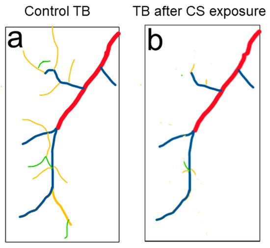
COPD, a chronic obstructive pulmonary disease, is one of the leading causes of death worldwide. Clinical studies and research in rodent models demonstrated that failure of repair mechanisms to cope with increased ROS and inflammation in the lung leads to COPD. Despite this progress, the molecular mechanisms underlying the development of COPD remain poorly understood, resulting in a lack of effective treatments. Thus, an informative, simple model is highly valued and desired. Recently, the cigarette smoke-induced Drosophila COPD model showed a complex set of pathological phenotypes that resemble those seen in human COPD patients. The Drosophila trachea has been used as a premier model to reveal the mechanisms of tube morphogenesis. The association of these mechanisms to structural changes in COPD can be analyzed by using Drosophila trachea. 

COPD Drosophila trachea model

1. Urgent Need to Reveal Novel Mechanisms of COPD

Chronic obstructive pulmonary disease (COPD) diminishes lung function and causes breathing difficulty and is one of the leading causes of death in the United States and worldwide [1]. Various natural and anthropogenic sources of chemical products (e.g., cigarette smoking, wood combustion, vehicle pollution, dust) are the causes of different types of air pollution. As the location of gas exchange, the respiratory system is most susceptible to air pollutants as it is directly exposed to atmospheric toxins. In the past few decades, the incidence of respiratory diseases, such as COPD, has sharply increased [2]. For example, long term exposure to pollutants such as NO2, SO2, and ozone (O3) leads to COPD [3][4][5]. Even exposure to low-level air pollution, when particulate matter (PM2.5), nitrogen dioxide (NO2), and black carbon (BC) are below current EU and US limits, has been linked to the development of COPD [3]. Clinically, COPD is characterized by progressive airflow limitations and the development of emphysema, defined by the loss of parenchymal lung tissue [6]. Thus, the loss of surface area for gas exchange in functional alveolar structures is a major hallmark of COPD. According to the Global Disease Burden Study of 2017, an estimated 272 million people worldwide are afflicted with COPD. In 2040, it is expected to be the fourth leading cause of death [1][7].
For the past 30 years, clinical studies and research in cigarette smoke (CS)-induced mouse models consistently showed that reactive oxygen species (ROS)-induced (e.g., O2•−, H2O2, NO) oxidative stress is a significant driver of COPD [8]. In COPD patients, the ROS derives from CS per se and/or is triggered by inflammatory and immune stimuli in epithelial cells of the airways [9][10]. The robust production of intracellular ROS is likely due to defects in oxidative phosphorylation caused by mitochondrial fragmentation due to CS exposure [11]. Incremented oxidative stress further enhances pulmonary inflammation with increased production of inflammatory mediators such as TNFα and Interleukins [9][12][13]. A vicious cycle of persistent inflammation, accompanied by chronic oxidative stress, leads to tissue damage and the progression of COPD [6][14]. Although treating inflammation and ROS is helpful, lung damage remains and continues to be irreversible [15].
The airway epithelium is the first line of defense against pollutants that enter the airways, yet this epithelium must maintain a barrier that is selectively permeable. A recent study shows that ozone-induced damage in airway epithelium structure occurs before the initiation of the inflammation pathways and the production of intracellular ROS [16]. This damage to the cellular junction is due to reduced junction proteins and the increased assembly of actin cytoskeleton that is linked to the tight junction [17][18]. Therefore, systematic studies on the timeline of early and late structural damage, ROS production, and inflammation will greatly help us to understand the developmental process of COPD. Understanding the mechanisms of these changes could provide an opportunity to develop effective therapeutic options that can prevent disease progression. Therefore, a fast, flexible, genetically tractable model organism is crucial to reveal novel mechanisms of COPD that will greatly enhance our understanding of COPD, thereby facilitating the discovery and development of novel and effective treatments.
The Drosophila respiratory system (i.e., the trachea) is an excellent system that is complementary to studies in mouse models. The Drosophila trachea is comparable to the mammalian lung system with many similarities in development and function. Compared to animal models, the fly trachea model has several advantages: (1) The timeline of early structural damage in the airway, production of ROS, and inflammation due to pollutant exposure can be easily observed through fluorescently-tagged reporter lines without invasive procedures. The Drosophila trachea provides the opportunity to observe these changes within live organisms; (2) the involved signaling pathways and cellular processes can be manipulated genetically or pharmacologically to analyze their relevance to the development of COPD phenotypes; (3) it has been estimated that over 60% of human genes associated with diseases have fly homologs [19][20]. Human COPD-associated genes that were recently identified through genome-wide association studies (GWAS) can be screened through GWAS in the Drosophila model for their association to COPD; (4) Drosophila have been successfully used as a drug screening platform to identify novel drugs for human diseases. Similarly, the Drosophila trachea can be used as a drug screening platform to identify novel treatments for COPD; (5) an increased life span can be used as a readout for the effectiveness of the manipulation, which is not suitable in most animal models. These advantages make Drosophila an ideal and indispensable model system to identify novel mechanisms of COPD and to explore innovative treatments for the disease that are not currently suitable for mammalian study.

2. Drosophila Trachea as a Model to Reveal Underlying Mechanisms of Tube Morphogenesis

The Drosophila trachea is a ramifying interconnected network of epithelial tubes with a monolayer of tightly-adhered polarized cells surrounding a central lumen [21] (Figure 1A larval trachea). Following the specification of branch identities, fibroblast growth factor receptor (FGFR) signaling guides the migration of tracheal cells in typical directions to form distinct tubes [22]. There are a total of four different types of tubes [23]. Type I multicellular branches are formed by multiple cells connecting together via intracellular junctions, such as the major branch of the trachea, the dorsal trunk (DT). Type II unicellular branches are formed by a linear arrangement of single cells through autocellular junctions, such as the lateral branch (LT) and dorsal branch (DB). Type III tubes are formed by two fusion cells of the adjacent tracheal metameres, with their apical surfaces spanning the inner wall of the ring or donut shape, resulting in seamless tubes without intracellular junctions. Type IV tubes are highly branched intracellular cytoplasmic extensions that form in terminal cells at the tips of the unicellular tubes, such as the branches formed in larval terminal cells. Tube morphogenesis has been extensively studied in Type I multicellular branches such as the DT (Figure 1B) and Type IV larval terminal branches (Figure 1C). In the DT, neighboring cells are connected together by adherens junctions (AJs) and septate junctions (SJs), which are equivalent to tight junctions (TJs) in mammals. SJs control the paracellular barrier function of the trachea while AJs stabilize cell–cell adhesion through the actin cytoskeleton. In Drosophila, the SJ is basal to AJ. In addition, cortical actin is highly concentrated beneath the apical membrane as well as the AJ and SJ and to a lesser degree at the basolateral membrane.
Figure 1. Drosophila tracheal branches. (A) Schematic larval trachea, an interconnected tubular network. The dorsal trunk (DT) is the multicellular major branch of the trachea. The lateral trunk (LT) and dorsal branch (DB) are unicellular branches. Terminal branches (TB) are intracellular branches, which are formed at the tip of unicellular branches. (B) Neighboring cells in DT are connected together by adherens junctions (green AJ) and septate junctions (yellow SJ), the latter of which is equivalent to the tight junction in mammalian systems. Cortical actin (blue) is highly concentrated beneath the apical membrane (red) as well as at the AJ and SJ, and relatively weaker at the basolateral membrane (black). Tracheal cells secrete apical luminal matrix (pink aECM), together with actin (blue), which forms a barrier to protect the apical membrane (red). (C). Schematic terminal branches (TB), which are formed in terminal cells, residing at the tip of the unicellular branch such as DB. There are 4 types of TB branches, including Type I (red), Type II (blue), Type III (yellow) and Type IV (green). (A) has been adapted from [24].
During tracheal development, the tracheal cells secrete material apically to form a transient apical luminal cable at the mid embryonic stage. This apical extracellular matrix (aECM) coordinates apical membrane growth and cell contractility to control tube growth [25][26]. This aECM cable is then degraded and absorbed by tracheal cells [27][28]. From the late embryonic stages through the larval stages, the tracheal cells secrete aECM material to form taenidial ridges that cover the apical membrane. This aECM is soft and flexible to provide ventilation but is also tight enough to function as a protective barrier that shields the tubes from dehydration, infections, and environmental stresses [29][30]
The formation of functional tracheal tubes depends on the sufficient transport of apical proteins, controlled plasma membrane expansion, effective cell junction maintenance, proper connection between the aECM and apical membrane, and coordinated cortical cytoskeleton reorganization. For example, defects in components of the apical secretion pathway such as COPI and COPII complex components Gartenzwerg (garz) and Sec24CD, respectively, lead to the defective secretion of luminal proteins [31][32]; mutations in lachesin or sinuous, which encode SJ components, lead to the defective secretion of aECM proteins [33][34]; mutations in dumpy or uninflated (uif) disrupt the connection between apical membrane and aECM [35][36]; the formin DAAM regulated actin nucleation and the subsequent polymerization through RhoA is required for actin ring formation, which is critical for the formation of taenidial folds [37][38]; mutations in either tramtrack or grainy head cause defective apical membrane expansion [39][40]. Taken together, disruption in any of these processes will lead to malformations in the trachea.
In addition to Type I multicellular tubes, such as the DT, tube morphogenesis has also been extensively studied in Type IV intracellular branches in larval terminal cells. Type IV terminal branches contact target tissues for gas exchange, which is similar to alveoli in human lung. The differentiation of terminal cell involves two processes, branching and subcellular lumen formation. Branching in the terminal cells heavily relies on cell migration. FGFR signaling, likely through the downstream GTPases Rac1 and Rac, allows cytoskeleton remodeling and filopodia formation for terminal cells to start branching [41]. Following the branching morphogenesis, it is essential to form a lumen within these branched structures primarily for the transport of gases.
Terminal cell branching and subcellular lumen formation are intimately associated. Subcellular lumen formation involves the structured expansion of the apical plasma membrane through the dynamic modulation of vesicle transport, which depends on the reorganization of the cytoskeleton. The initial subcellular lumen develops by invagination of the apical membrane inside the cell. The ingrowing apical membrane accumulates apical markers such as the PAR-polarity complex components aPKC/Par6/-Baz and Crumbs (Crb) [42][43]. Following apical membrane formation, aECM material is accumulated inside the lumen creating the aECM [44]. The growth of the subcellular lumen depends on intracellular trafficking of membrane and lumen material [41]. For example, Rab11 is involved in the trafficking of recycled and newly synthesized proteins to the apical plasma membrane [45]. In addition, other endosomes coordinate plasma membrane redistribution. For example, organelles carrying late endosomes and multivesicular bodies (MVBs) markers act as a transit station to redistribute the membrane apically and basally in terminal cells [46]. Therefore, coordination between cytoskeleton reorganization and vesicular trafficking is critical for terminal branch formation.
Unlike the embryonic tracheal system, which develops in a stereotypical and genetically controlled manner, the development of the larval trachea exhibits plasticity and also adapts to particular oxygen needs of the different tissues of the body. Insufficient oxygen levels activate the hypoxia signaling pathway, which is largely regulated by hypoxia-inducible factor 1α (HIF-1α) [47]. HIF-1α is a transcription factor that is considered to be a master transcriptional regulator of O2 homeostasis [48]. HIF-1α induces the transcription of genes related to angiogenesis, cell proliferation/survival, as well as inflammation [49][50]. The HIF-1α signaling pathway is activated in smokers with COPD. Thus, the increased expression of HIF-1α, VEGF, and VEGF receptor 2 were associated with decreased lung function, reduced quality of life, and progression of COPD [51]. The Drosophila functional homolog of HIF-1α is Similar [52]. Hypoxia-induced activation of Similar leads to over branching caused by elevated Drosophila FGF, Branchless (Bnl) [53] as well as increased ROS levels, similar to what was observed in COPD patients [54].
The Drosophila adult tracheal system, called air sacs, forms during the pupal period. The development of the air sacs starts from the third instar larval stage. The air sac precursor cells bud from the larval trachea, proliferate, and migrate towards the wing imaginal disc to form air sac primordia (ASPs). During pupal stages, the cells in ASPs migrate to form branches. Thereafter, they cease migrations and begin to elaborate into air sacs, which will further expand in adults. The air sacs are associated with numerous bundles of trachea, which extensively interdigitate with flight muscle to supply oxygen [55]. The development of ASPs involves three processes: cell proliferation, downregulation of adhesion molecules at tip cells, and extracellular matrix remodeling.
The morphogenesis of ASP is directed by FGFR signaling-induced outward migration of distal tip cells towards the wing imaginal disc, which secretes the Drosophila FGF, Bnl [55][56]. In addition, FGFR signaling induces the expression of epithelial growth factor (EGF)/vein, which activates epithelial growth factor receptor (EGFR) signaling to stimulate cell proliferation and survival [56][57]. The turnover of EGFR and FGFR is mediated by endosomes. For example, compromised endosomal sorting complex leads to impaired ASP development [58]. For the proper cell migration during ASP development, down-regulation of cell adhesion molecules such as Drosophila E-cadherin (shotgun), and Drosophila β-catenin (armadillo) is required in the tip cells [59]. Furthermore, remodeling of the ECM is necessary to facilitate the growth of ASPs. ASPs arise from a region of a tracheal branch that is directly juxtaposed to the wing disc [55]. This tracheal region lacks a visible tracheal-specific ECM, called basal lamina (BL), which is proteolyzed by the endopeptidase Mmp2 [60][61]. It was reported that elevated BL in mmp2 mutants leads to stunted ASP growth and failed formation of functional air sacs [61]. Another class of proteases, cathepsins, have also been implicated in ECM remodeling around the ASP [59]. The reduced BL thickness allows for greater FGF signaling response, similar to what is observed during mammalian lung development [62][63].

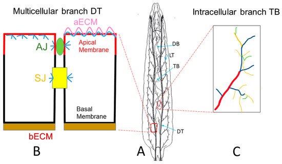
3. Drosophila Trachea as a COPD Model to Systematically Study the Development of COPD

Long-term CS exposure is by far the most important risk factor for COPD. Thus, chronic CS exposure has been used to investigate the mechanisms of COPD in mouse models [64][65]. Overall, for COPD, the failure of repair mechanisms to cope with the increased ROS and inflammation in the lungs leads to the loss of functional alveolar structure. As such, signaling pathways associated with tissue repair and ROS production have been extensively studied. For example, tissue repair relevant Wnt signaling [11] and JAK/STAT signaling [66], as well as cytokines (IL5 and IL6) that activate the JAK/STAT signaling, have been identified as potential drug targets [66]. In addition, the Nrf2 (NF-E2-related factor 2) pathway is also strongly activated by CS exposure and linked to COPD development [67]. The Nrf2 pathway is a potential drug target due to its function in controlling the expression of antioxidant genes that ultimately exert anti-inflammatory functions [68]. Despite the progress made towards COPD research, the molecular mechanisms underlying the development of COPD are still not well understood. This is reflected by the lack of effective treatments for this disease [15][69]. Thus, informative and druggable animal models are highly valued and desired. A simple model, such as the Drosophila trachea, may provide promising new candidates that are complementary to the current knowledge of COPD.
CS exposure affects functions in various organs in Drosophila. CS exposure has been shown to increase heart rate and cause alterations in the dynamics of the transient increase in intracellular calcium in myocardial cells [70]. CS and nicotine exposure have also been linked to changes in Drosophila sexual behavior, larval brain size, and the adult fly dopaminergic system [71][72]. Recently, the CS-induced Drosophila COPD model showed a complex set of pathological phenotypes that resemble those seen in human COPD patients [73]. These phenotypes include premature death, reduced physical activity, enhanced metabolic rates, and reduced respiratory surfaces in Drosophila trachea. Due to the short life span of fruit flies, the survival rate was measured within 2 weeks, compared to a survival rate of months in the mouse model. The physical activity of a large quantity of flies was automatically evaluated by the Drosophila activity monitoring system [74]. Metabolic rate was measured by body fat content using ELISA [75]. The reduced respiratory surface was analyzed in live 2nd instar larval terminal cells by using a btl:GFP transgenic line without an invasive procedure (Figure 2). Similar to the reduced alveolar surface area observed in human COPD lungs, Type III and Type IV branches in CS-induced COPD Drosophila larvae show the most obvious COPD phenotype: reduced numbers and overall length [73]. Comparably, as chronic CS exposure in the mouse model leads to the development of COPD, chronic CS exposure also leads to the development of COPD-like phenotypes in Drosophila larval trachea.
Figure 2. Tracheal morphology changes were observed in 2nd instar larvae upon CS exposure. (a) Schematic of a terminal cell of the dorsal branch in the third segment of Drosophila 2nd instar larvae. Terminal branches (TB) include Type I (red), Type II (blue), Type III (yellow) and Type IV (green) branches. (b) Type III (yellow) and Type IV (green) TBs are significantly reduced upon CS exposure.
The early junctional defects upon pollutant exposure in the mouse model suggest other early structural damages. These damages can be studied in the Drosophila larval multicellular DT. Various structures, including the aECM, basal ECM (bECM), apical membrane, basal membrane, junctions, and cytoskeleton in DT, can be easily observed using available transgenic lines with fluorescently-tagged structural proteins in live organisms (Figure 1B). These lines include the aECM (Vermiform:RFP and Serpentine:GFP [76] and bECM (type IV collagen Col41A:GFP [77]), apical membrane (Crumb:RFP and Crumb:GFP [78]), basal membrane (αSpectrin:GFP [79]) actin cytoskeleton (actin:GFP [80], AJ junction (DEcad:GFP, RFP [81]), SJ (Discs Large:GFP [76]). The later structural damage, reduced numbers, and length in terminal branches can be observed in larval trachea using btl:GFP lines [82]. Therefore, the Drosophila larval trachea can be used as a COPD model to systematically study the timeline of early- and late-stage structural damage upon chronic pollutant exposure.
In addition to the timeline of structural changes in the Drosophila COPD model, their relevance to known molecular mechanisms and components of tube morphology can be tested. For example, the activation of the MAPK/ERK pathway leads to the disruption of the tight junction [83]. The RhoA/ROCK signaling pathway leads to disruption of the tight junction through the aggregation of cytoskeletal actin [84]. Pollutant exposure could also cause structural damages through other mechanisms such as defective apical membrane expansion, aECM formation, or tube size maintenance. Similarly, the involvement of the underlying mechanisms, such as the protein trafficking pathway and cytoskeleton reorganization, in the development of COPD can be further studied in the larval trachea.
Along with structural damage, the intracellular production of ROS can be indirectly measured by ROS-inducible gstD (glutathione S transferase D)-GFP reporter, gstD-GFP [85]. Overall levels of peroxides can be detected using 2′,7′-dichlorofluorescein (H2DCF) followed by imaging as described [86]. Furthermore, increased concentrations of multiple cytokines, such as TNFα and interleukins, orchestrate chronic inflammation in COPD [12][13][87]. The only Drosophila TNF superfamily member is Eiger (Egr) [88][89]. Three cytokine molecules, namely Unpaired (Upd), Upd2 and Upd3, function similarly as interleukins [90][91][92]. Reporter lines egr-lacZ and upd-lacZ lines are available to measure the expression of these inflammatory cytokines [73]. It is also possible to observe the production of these inflammatory cytokines in vivo by generating fluorescent protein-tagged reporter lines. Previous studies in Drosophila trachea have revealed genes, pathways, and cellular processes involved in tube morphogenesis. The relevance of these mechanisms to the development of COPD can be further studied in the Drosophila trachea. Thus, the larval trachea provides an excellent model to systematically study the timeline of early and late structural damage, production of ROS, and inflammation upon pollutant exposure in vivo. However, the larval trachea is roughly equivalent to adolescence, while the adult trachea is comparable to adult human lungs. A recent study showed that early COPD in young adults is associated with clinical COPD 10 years later [93]. Therefore, the mechanisms of COPD revealed by studies of Drosophila larval trachea would be more relevant to the initiation of COPD in young adults. Although the current knowledge of the Drosophila adult trachea is still limited, studies of the development of air sacs provide opportunities to unveil mechanisms of COPD in relation to the disease progression in adult trachea. For example, the morphological changes in the air sac (btlenhancer-mRFP1moesin [57]), tracheal BL (collagen IV:GFP and Perlecan:GFP [61]), and junction proteins (Dα-cat-GFP, RFP [81]) can be visualized through fluorescently-tagged structural proteins in the late pupal stage, when air sacs form.

References

  1. Viegi, G.; Maio, S.; Fasola, S.; Baldacci, S. Global Burden of Chronic Respiratory Diseases. J. Aerosol Med. Pulm. Drug Deliv. 2020, 33, 171–177.
  2. Terzikhan, N.; Verhamme, K.M.; Hofman, A.; Stricker, B.H.; Brusselle, G.G.; Lahousse, L. Prevalence and incidence of COPD in smokers and non-smokers: The Rotterdam Study. Eur. J. Epidemiol. 2016, 31, 785–792.
  3. Liu, S.; Jørgensen, J.T.; Ljungman, P.; Pershagen, G.; Bellander, T.; Leander, K.; Magnusson, P.K.E.; Rizzuto, D.; Hvidtfeldt, U.A.; Raaschou-Nielsen, O.; et al. Long-term exposure to low-level air pollution and incidence of chronic obstructive pulmonary disease: The ELAPSE project. Environ. Int. 2021, 146, 106267.
  4. Hendryx, M.; Luo, J.; Chojenta, C.; Byles, J.E. Air pollution exposures from multiple point sources and risk of incident chronic obstructive pulmonary disease (COPD) and asthma. Environ. Res. 2019, 179, 108783.
  5. Wiegman, C.H.; Li, F.; Ryffel, B.; Togbe, D.; Chung, K.F. Oxidative Stress in Ozone-Induced Chronic Lung Inflammation and Emphysema: A Facet of Chronic Obstructive Pulmonary Disease. Front. Immunol. 2020, 11, 1957.
  6. Tuder, R.M.; Petrache, I. Pathogenesis of chronic obstructive pulmonary disease. J. Clin. Investig. 2012, 122, 2749–2755.
  7. López-Campos, J.L.; Tan, W.; Soriano, J.B. Global burden of COPD. Respirology 2016, 21, 14–23.
  8. Kluchova, Z.; Petrasova, D.; Joppa, P.; Dorkova, Z.; Tkacova, R. The association between oxidative stress and obstructive lung impairment in patients with COPD. Physiol. Res. 2007, 56, 51–56.
  9. Barnes, P.J. Inflammatory mechanisms in patients with chronic obstructive pulmonary disease. J. Allergy Clin. Immunol. 2016, 138, 16–27.
  10. Zuo, L.; He, F.; Sergakis, G.G.; Koozehchian, M.S.; Stimpfl, J.N.; Rong, Y.; Diaz, P.T.; Best, T.M. Interrelated role of cigarette smoking, oxidative stress, and immune response in COPD and corresponding treatments. Am. J. Physiol. Lung Cell. Mol. Physiol. 2014, 307, 205.
  11. Baarsma, H.A.; Skronska-Wasek, W.; Mutze, K.; Ciolek, F.; Wagner, D.E.; John-Schuster, G.; Heinzelmann, K.; Gunther, A.; Bracke, K.R.; Dagouassat, M.; et al. Noncanonical WNT-5A signaling impairs endogenous lung repair in COPD. J. Exp. Med. 2017, 214, 143–163.
  12. Barnes, P.J. The cytokine network in asthma and chronic obstructive pulmonary disease. J. Clin. Investig. 2008, 118, 3546–3556.
  13. Barnes, P.J. Targeting cytokines to treat asthma and chronic obstructive pulmonary disease. Nat. Rev. Immunol. 2018, 18, 454–466.
  14. Tuder, R.M.; McGrath, S.; Neptune, E. The pathobiological mechanisms of emphysema models: What do they have in common? Pulm. Pharmacol. Ther. 2003, 16, 67–78.
  15. Martinez, F.J.; Donohue, J.F.; Rennard, S.I. The future of chronic obstructive pulmonary disease treatment--difficulties of and barriers to drug development. Lancet 2011, 378, 1027–1037.
  16. Michaudel, C.; Mackowiak, C.; Maillet, I.; Fauconnier, L.; Akdis, C.A.; Sokolowska, M.; Dreher, A.; Tan, H.T.; Quesniaux, V.F.; Ryffel, B.; et al. Ozone exposure induces respiratory barrier biphasic injury and inflammation controlled by IL-33. J. Allergy Clin. Immunol. 2018, 142, 942–958.
  17. Nishida, K.; Brune, K.A.; Putcha, N.; Mandke, P.; O’Neal, W.K.; Shade, D.; Srivastava, V.; Wang, M.; Lam, H.; An, S.S.; et al. Cigarette smoke disrupts monolayer integrity by altering epithelial cell-cell adhesion and cortical tension. Am. J. Physiol. Lung Cell. Mol. Physiol. 2017, 313, L581–L591.
  18. Aghapour, M.; Raee, P.; Moghaddam, S.J.; Hiemstra, P.S.; Heijink, I.H. Airway Epithelial Barrier Dysfunction in Chronic Obstructive Pulmonary Disease: Role of Cigarette Smoke Exposure. Am. J. Respir. Cell Mol. Biol. 2018, 58, 157–169.
  19. Wangler, M.F.; Yamamoto, S.; Chao, H.T.; Posey, J.E.; Westerfield, M.; Postlethwait, J.; Members of the Undiagnosed Diseases Network, (UDN); Hieter, P.; Boycott, K.M.; Campeau, P.M.; et al. Model Organisms Facilitate Rare Disease Diagnosis and Therapeutic Research. Genetics 2017, 207, 9–27.
  20. Schneider, D. Using Drosophila as a model insect. Nat. Rev. Genet. 2000, 1, 218–226.
  21. Ghabrial, A.; Luschnig, S.; Metzstein, M.M.; Krasnow, M.A. Branching morphogenesis of the Drosophila tracheal system. Annu. Rev. Cell Dev. Biol. 2003, 19, 623–647.
  22. Ohshiro, T.; Emori, Y.; Saigo, K. Ligand-dependent activation of breathless FGF receptor gene in Drosophila developing trachea. Mech. Dev. 2002, 114, 3–11.
  23. Zuo, L.; Iordanou, E.; Chandran, R.R.; Jiang, L. Novel mechanisms of tube-size regulation revealed by the Drosophila trachea. Cell Tissue Res. 2013, 354, 343–354.
  24. Ruhle, H. Das larvale Tracheensystem von Drosophila melanogaster Meigen und seine Variabilita. Z. Wiss. Zool. 1932, 159–245.
  25. Devine, W.P.; Lubarsky, B.; Shaw, K.; Luschnig, S.; Messina, L.; Krasnow, M.A. Requirement for chitin biosynthesis in epithelial tube morphogenesis. Proc. Natl. Acad. Sci. USA 2005, 102, 17014–17019.
  26. Tonning, A.; Hemphala, J.; Tang, E.; Nannmark, U.; Samakovlis, C.; Uv, A. A transient luminal chitinous matrix is required to model epithelial tube diameter in the Drosophila trachea. Dev. Cell 2005, 9, 423–430.
  27. Behr, M.; Wingen, C.; Wolf, C.; Schuh, R.; Hoch, M. Wurst is essential for airway clearance and respiratory-tube size control. Nat. Cell Biol. 2007, 9, 847–853.
  28. Tsarouhas, V.; Senti, K.A.; Jayaram, S.A.; Tiklova, K.; Hemphala, J.; Adler, J.; Samakovlis, C. Sequential pulses of apical epithelial secretion and endocytosis drive airway maturation in Drosophila. Dev. Cell 2007, 13, 214–225.
  29. Petkau, G.; Wingen, C.; Jussen, L.C.; Radtke, T.; Behr, M. Obstructor-A is required for epithelial extracellular matrix dynamics, exoskeleton function, and tubulogenesis. J. Biol. Chem. 2012, 287, 21396–21405.
  30. Dong, B.; Hayashi, S. Shaping of biological tubes by mechanical interaction of cell and extracellular matrix. Curr. Opin. Genet. Dev. 2015, 32, 129–134.
  31. Armbruster, K.; Luschnig, S. The Drosophila Sec7 domain guanine nucleotide exchange factor protein Gartenzwerg localizes at the cis-Golgi and is essential for epithelial tube expansion. J. Cell Sci. 2012, 125, 1318–1328.
  32. Wang, S.; Meyer, H.; Ochoa-Espinosa, A.; Buchwald, U.; Onel, S.; Altenhein, B.; Heinisch, J.J.; Affolter, M.; Paululat, A. GBF1 (Gartenzwerg)-dependent secretion is required for Drosophila tubulogenesis. J. Cell Sci. 2012, 125, 461–472.
  33. Llimargas, M.; Strigini, M.; Katidou, M.; Karagogeos, D.; Casanova, J. Lachesin is a component of a septate junction-based mechanism that controls tube size and epithelial integrity in the Drosophila tracheal system. Development 2004, 131, 181–190.
  34. Wu, V.M.; Schulte, J.; Hirschi, A.; Tepass, U.; Beitel, G.J. Sinuous is a Drosophila claudin required for septate junction organization and epithelial tube size control. J. Cell Biol. 2004, 164, 313–323.
  35. Wilkin, M.B.; Becker, M.N.; Mulvey, D.; Phan, I.; Chao, A.; Cooper, K.; Chung, H.J.; Campbell, I.D.; Baron, M.; MacIntyre, R. Drosophila dumpy is a gigantic extracellular protein required to maintain tension at epidermal-cuticle attachment sites. Curr. Biol. 2000, 10, 559–567.
  36. Zhang, L.; Ward, R.E. uninflatable encodes a novel ectodermal apical surface protein required for tracheal inflation in Drosophila. Dev. Biol. 2009, 336, 201–212.
  37. Hannezo, E.; Dong, B.; Recho, P.; Joanny, J.F.; Hayashi, S. Cortical instability drives periodic supracellular actin pattern formation in epithelial tubes. Proc. Natl. Acad. Sci. USA 2015, 112, 8620–8625.
  38. Matusek, T.; Djiane, A.; Jankovics, F.; Brunner, D.; Mlodzik, M.; Mihaly, J. The Drosophila formin DAAM regulates the tracheal cuticle pattern through organizing the actin cytoskeleton. Development 2006, 133, 957–966.
  39. Araujo, S.J.; Cela, C.; Llimargas, M. Tramtrack regulates different morphogenetic events during Drosophila tracheal development. Development 2007, 134, 3665–3676.
  40. Hemphala, J.; Uv, A.; Cantera, R.; Bray, S.; Samakovlis, C. Grainy head controls apical membrane growth and tube elongation in response to Branchless/FGF signalling. Development 2003, 130, 249–258.
  41. Gervais, L.; Casanova, J. In vivo coupling of cell elongation and lumen formation in a single cell. Curr. Biol. 2010, 20, 359–366.
  42. Best, B.T. Single-cell branching morphogenesis in the Drosophila trachea. Dev. Biol. 2019, 451, 5–15.
  43. Jones, T.A.; Metzstein, M.M. A novel function for the PAR complex in subcellular morphogenesis of tracheal terminal cells in Drosophila melanogaster. Genetics 2011, 189, 153–164.
  44. Ozturk-Colak, A.; Moussian, B.; Araujo, S.J. Drosophila chitinous aECM and its cellular interactions during tracheal development. Dev. Dyn. 2016, 245, 259–267.
  45. Jones, T.A.; Nikolova, L.S.; Schjelderup, A.; Metzstein, M.M. Exocyst-mediated membrane trafficking is required for branch outgrowth in Drosophila tracheal terminal cells. Dev. Biol. 2014, 390, 41–50.
  46. Mathew, R.; Rios-Barrera, L.D.; Machado, P.; Schwab, Y.; Leptin, M. Transcytosis via the late endocytic pathway as a cell morphogenetic mechanism. EMBO J. 2020, 39, e105332.
  47. Lee, J.W.; Ko, J.; Ju, C.; Eltzschig, H.K. Hypoxia signaling in human diseases and therapeutic targets. Exp. Mol. Med. 2019, 51, 1–13.
  48. Iyer, N.V.; Kotch, L.E.; Agani, F.; Leung, S.W.; Laughner, E.; Wenger, R.H.; Gassmann, M.; Gearhart, J.D.; Lawler, A.M.; Yu, A.Y.; et al. Cellular and developmental control of O2 homeostasis by hypoxia-inducible factor 1 alpha. Genes Dev. 1998, 12, 149–162.
  49. Lee, J.W.; Bae, S.H.; Jeong, J.W.; Kim, S.H.; Kim, K.W. Hypoxia-inducible factor (HIF-1)alpha: Its protein stability and biological functions. Exp. Mol. Med. 2004, 36, 1–12.
  50. Corrado, C.; Fontana, S. Hypoxia and HIF Signaling: One Axis with Divergent Effects. Int. J. Mol. Sci. 2020, 21, 5611.
  51. Fu, X.; Zhang, F. Role of the HIF-1 signaling pathway in chronic obstructive pulmonary disease. Exp. Ther. Med. 2018, 16, 4553–4561.
  52. Bacon, N.C.; Wappner, P.; O’Rourke, J.F.; Bartlett, S.M.; Shilo, B.; Pugh, C.W.; Ratcliffe, P.J. Regulation of the Drosophila bHLH-PAS protein Sima by hypoxia: Functional evidence for homology with mammalian HIF-1 alpha. Biochem. Biophys. Res. Commun. 1998, 249, 811–816.
  53. Centanin, L.; Dekanty, A.; Romero, N.; Irisarri, M.; Gorr, T.A.; Wappner, P. Cell Autonomy of HIF Effects in Drosophila: Tracheal Cells Sense Hypoxia and Induce Terminal Branch Sprouting. Dev. Cell 2008, 14, 547–558.
  54. Habib, P.; Jung, J.; Wilms, G.M.; Kokott-Vuong, A.; Habib, S.; Schulz, J.B.; Voigt, A. Posthypoxic behavioral impairment and mortality of Drosophila melanogaster are associated with high temperatures, enhanced predeath activity and oxidative stress. Exp. Mol. Med. 2021, 53, 264–280.
  55. Sato, M.; Kornberg, T.B. FGF is an essential mitogen and chemoattractant for the air sacs of the drosophila tracheal system. Dev. Cell 2002, 3, 195–207.
  56. Cruz, J.; Bota-Rabassedas, N.; Franch-Marro, X. FGF coordinates air sac development by activation of the EGF ligand Vein through the transcription factor PntP2. Sci. Rep. 2015, 5, 17806.
  57. Cabernard, C.; Affolter, M. Distinct Roles for Two Receptor Tyrosine Kinases in Epithelial Branching Morphogenesis in Drosophila. Dev. Cell 2005, 9, 831–842.
  58. Chanut-Delalande, H.; Jung, A.C.; Baer, M.M.; Lin, L.; Payre, F.; Affolter, M. The Hrs/Stam Complex Acts as a Positive and Negative Regulator of RTK Signaling during Drosophila Development. PLoS ONE 2010, 5, e10245.
  59. Dong, Q.; Brenneman, B.; Fields, C.; Srivastava, A. A Cathepsin-L is required for invasive behavior during Air Sac Primordium development inDrosophila melanogaster. FEBS Lett. 2015, 589, 3090–3097.
  60. Llano, E.; Adam, G.; Pendás, A.M.; Quesada, V.; Sánchez, L.M.; Santamaría, I.; Noselli, S.; López-Otín, C. Structural and Enzymatic Characterization of Drosophila Dm2-MMP, a Membrane-bound Matrix Metalloproteinase with Tissue-specific Expression. J. Biol. Chem. 2002, 277, 23321–23329.
  61. Guha, A.; Lin, L.; Kornberg, T.B. Regulation of Drosophila matrix metalloprotease Mmp2 is essential for wing imaginal disc:trachea association and air sac tubulogenesis. Dev. Biol. 2009, 335, 317–326.
  62. Park, W.Y.; Miranda, B.; Lebeche, D.; Hashimoto, G.; Cardoso, W.V. FGF-10 Is a Chemotactic Factor for Distal Epithelial Buds during Lung Development. Dev. Biol. 1998, 201, 125–134.
  63. Abler, L.L.; Mansour, S.L.; Sun, X. Conditional gene inactivation reveals roles forFgf10andFgfr2in establishing a normal pattern of epithelial branching in the mouse lung. Dev. Dyn. 2009, 238, 1999–2013.
  64. Brandsma, C.A.; de Vries, M.; Costa, R.; Woldhuis, R.R.; Konigshoff, M.; Timens, W. Lung ageing and COPD: Is there a role for ageing in abnormal tissue repair? Eur. Respir. Rev. 2017, 26, 170073.
  65. McDonough, J.E.; Yuan, R.; Suzuki, M.; Seyednejad, N.; Elliott, W.M.; Sanchez, P.G.; Wright, A.C.; Gefter, W.B.; Litzky, L.; Coxson, H.O.; et al. Small-airway obstruction and emphysema in chronic obstructive pulmonary disease. N. Engl. J. Med. 2011, 365, 1567–1575.
  66. Yew-Booth, L.; Birrell, M.A.; Lau, M.S.; Baker, K.; Jones, V.; Kilty, I.; Belvisi, M.G. JAK-STAT pathway activation in COPD. Eur. Respir. J. 2015, 46, 843–845.
  67. Suzuki, M.; Betsuyaku, T.; Ito, Y.; Nagai, K.; Nasuhara, Y.; Kaga, K.; Kondo, S.; Nishimura, M. Down-regulated NF-E2-related factor 2 in pulmonary macrophages of aged smokers and patients with chronic obstructive pulmonary disease. Am. J. Respir. Cell. Mol. Biol. 2008, 39, 673–682.
  68. Saha, S.; Buttari, B.; Panieri, E.; Profumo, E.; Saso, L. An Overview of Nrf2 Signaling Pathway and Its Role in Inflammation. Molecules 2020, 25, 5474.
  69. Vogelmeier, C.F.; Roman-Rodriguez, M.; Singh, D.; Han, M.K.; Rodriguez-Roisin, R.; Ferguson, G.T. Goals of COPD treatment: Focus on symptoms and exacerbations. Respir. Med. 2020, 166, 105938.
  70. Santalla, M.; Pagola, L.; Gómez, I.; Balcazar, D.; Valverde, C.A.; Ferrero, P. Smoking flies: Testing the effect of tobacco cigarettes on heart function of Drosophila melanogaster. Biol. Open 2021, 10, bio055004.
  71. Giannopoulos, A.; Giannakou, L.; Gourgouliani, N.; Lüpold, S.; Rouka, E.; Jagirdar, R.; Pitaraki, E.; Hatzoglou, C.; Gourgoulianis, K.; Blanckenhorn, W.; et al. Exposure of Drosophila melanogaster to cigarette smoke extract changes its sexual behavior. Eur. Respir. J. 2020, 56, 1326.
  72. Morris, M.; Shaw, A.; Lambert, M.; Perry, H.H.; Lowenstein, E.; Valenzuela, D.; Velazquez-Ulloa, N.A. Developmental nicotine exposure affects larval brain size and the adult dopaminergic system of Drosophila melanogaster. BMC Dev. Biol. 2018, 18, 13–16.
  73. Prange, R.; Thiedmann, M.; Bhandari, A.; Mishra, N.; Sinha, A.; Hasler, R.; Rosenstiel, P.; Uliczka, K.; Wagner, C.; Yildirim, A.O.; et al. A Drosophila model of cigarette smoke induced COPD identifies Nrf2 signaling as an expedient target for intervention. Aging 2018, 10, 2122–2135.
  74. Pfeiffenberger, C.; Lear, B.C.; Keegan, K.P.; Allada, R. Locomotor activity level monitoring using the Drosophila Activity Monitoring (DAM) System. Cold Spring Harb. Protoc. 2010, 2010, pdb.prot5518.
  75. Hoffmann, J.; Romey, R.; Fink, C.; Yong, L.; Roeder, T. Overexpression of Sir2 in the adult fat body is sufficient to extend lifespan of male and female Drosophila. Aging 2013, 5, 315–327.
  76. Wu, V.M.; Yu, M.H.; Paik, R.; Banerjee, S.; Liang, Z.; Paul, S.M.; Bhat, M.A.; Beitel, G.J. Drosophila Varicose, a member of a new subgroup of basolateral MAGUKs, is required for septate junctions and tracheal morphogenesis. Development 2007, 134, 999–1009.
  77. Kiss, M.; Kiss, A.A.; Radics, M.; Popovics, N.; Hermesz, E.; Csiszar, K.; Mink, M. Drosophila type IV collagen mutation associates with immune system activation and intestinal dysfunction. Matrix Biol. 2016, 49, 120–131.
  78. Ling, C.; Zheng, Y.; Yin, F.; Yu, J.; Huang, J.; Hong, Y.; Wu, S.; Pan, D. The apical transmembrane protein Crumbs functions as a tumor suppressor that regulates Hippo signaling by binding to Expanded. Proc. Natl. Acad. Sci. USA 2010, 107, 10532–10537.
  79. Khanna, M.R.; Mattie, F.J.; Browder, K.C.; Radyk, M.D.; Crilly, S.E.; Bakerink, K.J.; Harper, S.L.; Speicher, D.W.; Thomas, G.H. Spectrin tetramer formation is not required for viable development in Drosophila. J. Biol. Chem. 2015, 290, 706–715.
  80. Kessenbrock, K.; Holak, T.A.; Sixt, M.; Jenne, D.; Werb, Z.; Riedl, J.; Wedlich-Soldner, R.; Neukirchen, D.; Bista, M.; Crevenna, A.H.; et al. Lifeact: A versatile marker to visualize F-actin. Nat. Methods 2008, 5, 605–607.
  81. Chen, Y.J.; Huang, J.; Huang, L.; Austin, E.; Hong, Y. Phosphorylation potential of Drosophila E-Cadherin intracellular domain is essential for development and adherens junction biosynthetic dynamics regulation. Development 2017, 144, 1242–1248.
  82. Ghabrial, A.S.; Krasnow, M.A. Social interactions among epithelial cells during tracheal branching morphogenesis. Nature 2006, 441, 746–749.
  83. Liu, Y.; Wei, H.; Tang, J.; Yuan, J.; Wu, M.; Yao, C.; Hosoi, K.; Yu, S.; Zhao, X.; Han, Y.; et al. Dysfunction of pulmonary epithelial tight junction induced by silicon dioxide nanoparticles via the ROS/ERK pathway and protein degradation. Chemosphere 2020, 255, 126954.
  84. Feng, S.; Zou, L.; Wang, H.; He, R.; Liu, K.; Zhu, H. RhoA/ROCK-2 Pathway Inhibition and Tight Junction Protein Upregulation by Catalpol Suppresses Lipopolysaccaride-Induced Disruption of Blood-Brain Barrier Permeability. Molecules 2018, 23, 2371.
  85. Landis, G.; Shen, J.; Tower, J. Gene expression changes in response to aging compared to heat stress, oxidative stress and ionizing radiation in Drosophila melanogaster. Aging 2012, 4, 768–789.
  86. Dar, N.J.; Satti, N.K.; Dutt, P.; Hamid, A.; Ahmad, M. Attenuation of Glutamate-Induced Excitotoxicity by Withanolide-A in Neuron-Like Cells: Role for PI3K/Akt/MAPK Signaling Pathway. Mol. Neurobiol. 2018, 55, 2725–2739.
  87. Yao, Y.; Zhou, J.; Diao, X.; Wang, S. Association between tumor necrosis factor-alpha and chronic obstructive pulmonary disease: A systematic review and meta-analysis. Ther. Adv. Respir. Dis. 2019, 13, 1753466619866096.
  88. Moreno, E.; Yan, M.; Basler, K. Evolution of TNF signaling mechanisms: JNK-dependent apoptosis triggered by Eiger, the Drosophila homolog of the TNF superfamily. Curr. Biol. 2002, 12, 1263–1268.
  89. Igaki, T.; Kanda, H.; Yamamoto-Goto, Y.; Kanuka, H.; Kuranaga, E.; Aigaki, T.; Miura, M. Eiger, a TNF superfamily ligand that triggers the Drosophila JNK pathway. EMBO J. 2002, 21, 3009–3018.
  90. Boulay, J.L.; O’Shea, J.J.; Paul, W.E. Molecular phylogeny within type I cytokines and their cognate receptors. Immunity 2003, 19, 159–163.
  91. Brown, S.; Hu, N.; Hombria, J.C. Identification of the first invertebrate interleukin JAK/STAT receptor, the Drosophila gene domeless. Curr. Biol. 2001, 11, 1700–1705.
  92. Chen, H.W.; Chen, X.; Oh, S.W.; Marinissen, M.J.; Gutkind, J.S.; Hou, S.X. mom identifies a receptor for the Drosophila JAK/STAT signal transduction pathway and encodes a protein distantly related to the mammalian cytokine receptor family. Genes Dev. 2002, 16, 388–398.
  93. Çolak, Y.; Afzal, S.; Nordestgaard, B.G.; Lange, P.; Vestbo, J. Importance of Early COPD in Young Adults for Development of Clinical COPD: Findings from the Copenhagen General Population Study. Am. J. Respir. Crit. Care Med. 2021, 203, 1245–1256.
More
Information
Contributor MDPI registered users' name will be linked to their SciProfiles pages. To register with us, please refer to https://encyclopedia.pub/register :
View Times: 428
Revisions: 2 times (View History)
Update Date: 08 Dec 2021
1000/1000
Video Production Service