Submitted Successfully!
To reward your contribution, here is a gift for you: A free trial for our video production service.
Thank you for your contribution! You can also upload a video entry or images related to this topic.
Version Summary Created by Modification Content Size Created at Operation
1 + 3948 word(s) 3948 2021-12-02 02:50:53

Video Upload Options

Do you have a full video?

Confirm

Are you sure to Delete?
Cite
If you have any further questions, please contact Encyclopedia Editorial Office.
De Mendoza Barberá, E. Surface Glucan Structures in Aeromonas spp.. Encyclopedia. Available online: https://encyclopedia.pub/entry/16795 (accessed on 17 May 2024).
De Mendoza Barberá E. Surface Glucan Structures in Aeromonas spp.. Encyclopedia. Available at: https://encyclopedia.pub/entry/16795. Accessed May 17, 2024.
De Mendoza Barberá, Elena. "Surface Glucan Structures in Aeromonas spp." Encyclopedia, https://encyclopedia.pub/entry/16795 (accessed May 17, 2024).
De Mendoza Barberá, E. (2021, December 06). Surface Glucan Structures in Aeromonas spp.. In Encyclopedia. https://encyclopedia.pub/entry/16795
De Mendoza Barberá, Elena. "Surface Glucan Structures in Aeromonas spp.." Encyclopedia. Web. 06 December, 2021.
Surface Glucan Structures in Aeromonas spp.
Edit

Aeromonas spp. are generally found in aquatic environments, although they have also been isolated from both fresh and processed food. These Gram-negative, rod-shaped bacteria are mostly infective to poikilothermic animals, although they are also considered opportunistic pathogens of both aquatic and terrestrial homeotherms, and some species have been associated with gastrointestinal and extraintestinal septicemic infections in humans. Several cell-surface glucans have been shown to contribute to colonization and survival of Aeromonas pathogenic strains in different hosts, playing important roles in bacterial–host interactions related to pathogenesis These include lipopolysaccharide (LPS), capsule, α-glucan, and glycosylated polar and lateral flagella.

Aeromonas capsule polysaccharide α-glucan glycosylation O-antigen LPS

1. Introduction

The Aeromonadaceae family comprises Gram-negative, facultative anaerobic, and chemoorganotrophic bacteria, with an optimal growing temperature of 22–37 °C [1]Members of this family are generally motile by polar flagellation, and are able to reduce nitrates to nitrites. They also have the ability to catabolize glucose and other carbohydrates, producing acids and often gases. In particular, the genus Aeromonas is comprised of water-borne bacteria, ubiquitously found in aquatic environments, and also identified in different types of food [1][2]Generally, the members of this genus are classified in two different groups: the non-motile, psychrophilic species (e.g., A. salmonicida) and the motile, mesophilic species (e.g., A. hydrophilaA. caviae, and A. sobria[2]Aeromonas spp. could potentially pose a serious risk to public health, as many strains are able to grow and produce exotoxins at low temperatures and high salt concentrations [3][4]In fact, they are emerging as the causative agents of gastrointestinal and extraintestinal diseases in a wide range of animals [2]In fish, they are responsible for major economic losses in aquaculture [5][6]In humans, they are implicated in several septicemic infections, ranging from relatively mild illnesses (e.g., acute gastroenteritis or superficial wound infections) to more complicated pathologies (e.g., respiratory-tract or eye infections), or even life-threatening conditions (e.g., septicemia, meningitis, myonecrosis or osteomyelitis) [7][8]The number of reported Aeromonas spp. infections in humans has been steadily rising in recent years, presenting a serious threat to the increasing population of immunocompromised patients [9][10], and will remain a human health problem in the near future, considering the increased and more susceptible population of elderly people with potential underlying diseases [11]Among the different pathogenic factors associated with virulence of Aeromonas spp., cell-surface glucans have been reported to play important roles in host–pathogen interactions, contributing to adherence, colonization, and overall survival of pathogenic strains in different hosts [12].

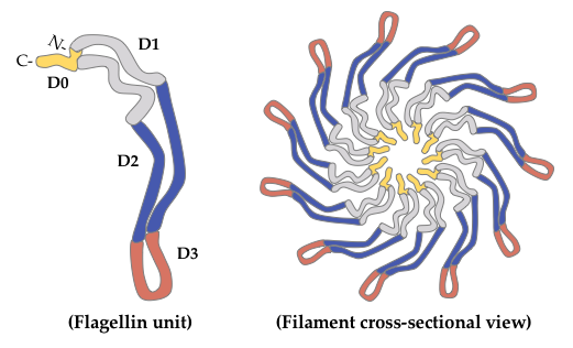
2. Glycosylated Flagella

Bacterial flagella are protein structures whose main function is motility, both in liquid and solid environments. Mesophilic Aeromonas spp. constitutively express a single polar flagellum for movement in liquid environments, and 50–60% of clinical isolates also possess an additional inducible lateral-flagella system, expressed for motility in solid or viscous conditions [13]. Structurally, the flagellar system is comprised of the basal body, embedded in the bacterial surface, and the hook and the filament, constituting the external part. The flagellar filament is in turn composed of flagellin proteins, assembled into 11 protofilaments [14], whose expression differs in number and diversity among different Gram-negative species. Mesophilic Aeromonas spp. have been shown to generally express two polar (FlaA and FlaB) and one lateral (LafA) flagellin [15][16], although a few strains (e.g., A. caviae Sch3N) express two different flagellin proteins (LafA1 and LafA2) in their lateral-flagella filaments [17]. Structurally, as determined in S. enterica serovar Typhimurium, functional flagellins are comprised of four domains: D0, D1, D2 and D3 (Figure 1) [18]. Domains D0 and D1 present an α-helix conformation, and are comprised by the well-conserved N- and C-terminal ends of the flagellin protein. These domains are embedded in the inner core of the filament, and are necessary for filament architecture and motility functions. On the other hand, the central domains D2 and D3 present a variable β-sheet conformation, and are exposed to the outer surface, where they can be subjected to glycosylation [19]. In A. piscicola (previously known as A. hydrophila), this D2/D3 region seems to be involved in the adhesin-like behavior of flagella [20].
Figure 1. Schematic representation of flagellin structure and arrangement into the flagellar filament. Flagellins are arranged into 11 protofilaments in a way that domains D0 (yellow) and D1 (grey) remain in the interior of the flagellar filament, while domains D2 (blue) and D3 (red) are exposed to the outer surface.
In Aeromonas spp., only O-glycosylation (which entails the covalent attachment of glycans to the hydroxyl oxygen of Ser, Thr or Tyr residues) has been reported to date, and it has been shown to be required for flagellar assembly and motility, in A. caviae and A. piscicola AH-3 [21][22]. Polar-flagella glycosylation, in particular, has been shown to be required for adherence to human epithelial (HEp-2) cells, and to be involved in the immune stimulation of IL-8 production via TLR5 [23][20]. Similarly, in A. hydrophila AH-1, polar-flagella glycosylation has been shown to be involved in adherence, biofilm formation, and stimulation of the host immune response [24]O-glycosylation was first described in A. caviae, where polar flagellins FlaA and FlaB were shown to be modified with pseudaminic acid at 5–8 Ser or Thr residues of their central, immunogenic D2/D3 domains [19]. As observed in A. caviae Sch3N, the glycan-modifying polar flagellin in A. hydrophila AH-1 has been shown to be a single monosaccharide, also speculated to be a pseudaminic-acid derivative [24]. In A. piscicola AH-3, both polar and lateral flagellins have been shown to be modified by the addition of sugars [21][25] and, although the same pseudaminic-acid derivative is used to modify these structures, notable differences have been observed in this case. For instance, O-glycosylation of the lateral flagellin LafA has been shown to occur at three flagellin sites, and always by the addition of a single pseudaminic-acid-derivative molecule of 376 Da. On the other hand, polar flagellins FlaA and FlaB have been shown to be glycosylated at one and six flagellin sites, respectively, and the pseudaminic-acid-derivative molecule can be found either alone or as part of a more complex glycan, composed of up to seven different sugar molecules. The longest form of this glycan (a 1679-Da heptasaccharide) is the only one identified in both A. piscicola AH-3 polar flagellins, and it is composed of two hexoses, three N-acetylhexosamines with a variable number of phosphate and methyl groups, one 102-Da monosaccharide, and the pseudaminic-acid-derivative molecule that links the glycan to the peptide [21].
Several genes involved in Aeromonas spp. flagellin O-glycosylation have been identified in the genomes of mesophilic species. In A. piscicola AH-3, genes involved in sugar biosynthesis are located near the polar-flagellin structural genes, and their mutation has been shown to affect both polar and lateral structures [25]. On the other hand, genes coding for glycosyltransferases are found near the structural region of each flagellar system, and their mutation only affects one flagellar structure [15][16]. Interestingly, in A. caviae Sch3N, mutation of genes involved in pseudaminic-acid biosynthesis has been shown to affect both flagellar biogenesis and LPS O-antigen biosynthesis, suggesting a shared glycosylation route for these structures [19]. However, in A. piscicola AH-3, mutation of these genes only affects flagellar expression, implying that a different sugar and glycosylation pathway is used for LPS biosynthesis [25]. Among the genes coding for glycosyltransferases are the motility accessory factor (maf) genes. In Aeromonas spp., particularly in A. caviae Sch3N, maf-1 is suggested to be responsible for the transfer of the pseudaminic-acid derivative to the polar-flagellin monomers [22], and homologous genes have been found in A. hydrophila ATCC 7966T, A. hydrophila AH-1, and A. piscicola AH-3 [26][27][28]. A second motility accessory factor (maf-2), shown to be required for both polar- and lateral-flagella production in A. piscicola AH-3 [25], has been identified in A. hydrophila ATCC 7966T and some other mesophilic species [29], and yet another gene of this family (maf-5) has been shown to be required for lateral-flagella production in A. piscicola AH-3 [16]. Interestingly, Aeromonas spp. that modify polar flagellins with heterogeneous glycans (e.g., A. piscicola AH-3) present larger glycosylation islands in their genomes than those that modify polar flagellins with a single pseudaminic-acid derivative (e.g., A. hydrophila AH-1 and A. caviae Sch3N), and contain a newly identified glycosyltransferase gene (fgi-1), responsible for transferring the first sugar of the heterogeneous glycan to the pseudaminic-acid derivative that links the glycan to the polar flagellin [29].

3. Lipopolysaccharide

The surface glycoconjugate LPS, exclusively found in Gram-negative bacteria, is a major component of the bacterial outer membrane. LPS interaction with host immune cells triggers host inflammatory and immune responses through TLR4-mediated signaling [30]. However, this primarily protective mechanism may become overshadowed by an acute pathophysiological response that leads to the overstimulation and release of proinflammatory cytokines, causing multiorgan dysfunction [31]. Structurally, LPS is comprised of three linked domains: a conserved and toxic lipid component, known as lipid A; the core oligosaccharide (core OS); and a highly variable O-specific-polysaccharide chain, known as the O-antigen (Figure 2) [32]. LPS molecules that contain all three regions are termed smooth (S)-LPS, while those lacking the O-antigen are referred to as rough (R)-LPSThe biological activity of LPS appears to depend on its specific conformation, which is in turn determined by the composition of each of its structural components. These components are initially synthesized at the cytosolic membrane and, once assembled, the whole LPS molecule is transferred to the most external part of the bacterial outer membrane by the Lpt proteins, where it becomes surface-exposed [33].
Figure 2. Schematic representation of the LPS molecule of Gram-negative bacteria. The lipid-A moiety consists of two parts: a lipid fraction composed of fatty-acid chains, which anchors LPS in the bacterial outer membrane, and a sugar backbone that links the molecule to the core oligosaccharide (core OS). The O-antigen chain, located at the most external part of the LPS molecule, is built of repetitive saccharide units that vary in number (n) among different bacterial cells. Smooth (S)-LPS is comprised of all three components, while rough (R)-LPS lacks the O-antigen subunit. The number and chemical structure of the acyl chains and sugar moieties represented in the figure can vary. NAG, N-acetylglucosamine. Kdo, 3-deoxy-D-manno-oct-2-ulosonic acid.
Lipid A
The highly conserved lipid-A structure constitutes the hydrophobic, membrane-anchoring region of LPS, and plays an important role in the immunogenic properties of this molecule. Its release from lysed bacteria can provoke a major systemic inflammation (known as septic or endotoxic shock) [34], and it has also been reported to induce B-cell polyclonal activation (which can lead to leukopenia, hemorrhagic necrosis of tumors, diarrhea, or even death) [35]. Structurally, lipid A consists of a phosphorylated N-acetylglucosamine (NAG) dimer, with 6 or 7 saturated fatty acids attached to it (Figure 2) [34]. Three major molecules differing in their acylation patterns (tetra-, penta- or hexa-acylated) have been identified in the lipid-A structure of A. salmonicida subsp. salmonicida [36]. These findings are of significant importance, as the level of lipid-A acylation has been shown to be related to the host ability to recognize LPS in other Gram-negative species. In Francisella novicida, in particular, reduced acylation of lipid A results in reduced LPS recognition by murine caspase 11, but not by human caspases 4/5 [37][38], suggesting that these lipid-A modifications could be critical for pathogenesis in certain mammals.
Core Oligosaccharide
The core OS is located between the lipid-A and O-antigen components of the LPS molecule (Figure 2). It is covalently attached to the lipid-A region, and it is therefore localized near the vicinity of the hydrophobic membrane. At the genetic level, three gene clusters have been associated with LPS core-OS biosynthesis in Aeromonas spp. Regions 2 and 3 are identical in A. piscicola AH-3 (O:34) and A. salmonicida strains A449 and A450. However, of the seven genes comprising region 1 in A. salmonicida A450, only three of them have been found to be identical in A. piscicola AH-3. Other three share high similarity, and the other one shows no homology whatsoever to any well-characterized gene [39][40]. In agreement with these observations, the comparison of the LPS core-OS structures of these two species renders a great similarity in the inner core [40][41], while some differences can be observed in the outer core [39][40]. As determined in A. salmonicida, the LPS core-OS is composed of 8–12 sugars, the inner and the outer core differing in their sugar composition [41]. The outer core is generally characterized by the presence of hexose sugars that provide an attachment site for the O-antigen structure. The inner core, on the other hand, is a highly phosphorylated region, and is therefore very anionic in nature. It is attached to the lipid-A molecule at the 6′ position of one NAG, and it contains 3-deoxy-D-manno-oct-2-ulosonic acid (Kdo), or the derivative residue 3-glycero-D-talo-oct-2-ulosonic acid [42].
O-Antigen
The O-antigen is the largest and most surface-exposed LPS component. It is usually attached to a terminal residue of the outer core, and it has been shown to mediate pathogenicity by protecting invading bacteria from the host immune response, particularly from the alternative complement cascade [43]. This structure constitutes the hydrophilic domain of the LPS molecule, and it is considered a major antigenic determinant of the Gram-negative cell wall. In Aeromonas spp., LPS molecules are mainly highly heterogeneous mixtures of S-LPS with a varying proportion of ubiquitously located R-LPS, lacking the O-antigen. These molecules have a different ability to induce oxidative burst in human granulocytes, and to activate the host complement system [44], as only S-LPS requires the involvement of the CD14 antigen [45]. In addition, in A. piscicola AH-3 (O:34), S-LPS has been shown to be prevalent at 20 °C (or 37 °C with high osmolarity), whereas R-LPS has been shown to be more common at 37 °C and low osmolarity [46][47]. This LPS thermoregulation has been linked to colonization, as cells grown at 20 °C show increased adherence to HEp-2 cells, and higher virulence in fish and mice than those grown at higher temperatures. In the same line, S-LPS has been shown to protect bacteria from the bactericide effects of the nonimmune serum, since the complement component C3b needs to bind to the long O-antigen chains far away from the membrane, and it is therefore unable to form the complement attack complex [48].

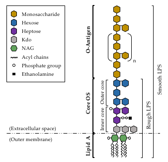
Prior to its attachment to the lipid-A-core-OS structure of LPS, before the whole complex is exported to the external side of the outer membrane, the O-antigen needs to be fully synthesized. O-antigen biosynthesis begins with the generation of the lipid-linked glycan intermediate undecaprenyl phosphate (UndP) [49] and, once generated, a sugar transfer reaction takes place. In this regard, A. piscicola AH-3 has been shown to use WecP to transfer N-acetyl-galactosamine [50]. Following this reaction, the O-antigen unit is flipped across the bacterial inner membrane by the Wzx protein [51], and assembled on the periplasmic side of the inner membrane by the Wzy O-antigen polymerase [52]. The O-antigen is elongated until it reaches the final polymer length, in a process regulated by the O-antigen chain length regulator Wzz [53]A. piscicolaAH-3 (O:34) has been shown to follow the Wzx/Wzy-dependent pathway for O-antigen assembly, as both wzy and wzx genes are found in the O-antigen gene cluster of this strain [54]. Interestingly, although the first sugar of the O-antigen repeating unit seems to be determinant for the generation of glycosidic bonds [55], the Wzy enzyme of A. piscicola AH-3 (O:34) has been shown to be permissive with this first sugar at the non-reducing end [56].

Structurally, O-antigens are composed of oligosaccharide polymers of various lengths, constituted by repeating subunits of 1–6 different sugars (Figure 2). The structural diversity of these O-polysaccharides leads to great variability among Gram-negative species, and even strains [57]. This variability (specially at the terminal sugar) confers immunological specificity to the O-antigen, giving rise to a large number of O-antigen groups or serogroups. To date, 97 different serogroups have been identified in the Aeromonas genus, and there are still many strains not belonging to a known serogroup yet [58]. Despite the high variability of their chemical composition, some aeromonad O-antigen structures have been determined by chemical analysis of their genome sequences, or inferred by bioinformatics [59][60][61]. In addition, the genes involved in O-antigen biosynthesis have been described in A. hydrophila PPD134/91 (O:18) and A. piscicola AH-3 (O:34) [54][62]. As reported for other clusters involved in polysaccharide biosynthesis, three classes of genes have been found: those that code for glycosyltransferases, those involved in the biosynthesis of activated sugars, and those whose products are necessary for O-antigen translocation and polymerization. Of particular note, sequence comparison of different Aeromonas spp. has shown epidemic strains to have larger O-antigen gene clusters than those previously reported for A. piscicola AH-3 and A. hydrophila PPD134/91 [63], and to contain genes for 3-acetamido-3,6-dideoxy-D-galactose biosynthesis, suggesting an important role of this sugar in the O-antigen structure of epidemic strains.

4. Capsular Polysaccharide

The bacterial capsule is a highly hydrated structure found on the cell surface of a broad range of bacterial species. It is composed of repeating monosaccharide units that are joined together by glycosidic bonds, forming homo- or heteropolymers. These large polysaccharides, usually negatively charged, extend far beyond the cell-wall components to produce a thick, protective coat around the entire bacterial cell [32]. Due to their location on the outermost layer of the bacterial cell, capsules are frequently involved in mediating direct interactions between bacteria and their environment, and are thus considered important virulence factors for many bacterial pathogens. Several functions have been assigned to these surface structures, including prevention of desiccation, adherence, biofilm formation, resistance to both specific and non-specific host immunity, and mediating the diffusion of molecules through the cell surface [64]. In Aeromonas spp., capsule biosynthesis has been shown to contribute to bacterial adhesion to different fish cell lines in A. salmonicida and A. piscicola, increasing the invasion and survival abilities of these species [65][66]Given that all hydroxyl groups present within each monosaccharide may be involved in the formation of a glycosidic bond, the union between any two monosaccharides constituting the polysaccharide chain can occur in numerous configurations, leading to a large structural diversity among bacterial capsules [64]. Despite this high variability, the chemical composition of some Aeromonas spp. capsules has actually been described. Mesophilic Aeromonas spp. belonging to serotypes O:11 (e.g., A. hydrophila TF7 and LL1) and O:34 (e.g., A. hydrophila Ba5 and A. piscicola AH3) have been shown to produce a capsule composed of D-glucose, D-mannose, L-rhamnose, D-mannuronic acid, and acetic acid heteropolymers (in distinct molar ratios, determined by the species serogroup) [67]. Regarding non-motile Aeromonas spp., A. salmonicida A449, A450 and A894 have been shown to produce a similar capsule, composed of D-glucose, D-mannose, L-rhamnose, N-acetylmannosamine, and mannuronic acid [68]. However, A. salmonicida 80204-1 produces a rather different CPS, composed of repeating units of a linear trisaccharide of 2-acetamido-2-deoxy-D-quinovose, 3-[(N-acetyl-L-alanyl) amido]-3-deoxy-D-quinovose, and 2-acetamido-2-deoxy-D-galacturonic acid [69], and yet another capsule of different chemical composition has been recently identified in the Aeromonas sp. strain AMG272, isolated from agricultural soil. In this case, the CPS is a large heteropolysaccharide, composed of repeating units of a branched pentasaccharide of D-galactose, N-acetyl-D-glucosamine, N-acetyl-D-galactosamine, and 3-acetamido-4-O-acetyl-3,6-dideoxy-D-galactose [70].
At the genetic level, the genes required for CPS biosynthesis and export in Aeromonas spp. were first described in A. hydrophila PPD134/91 and JCM3980 (O:18) [62][71]. In these species, the capsule clusters contain 13 genes and are arranged into three distinct regions. Genes of regions 1 and 3 are involved in capsule maturation and export, while those of region 2 are responsible for CPS biosynthesis, and are serotype-specific [62]. Interestingly, two different capsules have been identified in Aeromonas spp. region 2: 2A and 2B. The gene cluster of 2A capsules (mainly found in serogroups O:18 and O:34) is about 10 kb long and contains five ORFs, while that of 2B capsules (found in serogroups O:21 and O:27) is about 5 kb long and contains four ORFs [71]. Of particular note, although no clusters involved in capsule biosynthesis have been identified in A. hydrophila 1051-88 (O:34), two genes (orf1 and wcaJ) have been described as responsible for CPS production in this strain [72].

5. α-Glucan

Glucans are the most widespread polysaccharides in nature. They are composed of D-glucose monomers linked to each other by glycosidic bonds, and show a great chemical and structural diversity. According to their polymer conformation, bacterial glucans are divided into two major types: α-glucan and β-glucan. Out of the several different α-glucans described in Gram-negative bacteria, glycogen is the most studied one. This biopolymer serves as the major carbon- and energy-storage compound, and is thus typically accumulated under nutrient-depletion conditions [73]Structurally, glycogen is known to be comprised of α-D-glucosyl units connected by α-1,4-linkages, and branched through α-1,6-glycosidic bonds [74]. Depending on the source, glycogen molecules vary in chain length and branching frequency, which determines their rate of degradation, long and highly branched chains being more rapidly degraded than shorter and slightly branched ones [75]In Aeromonas spp., a similar surface glucan has been described in A. piscicola AH-3, and A. hydrophila strains AH-1 and PPD134/91 [76]This α-glucan, highly expressed at temperatures below 20 °C, is also comprised of α-D-glucosyl units connected by α-1,4-linkages, and branched through α-1,6-glycosidic bonds (Figure 3). It is synthesized via UDP-Glucose (UDP-Glc), with the help of the UDP-Glc pyrophosphorylase GlgC and the glycogen synthase GlgA. Interestingly, in these species, GlgA is able to use UDP-Glc to produce α-glucan, and probably ADP-Glc as well for glycogen biosynthesis. In addition, in A. piscicola AH-3, the absence of GlgC does not seem to affect either LPS O-antigen or α-glucan biosynthesis, while the absence of GlgA results in incorrect LPS core-OS formation and reduced α-glucan production. Moreover, A. piscicola AH-3 synthesizes UDP-Glc, also needed for the formation of the LPS inner core, via both GlgC and GalU. In the absence of GalU (which consequently leads to reduced levels of UDP-Glc), A. piscicola AH-3 establishes a preference for survival and pathogenesis, abolishing the formation of surface α-glucan and rather producing a complete LPS core. Unlike the LPS O-antigen, which has a predominant role in both cell adhesion and biofilm formation, α-glucan does not seem to have a significant role in Aeromonas spp. cell adhesion, which supports the preference of these bacteria to produce LPS rather than α-glucan [76]Surprisingly, A. piscicola AH-3 GalU mutants have been shown to lack the LPS O-antigen fraction, although this is suggested to occur because of their inability to incorporate the terminal galactose residue to the LPS core-OS structure [77]Of particular note, in this species, surface α-glucan and LPS O-antigen are both exported via WecP, and ligated to the bacterial surface through WaaL, despite being independent polysaccharides [76].
Figure 3. Structure representation of the surface α-glucan described in A. piscicola AH-3. The polysaccharide chain is comprised of α-D-glucosyl units connected by α-1,4-linkages, and branched through α-1,6-glycosidic bonds (m, n, and p stand for different numbers of repeated D-glucose monomers).

6. Future Perspectives and Concluding Remarks

Among the different pathogenic factors associated with Aeromonas spp. virulence, surface glucans have been shown to play an important role in host–pathogen interactions, contributing to adherence, colonization, and overall survival of pathogenic strains. Given that the number of reported infections caused by Aeromonas spp. has been steadily rising in recent years [9], fully understanding the mechanisms underlying the biogenesis and regulation of Aeromonas spp. surface glucan structures seems to be crucial for novel therapeutic strategies. In this regard, some advances have been made in recent years. LPS, for instance, has been shown to be a key elicitor of the host immune system [30], but also to provoke an acute pathophysiological response that causes damage to tissues and organs [31]. To overcome this problem, LPS variants that stimulate the immune response without toxic effects have been explored as immunotherapeutics. For instance, Salmonella minnesota LPS with chemical variations in its lipid-A molecule has been used as a vaccine adjuvant in mice, resulting in reduced activation of the MyD88-dependent response [78]. Similarly, other studies have bioengineered various Gram-negative species (e.g., Neisseria meningitidis and non-pathogenic E. coli strains) to produce different LPS glycoforms [79][80]Although a similar range of immune stimulation was obtained, not all trends observed for the immune recognition of E. coli lipid A held true for N. meningitidis, highlighting the importance of specifically studying the LPS-dependent immune modulation caused by Aeromonas spp. In the same line, bacterial capsules have been historically used as vaccine target antigens for some bacterial species [81][82], although the emergence of antibiotic-resistant bacteria in recent years makes it necessary to find alternative approaches to combat this public health crisis. In this regard, it will be critical to identify the signals that stimulate and/or repress CPS biosynthesis in Aeromonas spp., and unravel the detailed network of molecular receptors and effectors of such signals. Given that flagellin glycosylation seems to be involved in several biological functions related to Aeromonas spp. pathogenesis and virulence [23][20][24], fully understanding the molecular mechanisms underlying such process may also allow for the development of novel antimicrobial strategies. Similarly, in regard to α-glucan, several published reports indicate that the use of yeast β-glucans increases fish resistance to Aeromonas spp. infection, by enhancing the non-specific immune response [83][84]. In this regard, administration of Aeromonas spp. α-glucan, instead of yeast β-glucans, could represent an improved strategy for providing protection against Aeromonas spp. disease.

References

  1. Martin-Carnahan, A.; Joseph, S.W. Order XII. Aeromonadales ord. nov. In Bergey’s Manual® of Systematic Bacteriology; Brenner, D.J., Krieg, N.R., Staley, J.T., Eds.; Springer: Boston, MA, USA, 2005; pp. 556-587.
  2. Janda, J.M.; Abbott, S.L.; The Genus Aeromonas : Taxonomy, Pathogenicity, and Infection. Clinical Microbiology Reviews 2010, 23, 35-73, 10.1128/cmr.00039-09.
  3. Majeed, K.N.; Egan, A.F.; Rae, I.C. Mac; Production of exotoxins by Aeromonas spp. at 5°C. Journal of Applied Bacteriology 1990, 69, 332-337, 10.1111/j.1365-2672.1990.tb01524.x.
  4. Galindo, C.L.; Chopra, A.K. Aeromonas and Plesiomonas species. In Food Microbiology: Fundamentals and Frontiers; Doyle, M.P., Beuchat, L.R., Montville, T.J., Eds.; ASM Press: Washington, DC, USA, 2007; pp. 381-400.
  5. Monette, S.; Dallaire, A.D.; Mingelbier, M.; Groman, D.; Uhland, C.; Richard, J.P.; Paillard, G.; Johannson, L.M.; Chivers, D.P.; Ferguson, H.W.; et al. Massive Mortality of Common Carp (Cyprinus carpio carpio) in the St. Lawrence River in 2001: Diagnostic Investigation and Experimental Induction of Lymphocytic Encephalitis. Veterinary Pathology 2006, 43, 302-310, 10.1354/vp.43-3-302.
  6. Rahman, M.; Colque-Navarro, P.; Kühn, I.; Huys, G.; Swings, J.; Möllby, R.; Identification and Characterization of Pathogenic Aeromonas veronii Biovar Sobria Associated with Epizootic Ulcerative Syndrome in Fish in Bangladesh. Applied and Environmental Microbiology 2002, 68, 650-655, 10.1128/aem.68.2.650-655.2002.
  7. Janda, J.M.; Abbott, S.L.; Evolving Concepts Regarding the Genus Aeromonas: An Expanding Panorama of Species, Disease Presentations, and Unanswered Questions. Clinical Infectious Diseases 1998, 27, 332-344, 10.1086/514652.
  8. Teunis, P.; Figueras, M.J.; Reassessment of the Enteropathogenicity of Mesophilic Aeromonas Species. Frontiers in Microbiology 2016, 7, 1395, 10.3389/fmicb.2016.01395.
  9. Pal, M.; Ayele, Y.; Durglishvili, N.; Emerging Role of Aeromonas hydrophila as a Foodborne Pathogen of Public Health Concern. EC Microbiology 2020, 16, 55-58.
  10. Parker, J.L.; Shaw, J.G.; Aeromonas spp. clinical microbiology and disease. Journal of Infection 2011, 62, 109-118, 10.1016/j.jinf.2010.12.003.
  11. Fernández-Bravo, A.; Figueras, M.J.; An Update on the Genus Aeromonas: Taxonomy, Epidemiology, and Pathogenicity. Microorganisms 2020, 8, 129-167, 10.3390/microorganisms8010129.
  12. Schäffer, C.; Messner, P.; Emerging facets of prokaryotic glycosylation. FEMS Microbiology Reviews 2017, 41, 49-91, 10.1093/femsre/fuw036.
  13. Kirov, S.M.; Tassell, B.C.; Semmler, A.B.T.; O’Donovan, L.A.; Rabaan, A.A.; Shaw, J.G.; Lateral Flagella and Swarming Motility in Aeromonas Species. Journal of Bacteriology 2002, 184, 547-555, 10.1128/jb.184.2.547-555.2002.
  14. Macnab, R.M.; How Bacteria Assemble Flagella. Annual Review of Microbiology 2003, 57, 77-100, 10.1146/annurev.micro.57.030502.090832.
  15. Canals, R.; Ramirez, S.; Vilches, S.; Horsburgh, G.; Shaw, J.G.; Tomás, J.M.; Merino, S.; Polar Flagellum Biogenesis in Aeromonas hydrophila. Journal of Bacteriology 2006, 188, 542-555, 10.1128/jb.188.2.542-555.2006.
  16. Canals, R.; Altarriba, M.; Vilches, S.; Horsburgh, G.; Shaw, J.G.; Tomás, J.M.; Merino, S.; Analysis of the Lateral Flagellar Gene System of Aeromonas hydrophila AH-3. Journal of Bacteriology 2006, 188, 852-862, 10.1128/jb.188.3.852-862.2006.
  17. Gavín, R.; Rabaan, A.A.; Merino, S.; Tomás, J.M.; Gryllos, I.; Shaw, J.G.; Lateral flagella of Aeromonas species are essential for epithelial cell adherence and biofilm formation. Molecular Microbiology 2002, 43, 383-397, 10.1046/j.1365-2958.2002.02750.x.
  18. Yonekura, K.; Maki-Yonekura, S.; Namba, K.; Complete atomic model of the bacterial flagellar filament by electron cryomicroscopy. Nature 2003, 424, 643-650, 10.1038/nature01830.
  19. Tabei, S.M.B.; Hitchen, P.G.; Day-Williams, M.J.; Merino, S.; Vart, R.; Pang, P.-C.; Horsburgh, G.J.; Viches, S.; Wilhelms, M.; Tomás, J.M.; et al. An Aeromonas caviae Genomic Island Is Required for both O-Antigen Lipopolysaccharide Biosynthesis and Flagellin Glycosylation. Journal of Bacteriology 2009, 191, 2851-2863, 10.1128/jb.01406-08.
  20. Merino, S.; Wilhelms, M.; Tomás, J.M.; Role of Aeromonas hydrophila Flagella Glycosylation in Adhesion to Hep-2 Cells, Biofilm Formation and Immune Stimulation. International Journal of Molecular Sciences 2014, 15, 21935-21946, 10.3390/ijms151221935.
  21. Wilhelms, M.; Fulton, K.M.; Twine, S.M.; Tomás, J.M.; Merino, S.; Differential Glycosylation of Polar and Lateral Flagellins in Aeromonas hydrophila AH-3. Journal of Biological Chemistry 2012, 287, 27851-27862, 10.1074/jbc.m112.376525.
  22. Parker, J.L.; Day-Williams, M.J.; Tomas, J.M.; Stafford, G.P.; Shaw, J.G.; Identification of a putative glycosyltransferase responsible for the transfer of pseudaminic acid onto the polar flagellin of Aeromonas caviae Sch3N. Microbiology Open 2012, 1, 149-160, 10.1002/mbo3.19.
  23. Rabaan, A.A.; Gryllos, I.; Tomás, J.M.; Shaw, J.G.; Motility and the Polar Flagellum Are Required for Aeromonas caviae Adherence to HEp-2 Cells. Infection and Immunity 2001, 69, 4257-4267, 10.1128/iai.69.7.4257-4267.2001.
  24. Fulton, K.M.; Mendoza-Barberá, E.; Twine, S.M.; Tomas, J.; Merino, S.; Polar Glycosylated and Lateral Non-Glycosylated Flagella from Aeromonas hydrophila Strain AH-1 (Serotype O11). International Journal of Molecular Sciences 2015, 16, 28255-28269, 10.3390/ijms161226097.
  25. Canals, R.; Vilches, S.; Wilhelms, M.; Shaw, J.G.; Merino, S.; Tomás, J.M.; Non-structural flagella genes affecting both polar and lateral flagella-mediated motility in Aeromonas hydrophila. Microbiology 2007, 153, 1165-1175, 10.1099/mic.0.2006/000687-0.
  26. Seshadri, R.; Joseph, S.W.; Chopra, A.K.; Sha, J.; Shaw, J.; Graf, J.; Haft, D.; Wu, M.; Ren, Q.; Rosovitz, M.J.; et al. Genome Sequence of Aeromonas hydrophila ATCC 7966 T : Jack of All Trades. Journal of Bacteriology 2006, 188, 8272-8282, 10.1128/jb.00621-06.
  27. Forn-Cuní, G.; Tomás, J.M.; Merino, S.; Whole-Genome Sequence of Aeromonas hydrophila Strain AH-1 (Serotype O11). Genome Announcements 2016, 4, e00920-16, 10.1128/genomea.00920-16.
  28. Forn-Cuní, G.; Tomás, J.M.; Merino, S.; Genome Sequence of Aeromonas hydrophila Strain AH-3 (Serotype O34). Genome Announcements 2016, 4, e00919-16, 10.1128/genomea.00919-16.
  29. Forn-Cuní, G.; Fulton, K.M.; Smith, J.C.; Twine, S.M.; Mendoza-Barberà, E.; Tomás, J.M.; Merino, S.; Polar Flagella Glycosylation in Aeromonas: Genomic Characterization and Involvement of a Specific Glycosyltransferase (Fgi-1) in Heterogeneous Flagella Glycosylation. Frontiers in Microbiology 2021, 11, 595697, 10.3389/fmicb.2020.595697.
  30. Zamyatina, A.; Heine, H.; Lipopolysaccharide Recognition in the Crossroads of TLR4 and Caspase-4/11 Mediated Inflammatory Pathways. Frontiers in Immunology 2020, 11, 585146, 10.3389/fimmu.2020.585146.
  31. Gumenscheimer, M.; Mitov, I.; Galanos, C.; Freudenberg, M.A.; Beneficial or Deleterious Effects of a Preexisting Hypersensitivity to Bacterial Components on the Course and Outcome of Infection. Infection and Immunity 2002, 70, 5596-5603, 10.1128/iai.70.10.5596-5603.2002.
  32. Whitfield, C.; Valvano, M.A.; Biosynthesis and Expression of Cell-Surface Polysaccharides in Gram-Negative Bacteria. Advances in Microbial Physiology 1993, 35, 135-246, 10.1016/s0065-2911(08)60099-5.
  33. Silhavy, T.J.; Kahne, D.; Walker, S.; The Bacterial Cell Envelope. Cold Spring Harbor Perspectives in Biology 2010, 2, a000414, 10.1101/cshperspect.a000414.
  34. Lüderitz, O.; Galanos, C.; Lehmann, V.; Mayer, H.; Rietschel, E.T.; Weckesser, J.; Chemical structure and biological activities of lipid A's from various bacterial families. Naturwissenschaften 1978, 65, 578-585, 10.1007/bf00364907.
  35. Merino, S.; Rubires, X.; Knochel, S.; Tomas, J.M.; Emerging pathogens: Aeromonas spp.. International Journal of Food Microbiology 1995, 28, 157-168, 10.1016/0168-1605(95)00054-2.
  36. Wang, Z.; Li, J.; Altman, E.; Structural characterization of the lipid A region of Aeromonas salmonicida subsp. salmonicida lipopolysaccharide. Carbohydrate Research 2006, 341, 2816-2825, 10.1016/j.carres.2006.09.020.
  37. Hagar, J.A.; Powell, D.A.; Aachoui, Y.; Ernst, R.K.; Miao, E.A.; Cytoplasmic LPS Activates Caspase-11: Implications in TLR4-Independent Endotoxic Shock. Science 2013, 341, 1250-1253, 10.1126/science.1240988.
  38. Lagrange, B.; Benaoudia, S.; Wallet, P.; Magnotti, F.; Provost, A.; Michal, F.; Martin, A.; Di Lorenzo, F.; Py, B.F.; Molinaro, A.; et al. Human caspase-4 detects tetra-acylated LPS and cytosolic Francisella and functions differently from murine caspase-11. Nature Communications 2018, 9, 242, 10.1038/s41467-017-02682-y.
  39. Jimenez, N.; Lacasta, A.; Vilches, S.; Reyes, M.; Vazquez, J.; Aquillini, E.; Merino, S.; Regué, M.; Tomás, J.M.; Genetics and Proteomics of Aeromonas salmonicida Lipopolysaccharide Core Biosynthesis. Journal of Bacteriology 2009, 191, 2228-2236, 10.1128/jb.01395-08.
  40. Jimenez, N.; Canals, R.; Lacasta, A.; Kondakova, A.N.; Lindner, B.; Knirel, Y.A.; Merino, S.; Regué, M.; Tomás, J.M.; Molecular Analysis of Three Aeromonas hydrophila AH-3 (Serotype O34) Lipopolysaccharide Core Biosynthesis Gene Clusters. Journal of Bacteriology 2008, 190, 3176-3184, 10.1128/jb.01874-07.
  41. Wang, Z.; Li, J.; Vinogradov, E.; Altman, E.; Structural studies of the core region of Aeromonas salmonicida subsp. salmonicida lipopolysaccharide. Carbohydrate Research 2006, 341, 109-117, 10.1016/j.carres.2005.10.017.
  42. Knirel, Y.A.; Vinogradov, E.; Jimenez, N.; Merino, S.; Tomás, J.M.; Structural studies on the R-type lipopolysaccharide of Aeromonas hydrophila. Carbohydrate Research 2004, 339, 787-793, 10.1016/j.carres.2003.12.008.
  43. Raetz, C.R.H.; Whitfield, C.; Lipopolysaccharide Endotoxins. Annual Review of Biochemistry 2002, 71, 635-700, 10.1146/annurev.biochem.71.110601.135414.
  44. Kapp, A.; Freudenberg, M.; Galanos, C.; Induction of human granulocyte chemiluminescence by bacterial lipopolysaccharides. Infection and Immunity 1987, 55, 758-761, 10.1128/iai.55.3.758-761.1987.
  45. Huber, M.; Kalis, C.; Keck, S.; Jiang, Z.; Georgel, P.; Du, X.; Shamel, L.; Sovath, S.; Mudd, S.; Beutler, B.; et al. R-form LPS, the master key to the activation ofTLR4/MD-2-positive cells. European Journal of Immunology 2006, 36, 701-711, 10.1002/eji.200535593.
  46. Aguilar, A.; Merino, S.; Rubires, X.; Tomas, J.M.; Influence of osmolarity on lipopolysaccharides and virulence of Aeromonas hydrophila serotype O:34 strains grown at 37 degrees C. Infection and Immunity 1997, 65, 1245-1250, 10.1128/iai.65.4.1245-1250.1997.
  47. Merino, S.; Camprubí, S.; Tomás, J.M.; Effect of growth temperature on outer membrane components and virulence of Aeromonas hydrophila strains of serotype O:34. Infection and Immunity 1992, 60, 4343-4349, 10.1128/iai.60.10.4343-4349.1992.
  48. Merino, S.; Rubires, X.; Aguilar, A.; Albert, S.; Hernandez-Allé, S.; Bened, V.J.; Tomás, J.M.; Mesophilic Aeromonas sp. serogroup O:11 resistance to complement-mediated killing. Infection and Immunity 1996, 64, 5302-5309, 10.1128/iai.64.12.5302-5309.1996.
  49. Valvano, M. O Antigen Biosynthesis. In Comprehensive Natural Products II: Chemistry and Biology; Mander, L., Liu, H.-W., Eds.; Elsevier: Oxford, UK, 2010; pp. 297-314.
  50. Merino, S.; Jimenez, N.; Molero, R.; Bouamama, L.; Regué, M.; Tomás, J.M.; A UDP-HexNAc:Polyprenol-P GalNAc-1-P Transferase (WecP) Representing a New Subgroup of the Enzyme Family. Journal of Bacteriology 2011, 193, 1943-1952, 10.1128/jb.01441-10.
  51. Islam, S.T.; Lam, J.S.; Wzx flippase-mediated membrane translocation of sugar polymer precursors in bacteria. Environmental Microbiology 2013, 15, 1001-1015, 10.1111/j.1462-2920.2012.02890.x.
  52. Collins, L.V.; Hackett, J.; Molecular cloning, characterization, and nucleotide sequence of the rfc gene, which encodes an O-antigen polymerase of Salmonella typhimurium. Journal of Bacteriology 1991, 173, 2521-2529, 10.1128/jb.173.8.2521-2529.1991.
  53. Kalynych, S.; Valvano, M.A.; Cygler, M.; Polysaccharide co-polymerases: the enigmatic conductors of the O-antigen assembly orchestra. Protein Engineering Design and Selection 2012, 25, 797-802, 10.1093/protein/gzs075.
  54. Jimenez, N.; Canals, R.; Saló, M.T.; Vilches, S.; Merino, S.; Tomás, J.M.; The Aeromonas hydrophila wb * O34 Gene Cluster: Genetics and Temperature Regulation. Journal of Bacteriology 2008, 190, 4198-4209, 10.1128/jb.00153-08.
  55. Merino, S.; Gonzalez, V.; Tomás, J.M.; The first sugar of the repeat units is essential for the Wzy polymerase activity and elongation of the O-antigen lipopolysaccharide. Future Microbiology 2016, 11, 903-918, 10.2217/fmb-2015-0028.
  56. Merino, S.; Gonzalez, V.; Tomás, J.M.; The Polymerization of Aeromonas hydrophila AH-3 O-Antigen LPS: Concerted Action of WecP and Wzy. PLOS ONE 2015, 10, e0131905, 10.1371/journal.pone.0131905.
  57. Raetz, C.R.H. Bacterial lipopolysaccharides: A remarkable family of bioactive macroamphiphiles. In Escherichia Coli and Salmonella: Cellular and Molecular Biology; Neidhardt, F.C., Curtiss, R., III; Ingraham, J.L., Lin, E.C.C., Low, K.B., Magasanik, B., Reznikoff, W.S., Riley, M., Schaechter, M., Eds.; ASM Press: Washington, DC, USA, 1996; pp. 1035-1063.
  58. Thomas, L.V.; Gross, R.J.; Cheasty, T.; Rowe, B.; Extended serogrouping scheme for motile, mesophilic Aeromonas species. Journal of Clinical Microbiology 1990, 28, 980-984, 10.1128/jcm.28.5.980-984.1990.
  59. Merino, S.; Canals, R.; Knirel, Y.A.; Tomás, J.M.; Molecular and Chemical Analysis of the Lipopolysaccharide from Aeromonas hydrophila Strain AH-1 (Serotype O11). Marine Drugs 2015, 13, 2233-2249, 10.3390/md13042233.
  60. Knirel, Y.A.; Shashkov, A.S.; Senchenkova, S.N.; Merino, S.; Tomás, J.M.; Structure of the O-polysaccharide of Aeromonas hydrophila O:34; a case of random O-acetylation of 6-deoxy-L-talose. Carbohydrate Research 2002, 337, 1381-1386, 10.1016/s0008-6215(02)00136-2.
  61. Merino, S.; de Mendoza, E.; Canals, R.; Tomás, J.M.; Functional Genomics of the Aeromonas salmonicida Lipopolysaccharide O-Antigen and A-Layer from Typical and Atypical Strains. Marine Drugs 2015, 13, 3791-3808, 10.3390/md13063791.
  62. Zhang, Y.L.; Arakawa, E.; Leung, K.Y.; Novel Aeromonas hydrophila PPD134/91 Genes Involved in O-Antigen and Capsule Biosynthesis. Infection and Immunity 2002, 70, 2326-2335, 10.1128/iai.70.5.2326-2335.2002.
  63. Hossain, M.J.; Waldbieser, G.C.; Sun, D.; Capps, N.K.; Hemstreet, W.B.; Carlisle, K.; Griffin, M.J.; Khoo, L.; Goodwin, A.E.; Sonstegard, T.S.; et al. Implication of Lateral Genetic Transfer in the Emergence of Aeromonas hydrophila Isolates of Epidemic Outbreaks in Channel Catfish. PLOS ONE 2013, 8, e80943, 10.1371/journal.pone.0080943.
  64. Merino, S.; Tomás, J.M. Bacterial Capsules and Evasion of Immune Responses. In Encyclopedia of Life Sciences; John Wiley & Sons, Ltd: Chichester, UK, 2015; pp. 1-10.
  65. Merino, S.; Aguilar, A.; Rubires, X.; Simon-Pujol, D.; Congregado, F.; Tomás, J.M.; The role of the capsular polysaccharide of Aeromonas salmonicida in the adherence and invasion of fish cell lines. FEMS Microbiology Letters 1996, 142, 185-189, 10.1111/j.1574-6968.1996.tb08428.x.
  66. Merino, S.; Aguilar, A.; Rubires, X.; Abitiu, N.; Regué, M.; Tomás, J.M.; The role of the capsular polysaccharide of Aeromonas hydrophila serogroup O:34 in the adherence to and invasion of fish cell lines. Research in Microbiology 1997, 148, 625-631, 10.1016/s0923-2508(97)88086-2.
  67. Martínez, M.J.; Simon-Pujol, D.; Congregado, F.; Merino, S.; Rubires, X.; Tomás, J.M.; The presence of capsular polysaccharide in mesophilic Aeromonas hydrophila serotypes O:11 and O:34. FEMS Microbiology Letters 1995, 128, 69-73, 10.1111/j.1574-6968.1995.tb07502.x.
  68. Garrote, A.; Bonet, R.; Merino, S.; Simon-Pujol, M.D.; Congregado, F.; Occurrence of a capsule in Aeromonas salmonicida. FEMS Microbiology Letters 1992, 74, 127-131, 10.1016/0378-1097(92)90417-m.
  69. Wang, Z.; Larocque, S.; Vinogradov, E.; Brisson, J.R.; Dacanay, A.; Greenwell, M.; Brown, L.L.; Li, J.; Altman, E.; Structural studies of the capsular polysaccharide and lipopolysaccharide O-antigen of Aeromonas salmonicida strain 80204-1 produced under in vitro and in vivo growth conditions. European Journal of Biochemistry 2004, 271, 4507-4516, 10.1111/j.1432-1033.2004.04410.x.
  70. Contreras Sánchez-Matamoros, R.; Gil-Serrano, A.M.; Espuny, M.R.; Ollero, F.J.; Megías, M.; Rodríguez-Carvajal, M.A.; Structure of surface polysaccharides from Aeromonas sp. AMG272, a plant-growth promoting rhizobacterium isolated from rice rhizosphere. Carbohydrate Research 2018, 462, 1-6, 10.1016/j.carres.2018.03.012.
  71. Zhang, Y.L.; Lau, Y.L.; Arakawa, E.; Leung, K.Y.; Detection and genetic analysis of group II capsules in Aeromonas hydrophila. Microbiology 2003, 149, 1051-1060, 10.1099/mic.0.26144-0.
  72. Aguilar, A.; Merino, S.; Nogueras, M.M.; Regué, M.; Tomás, J.M.; Two genes from the capsule of Aeromonas hydrophila (serogroup O:34) confer serum resistance to Escherichia coli K12 strains. Research in Microbiology 1999, 150, 395-402, 10.1016/s0923-2508(99)80074-6.
  73. Preiss, J.; Romeo, T.; Molecular Biology and Regulatory Aspects of Glycogen Biosynthesis in Bacteria. Progress in Nucleic Acid Research and Molecular Biology 1994, 47, 299-329, 10.1016/s0079-6603(08)60255-x.
  74. Preiss, J.; Bacterial Glycogen Synthesis and its Regulation. Annual Review of Microbiology 1984, 38, 419-458, 10.1146/annurev.mi.38.100184.002223.
  75. Wang, L.; Wise, M.J.; Glycogen with short average chain length enhances bacterial durability. Naturwissenschaften 2011, 98, 719-729, 10.1007/s00114-011-0832-x.
  76. Merino, S.; Bouamama, L.; Knirel, Y.A.; Senchenkova, S.N.; Regué, M.; Tomás, J.M.; Aeromonas Surface Glucan Attached through the O-Antigen Ligase Represents a New Way to Obtain UDP-Glucose. PLoS ONE 2012, 7, e35707, 10.1371/journal.pone.0035707.
  77. Vilches, S.; Canals, R.; Wilhelms, M.; Saló, M.T.; Knirel, Y.A.; Vinogradog, E.; Merino, S.; Tomás, J.M.; Mesophilic Aeromonas UDP-glucose pyrophosphorylase (GalU) mutants show two types of lipopolysaccharide structures and reduced virulence. Microbiology 2007, 153, 2393-2404, 10.1099/mic.0.2007/006437-0.
  78. Casella, C.R.; Mitchell, T.C.; Putting endotoxin to work for us: Monophosphoryl lipid A as a safe and effective vaccine adjuvant. Cellular and Molecular Life Sciences 2008, 65, 3231-3240, 10.1007/s00018-008-8228-6.
  79. Zariri, A.; Pupo, E.; van Riet, E.; van Putten, J.P.M.; Modulating endotoxin activity by combinatorial bioengineering of meningococcal lipopolysaccharide. Scientific Reports 2016, 6, 36575, 10.1038/srep36575.
  80. Needham, B.D.; Carroll, S.M.; Giles, D.K.; Georgiou, G.; Whiteley, M.; Trent, M.S.; Modulating the innate immune response by combinatorial engineering of endotoxin. Proceedings of the National Academy of Sciences 2013, 110, 1464-1469, 10.1073/pnas.1218080110.
  81. Prevnar 13 . U.S. Food & Drug Administration. Retrieved 2021-12-6
  82. PNEUMOVAX 23—Pneumococcal Vaccine, Polyvalent . U.S. Food & Drug Administration. Retrieved 2021-12-6
  83. Rodríguez, I.; Chamorro, R.; Novoa, B.; Figueras, A.; β-Glucan administration enhances disease resistance and some innate immune responses in zebrafish (Danio rerio). Fish & Shellfish Immunology 2009, 27, 369-373, 10.1016/j.fsi.2009.02.007.
  84. Kumari, J.; Sahoo, P.K.; Dietary beta-1,3 glucan potentiates innate immunity and disease resistance of Asian catfish, Clarias batrachus (L.). Journal of Fish Diseases 2006, 29, 95-101, 10.1111/j.1365-2761.2006.00691.x.
More
Information
Subjects: Microbiology
Contributor MDPI registered users' name will be linked to their SciProfiles pages. To register with us, please refer to https://encyclopedia.pub/register :
View Times: 527
Revision: 1 time (View History)
Update Date: 06 Dec 2021
1000/1000