Submitted Successfully!
To reward your contribution, here is a gift for you: A free trial for our video production service.
Thank you for your contribution! You can also upload a video entry or images related to this topic.
Version Summary Created by Modification Content Size Created at Operation
1 + 3564 word(s) 3564 2021-11-08 02:57:21 |
2 format correct Meta information modification 3564 2021-11-22 02:02:20 |

Video Upload Options

Do you have a full video?

Confirm

Are you sure to Delete?
Cite
If you have any further questions, please contact Encyclopedia Editorial Office.
Zhang, L. Functional Nucleic-Acid Biosensors. Encyclopedia. Available online: https://encyclopedia.pub/entry/16220 (accessed on 27 July 2024).
Zhang L. Functional Nucleic-Acid Biosensors. Encyclopedia. Available at: https://encyclopedia.pub/entry/16220. Accessed July 27, 2024.
Zhang, Li. "Functional Nucleic-Acid Biosensors" Encyclopedia, https://encyclopedia.pub/entry/16220 (accessed July 27, 2024).
Zhang, L. (2021, November 20). Functional Nucleic-Acid Biosensors. In Encyclopedia. https://encyclopedia.pub/entry/16220
Zhang, Li. "Functional Nucleic-Acid Biosensors." Encyclopedia. Web. 20 November, 2021.
Functional Nucleic-Acid Biosensors
Edit

According to the latest Global Cancer Statistics, there are about 19.3 million new cancer cases worldwide and nearly 10 million people died of cancer in 2020. As the largest threat to human life, the early detection of cancer is an effective way to reduce its mortality. In addition, heavy metal poisoning and biological toxins also seriously endanger human health, and their detection methods still have some shortcomings. Against this backdrop, biosensors have been developed by integrating modern biotechnology and advanced physical technology. Biosensors are devices that are used for the rapid and sensitive detection of substances at the molecular level. The basic unit of the biosensor includes the identification element, transducer and detector, etc. The components of organisms with molecular recognition capabilities or the organism itself can be used as recognition elements.

biosensor biomedicine functional nucleic-acid mediator

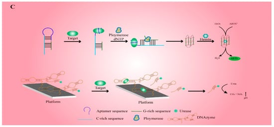
1. Working Principle for the Detection of Functional Nucleic-Acid Biosensors

There are many types of biosensors, which cannot function without signal transducers. Currently, transmitted signals consist primarily of electrical signals, optical signals, etc (Figure 1). FNA possesses the features of strong specificity, diverse functions and stable structure. Consequently, the detection performance of the FNA biosensors has been clearly improved as compared to conventional sensors.
Figure 1. Different working principle of functional nucleic-acid (FNA) biosensors. (A) Electrochemical FNA biosensor: The aptamer is screened in terms of the systematic evolution of ligands by exponential enrichment (SELEX) and fixed on the electrode to bind to the target. DNAzyme is immobilized on the electrode and then specifically recognizes the metal ion which activates it. After cutting the substrate chain, the signal is amplified by connecting the linker probe and the signal probe. (B) Fluorescent FNA biosensor: The turn-on principle and turn-off principle of the labeled fluorescent FNA biosensor designed by DNAzyme. The working principle of the label-free fluorescent FNA biosensor designed by DNAzyme and G-quadruplex. (C) Colorimetric FNA biosensor: The working principle of HRP mimics composed of G-quadruplex and hemin. The working principle of the colorimetric FNA biosensor composed of DNAzyme and urease.

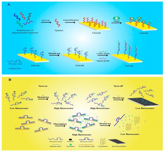
2. Electrochemical Functional Nucleic-Acid Biosensors

Electrochemical FNA biosensors are designed by applying FNA to an electrochemical transducer, which converts the analytical information generated by the electrochemical interaction of analytes on the electrode into measurable electrical signals [1]. The electrochemical analysis principle used by this kind of sensor has the characteristics of reusability, high stability, and high affinity. Therefore, it is widely used to detect ions, enzymes, proteins, viruses, and cells [2][3].
An aptamer is a kind of FNA that can be synthesized artificially in vitro. It is screened by the systematic evolution of ligands by exponential enrichment (SELEX) technology, which screens the oligonucleotide fragments that are specifically bound to the target substance from the oligonucleotide library that is synthesized in vitro [4]. Hence, an aptamer has the benefit of customization, and can theoretically be used to detect any target. Aptamers have been widely used as an important recognition element in electrochemical sensors. Using SELEX, Zhang et al screened the H37Rv aptamer of Mycobacterium tuberculosis through SELEX, hybridized it with treated oligonucleotides (AuNPs-DNA) and fixed it on a gold electrode for the detection of H37Rv [5]. When the test sample contains H37Rv, it can specifically bind with the target to release AuNPs-DNA, resulting in a change in the electrode conductivity and the ability to detect H37Rv. Compared with previous studies, FNA sensors can quickly detect H37Rv without culturing or special labeling. Based on a similar principle, electrochemical FNA sensors containing aptamers are also used to detect thrombin, bisphenol A, human papillomavirus, and other substances [6][7][8].
Mucin 1 (MUC1) is an important target that is related to various cancer types, and the development of detection methods has always attracted people’s attention [9][10]. Because surface-enhanced Raman scattering (SERS) has some problems such as poor detection specificity and high equipment requirements [11], a team fixed the MUC1 aptamer on a special electrode that was designed as an electrochemical FNA biosensor for detecting MUC1 [12]. Compared with the SERS method, which is commonly used in hospitals, the sensor offers prominent improvements in the detection range and the detection limit. The detection range of the system is 50–1000 nM, and the limit of detection (LOD) is reduced to 24 nM. It is applied to the detection of human serum samples, and other researchers have further developed many electrochemical FNA sensors for specific tumor cell detection on this basis [13]. Past studies have shown that carbon nanospheres (CNSs) have characteristics such as good chemical stability, bioaffinity, electrical conductivity, and non-biological toxicity [14]. Therefore, they are considered to be ideal materials for electrochemical FNA-sensor reaction platforms. Cao et al. used the MUC1 glycoprotein, which is expressed on the surface of colon cancer DLD-1 cells, as a target to screen specific aptamers and fix them on the CNS reaction platform in order to produce a sensor [13]. Compared with past research, this method greatly reduces the experimental difficulty and has similar detection effects [11]. Furthermore, the CNSs used in the system are non-biotoxic materials, which indicates that this sensor has the potential to be used for real-time detection in the human body. Karpik et al. applied a similar method to design an electrochemical FNA biosensor for detecting prostate cancer cells MUC1 [15]. In the experiment, the sensor detects the MUC1 expression patterns of different prostate cancer cell line models and normal prostate cells, and is able to distinguish between normal prostate cells and different types of prostate cancer cell in clinical testing. Compared with the existing methods such as ELISA and immunoassay, the sensor can resist the interference of a variety of substances, and also has higher detection sensitivity [16][17].
Another major type of FNA is DNAzyme, which has the ability to catalyze nucleic-acid cleavage. A detailed introduction to DNAzyme will be given later. As a result of the extraction of ores and the use of fossil fuels, heavy metal ions, as major environmental pollutants, pose serious threats to human life and health. Recent studies have shown that the electrochemical FNA biosensors developed by DNAzyme have high sensitivity and specificity for heavy metal ions, and thus, they have attracted extensive attention in the field of environmental detection [18][19][20][21]. Tang et al. selected 8–17 DNAzyme for the specific detection of Pb2+ [22]. Firstly, the designed substrate chain was hybridized with DNAzyme to form double stranded DNA (dsDNA). Then, DNAzyme was specifically activated by Pb2+, which cut and released the substrate chain. Subsequently, the connection probe and signal probe, which can generate electrical signals, were added and cyclically hybridized with DNAzyme to form long DNA structures. Therefore, the long-stranded DNA formed by a DNAzyme was able to contain multiple signal probes, and thus, could significantly amplify the electrical signal. The LOD of Pb2+ detected by the electrochemical FNA sensors was reduced to 6.1 pM, which is only one-tenth of that detected by previously examined electrochemical sensors [23][24]. As a result of these improvements in the detection sensitivity and biocompatibility of FNA, the sensor has great application prospects in the detection of human heavy metals.
In biomedical research and food safety investigations, it is often necessary to detect many important small molecular substances, such as adenosine triphosphate (ATP), pesticide residues, etc. However, due to the weight of small molecules, the detection conditions are difficult to ensure with accuracy. By combining aptamers and DNAzyme in aptazymes, one can harness the catalytic cleavage activity of DNAzyme when it has the recognition ability of the aptamer. In addition, aptazymes will only produce catalytic activity when aptamers bind to small molecular targets and cause structural changes [25]. Researchers used the aptamer that specifically recognizes ATP and 10–23 DNAzyme, which are sensitive to Mg2+, to form an aptazyme [24]. In addition, the target sequence of DNAzyme was connected with the signal probe to form a catalytic hairpin assembly (CHA). In the experiment, the ATP and Mg2+ in the system activated the enzyme activity of aptazyme, and then catalyzed the cleavage of CHA to release DNAzyme and a signal probe. The released Mg2+-sensitive DNAzyme continued to participate in the cutting of CHA, resulting in a large number of signal probes being released and bonding to the electrode, which caused an amplification of electrical signals. The electrochemical FNA sensors could accurately detect 0.6 nM ATP in the human serum samples.

3. Fluorescent Functional Nucleic-Acid Biosensors

Fluorescent FNA biosensors are composed of a recognition part, a transformation part, and a fluorescent material part. As FNA can be modified by fluorescent groups and quenchants and has strong a recognition ability, fluorescent FNA biosensors are widely used and comprise many types [26]. However, they are mainly divided into labeled fluorescent FNA biosensors and label-free fluorescent FNA biosensors [27].
The first report on labeled fluorescent FNA biosensors was published in 2000 [28]. The researchers used a TAMRA fluorophore and a Dabcyl quencher to label the dsDNA, which was composed of a substrate chain hybridized with 8–17 DNAzyme. In a natural state, the quenching effect of the quencher leads to the low-fluorescence reaction of the system. However, in the presence of Pb2+, in the above study, 8–17 DNAzyme was activated to cut the substrate chain and then release the substrate chain, which was labeled with fluorescent groups, resulting in the enhancement of the fluorescence signal. Finally, the concentration of Pb2+ was obtained by detecting the fluorescence signal intensity, but this method required the labelling of two groups on the enzyme chain, resulting in high experimental cost and time. In recent years, sensors for the detection of metal ions have been further developed. Fluorescein amidite (FAM) was used as a fluorescent group to label the substrate chain, and then hybridized with DNAzyme to form a bulged structure [29]. Then, ethidium bromide (EB) was embedded into the bulged structure as a quencher to quench the fluorescence signal. When DNAzyme was activated by Pb2+, the substrate was cut to release EB, so that the fluorescence signal was restored. The sensors made according to this fluorescence turn-on principle did not require the modification of the quencher on the DNAzyme, so the experimental cost was reduced and time was saved.
In contrast, the FNA sensors designed according to the opposite principle of fluorescence turn-off also showed good performance. Zhang et al used FAM to modify the substrate chain, and then hybridized it with UO22+-sensitive DNAzyme to form dsDNA [30]. In the experiment, the fluorescently labeled single-stranded DNA (ssDNA) released by the activated DNAzyme was adsorbed on a special reaction platform, causing the fluorescence signal to be quenched. Compared with the common colorimetric sensor, the FNA sensors designed using this method showed a 200-fold increase in sensitivity to UO22+. In addition, the researchers also designed a labeled fluorescent FNA sensor for the detection of H2S in the air based on the principle of fluorescence turn-off [31]. Up to now, a variety of metal ions have been detected by labeled fluorescent FNA sensors [32][33][34][35][36][37][38].
Label-free fluorescent FNA biosensors are usually designed by using intercalating fluorescent dyes to intercalate FNA, and then changing the fluorescence response through the interaction of the target with FNA, the most common of which is the G-quadruplex structure of FNA [39]. Guanine nucleotides can form a tetrad structure through hydrogen bonds, and multiple such tetrad structures can be stacked to form a G-quadruplex structure [40]. This type of sensor has a similar accuracy to the labeled type, but does not affect the functional activity of FNA. Therefore, it is widely used in many fields. Zhu et al. designed a dsDNA containing a G-rich substrate chain and a DNAzyme that is sensitive to UO22+, and used the nucleic-acid-intercalating dye SYBR Green I (SG I) for fluorescent labeling. When DNAzyme was not activated, the fluorescent dye was embedded in dsDNA and showed high fluorescence intensity. When UO22+ activated the DNAzyme, the substrate chain was cleaved to form a G-quadruplex, releasing fluorescent dyes, which resulted in a decrease in the fluorescence signal. Compared with the labeled type, the sensors had a similar sensitivity while reducing the difficulty of production [41]. Researchers also applied this principle to the detection of Pb2+ environmental samples [42]. As we all know, metal ion pollution in environmental pollution has always been one of the main threats to people’s health and safety. Therefore, a variety of types of label-free fluorescent FNA biosensors have developed that can be used for the detection of K+, Na+, Ba2+, Ir3+, Hg2+, Tl+, Tb3+ and other metal ions [43][44][45][46][47][48][49]. The G-quadruplex sequence and the metal cation will form a stable G-quadruplex structure. Screening of the aptamer with the G-quadruplex sequence (oligo-3), which has high selectivity for K+, can be applied to K+ detection [43]. It is then necessary to select the fluorophore which binds to the generated G-quadruplex. When K+ is present, the aptamer is induced to form a G-quadruplex and binds to a fluorophore to generate a high fluorescence reaction. In addition, the G-quadruplex can also be used for Hg2+ detection. Hg2+ produces a T-Hg2+-T mismatch with the DNA sequence [47]. Therefore, the G-quadruplex structure is destroyed, and the fluorescence is turned off. Highly specific aptamers with G-quadruplex sequences and fluorescent groups can be selected for the detection of other metal cations.
In addition, food safety is another major factor affecting human health; therefore, the development and application of label-free fluorescent FNA sensors are also of great value. Taghdisi et al. used aflatoxin B1 (AFB1)-specific aptamers containing a G-quadruplex-forming sequence to form a hairpin structure and used N-methyl mesoporphyrin IX (NMM) as a fluorescent reporter group [50]. It was found that when the aptamer binds to AFB1, it causes the hairpin structure to disintegrate and produce a G-quadruplex structure, and then combine with NMM to produce a strong fluorescent signal. The sensor was able to detect 30–900 pg/mL AFB1 within 30 min. In addition, it was successfully applied in the detection of actual products such as grape juice.
In recent years, in vivo imaging of tumor cells using fluorescent FNA biosensors has gradually become a research hot spot. This technological breakthrough has significantly promoted the development of the biomedical field. Studies have shown that quantum dots (QDs) are a type of nanomaterial with light stability and small size, and are good tools for cell imaging [51]. Chu et al. combined prostate-specific membrane antigen (PSMA) aptamers with luminescent QDs for prostate cancer cellular imaging [52]. The conjugate can target and label living cells, which has important application prospects in the research of human prostate cancer. However, the use of QDs is limited because of their potential biological toxicity and background interference. Therefore, in recent studies, researchers have used carbon dots (CDs) as fluorescent signals to replace QDs. CDs, as a novel member of carbon family, are a type of nanomaterial with good biocompatibility, low biotoxicity and facile preparation. They have become more and more popular in the field of biological detection [53]. CA125, which is an important characteristic antigen of ovarian cancer, was chosen by one group of researchers as the detection target. The team prepared CA125 aptamer-functionalized CDs (CDs-Apta) for cellular imaging of ovarian cancer cells [54]. The experimental results show that the specificity and sensitivity of this method are significantly improved compared with other studies [55][56]. In addition, the cell survival rate was good after 48 h of treatment, indicating very low biotoxicity and the potential for use in patient bioassays. FNA biosensors for tumor cell imaging are being developed towards non-toxicity, high precision and high flexibility. In the future, they will play a key role in tumor mechanism research and drug development.

4. Colorimetric Functional Nucleic-Acid Biosensors

The colorimetric FNA biosensors are based on the color change reaction of a certain compound, which causes the color of the entire system to change, and the result of the change can be obtained using a spectrophotometer or by visual observation to obtain a quantitative or qualitative result [57]. In order to solve the problem of rapid and high-precision detection of target substances without special instruments, this type of sensor has received extensive attention. Li et al. used aptamers that specifically recognize miRNAs and G-rich DNA (GDNA) to form a hairpin structure [58]. When the aptamer is combined with miRNA, GDNA can combine with hemin to form a G-quadruplex/hemin DNAzyme. This is a common enzyme mimic with horseradish peroxidase (HRP) activity, which can be used to catalyze the conversion of colorless 2,2′-azino-bis (ABTS2−) into green ABTS. This design can achieve rapid and highly sensitive detection of miRNA.
Moreover, colorimetric FNA biosensors also show great application potential in the detection of human pathogens. In a recent study, researchers screened out DNAzymes activated by Helicobacter pylori (HP) protein and then hybridized the urease-linked substrate strand with the DNAzyme strand [59]. In the experiment, HP protein activated DNAzyme to cleave the substrate chain so that urease was released into the system, and then urease entered the detection area, which contained urea and phenol red, to hydrolyze urea into ammonia, causing pH changes and discoloration of the area. The sensor uses paper as a reaction platform and can be applied to detect HP in human feces, which has important application value in terms of protecting people’s intestinal health. The developed HP detection technologies are mainly divided into invasive and non-invasive tests [60][61][62]. Invasive tests include endoscopic biopsy-based histology, followed by a rapid urease test and molecular PCR. This will consume a lot of time and entail significant costs. Among the non-invasive tests, the urea breath test (UBT) and the stool antigen test are commonly used [63]. However, the result of the non-invasive test is not very reliable and may be affected by urease produced by other bacteria in the digestive tract.
In summary, colorimetric FNA biosensors effectively solve the problem of restricting experimentation due to equipment requirements in the fields of clinical diagnosis and food testing.

5. Nanotechnology in Functional Nucleic-Acid Biosensors

5.1. DNA Hydrogel

Benefiting from the development of self-assembled FNA materials, the same principles can be applied to the construction of DNA hydrogels. DNA hydrogels can be divided into pure DNA hydrogels and composite DNA hydrogels according to whether they contain other non-DNA components [64]. As hydrogels have good biocompatibility and structural variability, DNA hydrogels are widely used in FNA biosensors. Zhang et al. designed a DNA hydrogel formed via the self-assembly of Y-DNA and an aptamer, and embedded AuNPs in it for the detection of thrombin [65]. When thrombin is present, the aptamer binds to it, causing the hydrogel to dissolve. The negatively charged AuNPs and the positively charged fluorescent carrier in the detection system attract each other and produce a quenching effect. The protective effect of the hydrogel makes the FNA biosensors exhibit extremely high stability and specificity in complex samples such as serum. Based on the same principle, the researchers used this hydrogel to develop a colorimetric FNA biosensor for the detection of ochratoxin A in food [66].
In past research, most of the FNA biosensors designed have been disposable products. Improper handling after use will not only cause a huge waste of resources, but also serious harm to the environment. Therefore, in recent research, Mao et al. immobilized the designed soft DNA hydrogel on the surface of the designed reaction platform, and encapsulated HRP to design a set of catalytic systems that can be used for colorimetric analysis [67]. The biggest advantage of this system is that it can be quickly rebuilt after being placed in a buffer solution, and the sensor can be recycled after it is naturally dried. The results of this research have encouraged the development of subsequent recyclable FNA biosensors.

5.2. Metal Nanomaterials

Metal nanomaterials have many characteristics such as good biocompatibility, fluorescence performance, activation electrochemical performance, and biocatalytic performance [68]. In recent years, metal nanomaterials have been used more and more widely in the sensors field, which greatly improves the performance of the sensors. With the development of the chemical industry and other industries, Cd2+ and polychlorinated biphenyls (PCB), as toxic substances that can induce human cancer and a variety of serious diseases, have posed a major threat to human life and health; therefore, effective detection is of great significance. Recent studies have shown that AuNPs-modified electrodes are used in aptamer electrochemical FNA biosensors for the detection of Cd2+ and PCB. Finally, thanks to the advantages of AuNPs such as high dielectric properties and high electron density, the detection sensitivity of the sensors modified by AuNPs has been increased more than 10-fold [69][70]. In addition, silver nanoclusters (AgNCs) also showed great application prospects in environmental monitoring. Studies have shown that when two dark DNA-templated AgNCs are connected by complementary sequences, the fluorescence intensity will increase more than 500-fold [71].
Protein kinase is an enzyme that can transfer the phosphoric acid of ATP to polypeptide or protein [72]. The phosphorylation of protein that is regulated by protein kinase is of great significance for the normal physiological function of the protein. Therefore, protein kinase activity (PKA) has become an important physiological and biochemical indicator for serious diseases such as cancer and Alzheimer’s disease [73]. However, its detection process has long been subject to harsh conditions, cumbersome procedures, and high costs. Therefore, Wang et al. connected the ATP-specific aptamer to the dsDNA template (dsDNA-CuNCs) of copper nanoclusters (CuNCs) and used graphene oxide (GO) as the reaction platform [74]. As dsDNA-CuNCs have the characteristics of fluorescence and, due to the π–π stacking effect, GO and ssDNA will spontaneously adsorb and quench the fluorescence reaction, when ATP is combined with the aptamer, the aptamer forms a dsDNA structure so that the system maintains a strong fluorescence reaction. When ATP is converted to ADP, the aptamer is adsorbed to GO because of its ssDNA structure and the fluorescence of CuNCs is quenched. This fluorescent FNA biosensor significantly reduces the experimental difficulty and experimental cost, and the sensitivity meets the requirements of clinical diagnosis.

References

  1. Yin, X.-B. Functional nucleic acids for electrochemical and electrochemiluminescent sensing applications. TrAC Trends Anal. Chem. 2012, 33, 81–94.
  2. Li, X.-M.; Ju, H.-Q.; Ding, C.-F.; Zhang, S.-S. Nucleic acid biosensor for detection of hepatitis B virus using 2,9-dimethyl-1,10-phenanthroline copper complex as electrochemical indicator. Anal. Chim. Acta 2007, 582, 158–163.
  3. Wang, C.-F.; Sun, X.-Y.; Su, M.; Wang, Y.-P.; Lv, Y.-K. Electrochemical biosensors based on antibody, nucleic acid and enzyme functionalized graphene for the detection of disease-related biomolecules. Analyst 2020, 145, 1550–1562.
  4. Tuerk, C.; Gold, L. Systematic evolution of ligands by exponential enrichment: RNA ligands to bacteriophage T4 DNA polymerase. Science 1990, 249, 505–510.
  5. Zhang, X.; Feng, Y.; Duan, S.; Su, L.; Zhang, J.; He, F. Mycobacterium tuberculosis strain H37Rv electrochemical sensor mediated by aptamer and AuNPs–DNA. ACS Sens. 2019, 4, 849–855.
  6. Chung, S.; Moon, J.-M.; Choi, J.; Hwang, H.; Shim, Y.-B. Magnetic force assisted electrochemical sensor for the detection of thrombin with aptamer-antibody sandwich formation. Biosens. Bioelectron. 2018, 117, 480–486.
  7. Beiranvand, S.; Azadbakht, A. Electrochemical switching with a DNA aptamer-based electrochemical sensor. Mater. Sci. Eng. C 2017, 76, 925–933.
  8. Teengam, P.; Siangproh, W.; Tuantranont, A.; Henry, C.S.; Vilaivan, T.; Chailapakul, O. Electrochemical paper-based peptide nucleic acid biosensor for detecting human papillomavirus. Anal. Chim. Acta 2017, 952, 32–40.
  9. Raina, D.; Kosugi, M.; Ahmad, R.; Panchamoorthy, G.; Rajabi, H.; Alam, M.; Shimamura, T.; Shapiro, G.I.; Supko, J.; Kharbanda, S. Dependence on the MUC1-C oncoprotein in non–small cell lung cancer cells. Mol. Cancer Ther. 2011, 10, 806–816.
  10. Besmer, D.M.; Curry, J.M.; Roy, L.D.; Tinder, T.L.; Sahraei, M.; Schettini, J.; Hwang, S.-I.; Lee, Y.Y.; Gendler, S.J.; Mukherjee, P. Pancreatic ductal adenocarcinoma mice lacking mucin 1 have a profound defect in tumor growth and metastasis. Cancer Res. 2011, 71, 4432–4442.
  11. Wu, P.; Gao, Y.; Zhang, H.; Cai, C. Aptamer-guided silver–gold bimetallic nanostructures with highly active surface-enhanced raman scattering for specific detection and near-infrared photothermal therapy of human breast cancer cells. Anal. Chem. 2012, 84, 7692–7699.
  12. Song, J.; Zhou, Y.; Chen, B.; Lou, W.; Gu, J. Development of electrochemical aptamer biosensor for tumor marker MUC1 determination. Int. J. Electrochem. Sci. 2017, 12, 5618–5627.
  13. Cao, H.; Ye, D.; Zhao, Q.; Luo, J.; Zhang, S.; Kong, J. A novel aptasensor based on MUC-1 conjugated CNSs for ultrasensitive detection of tumor cells. Analyst 2014, 139, 4917–4923.
  14. Sun, X.; Li, Y. Ga2O3 and GaN semiconductor hollow spheres. Angew. Chem. Int. Ed. 2004, 43, 3827–3831.
  15. Karpik, A.E.; Crulhas, B.P.; Rodrigues, C.B.; Castro, G.R.; Pedrosa, V.A. Aptamer-based biosensor developed to monitor MUC1 released by prostate cancer cells. Electroanalysis 2017, 29, 2246–2253.
  16. Day, E.S.; Riley, R.S.; Billingsley, M.M. Antibody-Nanoparticle Conjugates to Enhance the Sensitivity of ELISA-Based Detection Methods; Public Library of Science: Newark, NJ, USA, 2017.
  17. Jacobs, M.V.; Snijders, P.; Van Den Brule, A.; Helmerhorst, T.; Meijer, C.; Walboomers, J. A general primer GP5+/GP6 (+)-mediated PCR-enzyme immunoassay method for rapid detection of 14 high-risk and 6 low-risk human papillomavirus genotypes in cervical scrapings. J. Clin. Microbiol. 1997, 35, 791–795.
  18. Li, X.; Yang, J.; Xie, J.; Jiang, B.; Yuan, R.; Xiang, Y. Cascaded signal amplification via target-triggered formation of aptazyme for sensitive electrochemical detection of ATP. Biosens. Bioelectron. 2018, 102, 296–300.
  19. Ji, R.; Niu, W.; Chen, S.; Xu, W.; Ji, X.; Yuan, L.; Zhao, H.; Geng, M.; Qiu, J.; Li, C. Target-inspired Pb2+-dependent DNAzyme for ultrasensitive electrochemical sensor based on MoS2-AuPt nanocomposites and hemin/G-quadruplex DNAzyme as signal amplifier. Biosens. Bioelectron. 2019, 144, 111560.
  20. Liao, X.; Luo, J.; Wu, J.; Fan, T.; Yao, Y.; Gao, F.; Qian, Y. A sensitive DNAzyme-based electrochemical sensor for Pb2+ detection with platinum nanoparticles decorated TiO2/α-Fe2O3 nanocomposite as signal labels. J. Electroanal. Chem. 2018, 829, 129–137.
  21. Xiao, Y.; Rowe, A.A.; Plaxco, K.W. Electrochemical detection of parts-per-billion lead via an electrode-bound DNAzyme assembly. J. Am. Chem. Soc. 2007, 129, 262–263.
  22. Tang, S.; Lu, W.; Gu, F.; Tong, P.; Yan, Z.; Zhang, L. A novel electrochemical sensor for lead ion based on cascade DNA and quantum dots amplification. Electrochim. Acta 2014, 134, 1–7.
  23. Tagar, Z.A.; Memon, N.; Agheem, M.H.; Junejo, Y.; Hassan, S.S.; Kalwar, N.H.; Khattak, M.I. Selective, simple and economical lead sensor based on ibuprofen derived silver nanoparticles. Sens. Actuators B Chem. 2011, 157, 430–437.
  24. Li, F.; Yang, L.; Chen, M.; Li, P.; Tang, B. A selective amperometric sensing platform for lead based on target-induced strand release. Analyst 2013, 138, 461–466.
  25. Liu, J.; Lu, Y. Adenosine-dependent assembly of aptazyme-functionalized gold nanoparticles and its application as a colorimetric biosensor. Anal. Chem. 2004, 76, 1627–1632.
  26. Wang, X.X.; Zhu, L.J.; Li, S.T.; Zhang, Y.Z.; Liu, S.Y.; Huang, K.L.; Xu, W.T. Fluorescent Functional Nucleic Acid: Principles, Properties and Applications in Bioanalyzing. TrAC Trends Anal. Chem. 2021, 141, 116292.
  27. Lee, J.; Lin, L.; Li, Y. Functional nucleic acids for fluorescence-based biosensing applications. In Advanced Fluorescence Reporters in Chemistry and Biology III; Springer: Cham, Switzerland, 2011; pp. 201–221.
  28. Li, J.; Lu, Y. A highly sensitive and selective catalytic DNA biosensor for lead ions. J. Am. Chem. Soc. 2000, 122, 10466–10467.
  29. Guo, Y.; Li, J.; Zhang, X.; Tang, Y. A sensitive biosensor with a DNAzyme for lead (II) detection based on fluorescence turn-on. Analyst 2015, 140, 4642–4647.
  30. Zhang, H.; Ruan, Y.; Lin, L.; Lin, M.; Zeng, X.; Xi, Z.; Fu, F. A turn-off fluorescent biosensor for the rapid and sensitive detection of uranyl ion based on molybdenum disulfide nanosheets and specific DNAzyme. Spectrochim. Acta Part A Mol. Biomol. Spectrosc. 2015, 146, 1–6.
  31. Yue, G.; Huang, D.; Luo, F.; Guo, L.; Qiu, B.; Lin, Z.; Chen, G. Highly selective fluorescence sensor for hydrogen sulfide based on the Cu (II)-dependent DNAzyme. J. Lumin. 2019, 207, 369–373.
  32. Ji, X.; Wang, Z.; Niu, S.; Ding, C. DNAzyme-functionalized porous carbon nanospheres serve as a fluorescent nanoprobe for imaging detection of microRNA-21 and zinc ion in living cells. Microchim. Acta 2020, 187, 1–9.
  33. Torabi, S.-F.; Wu, P.; McGhee, C.E.; Chen, L.; Hwang, K.; Zheng, N.; Cheng, J.; Lu, Y. In vitro selection of a sodium-specific DNAzyme and its application in intracellular sensing. Proc. Natl. Acad. Sci. USA 2015, 112, 5903–5908.
  34. Saran, R.; Liu, J. A silver DNAzyme. Anal. Chem. 2016, 88, 4014–4020.
  35. Zhou, W.; Vazin, M.; Yu, T.; Ding, J.; Liu, J. In Vitro Selection of Chromium-Dependent DNAzymes for Sensing Chromium (III) and Chromium (VI); UWSpace: Waterloo, ON, Canada, 2016.
  36. Huang, P.-J.J.; Liu, J. Rational evolution of Cd2+-specific DNAzymes with phosphorothioate modified cleavage junction and Cd2+ sensing. Nucleic Acids Res. 2015, 43, 6125–6133.
  37. Huang, P.-J.J.; Lin, J.; Cao, J.; Vazin, M.; Liu, J. Ultrasensitive DNAzyme beacon for lanthanides and metal speciation. Anal. Chem. 2014, 86, 1816–1821.
  38. Huang, P.-J.J.; Vazin, M.; Liu, J. In vitro selection of a new lanthanide-dependent DNAzyme for ratiometric sensing lanthanides. Anal. Chem. 2014, 86, 9993–9999.
  39. Chen, Q.; Guo, Q.; Chen, Y.; Pang, J.; Fu, F.; Guo, L. An enzyme-free and label-free fluorescent biosensor for small molecules by G-quadruplex based hybridization chain reaction. Talanta 2015, 138, 15–19.
  40. Lipps, H.J.; Rhodes, D. G-quadruplex structures: In vivo evidence and function. Trends Cell Biol. 2009, 19, 414–422.
  41. Zhua, P.; Zhanga, Y.; Xub, S.; Zhanga, X. G-quadruplex-assisted enzyme strand recycling for amplified label-free fluorescent detection of UO2 2. Chin. Chem. Lett. 2019, 30, 58–62.
  42. Zhan, S.; Wu, Y.; Luo, Y.; Liu, L.; He, L.; Xing, H.; Zhou, P. Label-free fluorescent sensor for lead ion detection based on lead (II)-stabilized G-quadruplex formation. Anal. Biochem. 2014, 462, 19–25.
  43. Sun, X.; Li, Q.; Xiang, J.; Wang, L.; Zhang, X.; Lan, L.; Xu, S.; Yang, F.; Tang, Y. Novel fluorescent cationic benzothiazole dye that responds to G-quadruplex aptamer as a novel K+ sensor. Analyst 2017, 142, 3352–3355.
  44. Ma, G.; Yu, Z.; Zhou, W.; Li, Y.; Fan, L.; Li, X. Investigation of Na+ and K+ Competitively Binding with a G-Quadruplex and Discovery of a Stable K+–Na+-Quadruplex. J. Phys. Chem. B 2019, 123, 5405–5411.
  45. Xu, L.; Chen, Y.; Zhang, R.; Gao, T.; Zhang, Y.; Shen, X.; Pei, R. A highly Sensitive Turn-on Fluorescent Sensor for Ba2+ Based on G-Quadruplexes. J. Fluoresc. 2017, 27, 569–574.
  46. Wang, M.; Wang, W.; Kang, T.-S.; Leung, C.-H.; Ma, D.-L. Development of an Iridium (III) complex as a G-quadruplex probe and its application for the G-quadruplex-based luminescent detection of picomolar insulin. Anal. Chem. 2016, 88, 981–987.
  47. Zhu, Q.; Liu, L.; Xing, Y.; Zhou, X. Duplex functional G-quadruplex/NMM fluorescent probe for label-free detection of lead (II) and mercury (II) ions. J. Hazard. Mater. 2018, 355, 50–55.
  48. Hoang, M.; Huang, P.-J.J.; Liu, J. G-quadruplex DNA for fluorescent and colorimetric detection of thallium (I). Acs Sens. 2016, 1, 137–143.
  49. Chen, Q.; Zuo, J.; Chen, J.; Tong, P.; Mo, X.; Zhang, L.; Li, J. A label-free fluorescent biosensor for ultratrace detection of terbium (III) based on structural conversion of G-quadruplex DNA mediated by ThT and terbium (III). Biosens. Bioelectron. 2015, 72, 326–331.
  50. Taghdisi, S.M.; Danesh, N.M.; Ramezani, M.; Abnous, K. A new amplified fluorescent aptasensor based on hairpin structure of G-quadruplex oligonucleotide-Aptamer chimera and silica nanoparticles for sensitive detection of aflatoxin B1 in the grape juice. Food Chem. 2018, 268, 342–346.
  51. Michalet, X.; Pinaud, F.; Bentolila, L.; Tsay, J.; Doose, S.; Li, J.; Sundaresan, G.; Wu, A.; Gambhir, S.; Weiss, S. Quantum dots for live cells, in vivo imaging, and diagnostics. Science 2005, 307, 538–544.
  52. Chu, T.C.; Shieh, F.; Lavery, L.A.; Levy, M.; Richards-Kortum, R.; Korgel, B.A.; Ellington, A.D. Labeling tumor cells with fluorescent nanocrystal–aptamer bioconjugates. Biosens. Bioelectron. 2006, 21, 1859–1866.
  53. Lim, S.Y.; Shen, W.; Gao, Z. Carbon quantum dots and their applications. Chem. Soc. Rev. 2015, 44, 362–381.
  54. Hamd-Ghadareh, S.; Salimi, A.; Fathi, F.; Bahrami, S. An amplified comparative fluorescence resonance energy transfer immunosensing of CA125 tumor marker and ovarian cancer cells using green and economic carbon dots for bio-applications in labeling, imaging and sensing. Biosens. Bioelectron. 2017, 96, 308–316.
  55. Chen, S.; Yuan, R.; Chai, Y.; Xu, Y.; Min, L.; Li, N. A new antibody immobilization technique based on organic polymers protected Prussian blue nanoparticles and gold colloidal nanoparticles for amperometric immunosensors. Sens. Actuators B Chem. 2008, 135, 236–244.
  56. Wu, L.; Sha, Y.; Li, W.; Wang, S.; Guo, Z.; Zhou, J.; Su, X.; Jiang, X. One-step preparation of disposable multi-functionalized g-C3N4 based electrochemiluminescence immunosensor for the detection of CA125. Sens. Actuators B Chem. 2016, 226, 62–68.
  57. Zhu, D.; Liu, B.; Wei, G. Two-Dimensional Material-Based Colorimetric Biosensors: A Review. Biosensors 2021, 11, 259.
  58. Li, D.; Cheng, W.; Yan, Y.; Zhang, Y.; Yin, Y.; Ju, H.; Ding, S. A colorimetric biosensor for detection of attomolar microRNA with a functional nucleic acid-based amplification machine. Talanta 2016, 146, 470–476.
  59. Ali, M.M.; Wolfe, M.; Tram, K.; Gu, J.; Filipe, C.D.; Li, Y.; Brennan, J.D. A DNAzyme-Based Colorimetric Paper Sensor for Helicobacter pylori. Angew. Chem. 2019, 131, 10012–10016.
  60. Jiang, J.; Chen, Y.; Shi, J.; Song, C.; Zhang, J.; Wang, K. Population attributable burden of Helicobacter pylori-related gastric cancer, coronary heart disease, and ischemic stroke in China. Eur. J. Clin. Microbiol. Infect. Dis. 2017, 36, 199–212.
  61. Talebi Bezmin Abadi, A. Helicobacter pylori: Emergence of a Superbug. Front. Med. 2014, 1, 34.
  62. Backert, S.; Clyne, M. Pathogenesis of Helicobacter pylori infection. Helicobacter 2011, 16, 19–25.
  63. Ferwana, M.; Abdulmajeed, I.; Alhajiahmed, A.; Madani, W.; Firwana, B.; Hasan, R.; Altayar, O.; Limburg, P.J.; Murad, M.H.; Knawy, B. Accuracy of urea breath test in Helicobacter pylori infection: Meta-analysis. World J. Gastroenterol. WJG 2015, 21, 1305.
  64. Li, J.; Mo, L.; Lu, C.-H.; Fu, T.; Yang, H.-H.; Tan, W. Functional nucleic acid-based hydrogels for bioanalytical and biomedical applications. Chem. Soc. Rev. 2016, 45, 1410–1431.
  65. Zhang, L.; Lei, J.; Liu, L.; Li, C.; Ju, H. Self-assembled DNA hydrogel as switchable material for aptamer-based fluorescent detection of protein. Anal. Chem. 2013, 85, 11077–11082.
  66. Zhou, L.; Sun, N.; Xu, L.; Chen, X.; Cheng, H.; Wang, J.; Pei, R. Dual signal amplification by an “on-command” pure DNA hydrogel encapsulating HRP for colorimetric detection of ochratoxin A. Rsc Adv. 2016, 6, 114500–114504.
  67. Mao, X.; Chen, G.; Wang, Z.; Zhang, Y.; Zhu, X.; Li, G. Surface-immobilized and self-shaped DNA hydrogels and their application in biosensing. Chem. Sci. 2018, 9, 811–818.
  68. Xu, W.; He, W.; Du, Z.; Zhu, L.; Huang, K.; Lu, Y.; Luo, Y. Functional nucleic acid nanomaterials: Development, properties, and applications. Angew. Chem. Int. Ed. 2021, 60, 6890–6918.
  69. Fakude, C.T.; Arotiba, O.A.; Mabuba, N. Electrochemical aptasensing of cadmium (II) on a carbon black-gold nano-platform. J. Electroanal. Chem. 2020, 858, 113796.
  70. Yao, X.; Chadan Chen, L.C.; Wei, X.; Cui, H.; Xu, H.; Fan, H. A Novel PCB77 Electrochemical Sensor Based on Nano-functionalized Electrode and Selected Aptamer. J. New Mater. Electrochem. Syst. 2020, 23, 66–70.
  71. Yin, B.-C.; Ma, J.-L.; Le, H.-N.; Wang, S.; Xu, Z.; Ye, B.-C. A new mode to light up an adjacent DNA-scaffolded silver probe pair and its application for specific DNA detection. Chem. Commun. 2014, 50, 15991–15994.
  72. Sun, K.; Chang, Y.; Zhou, B.; Wang, X.; Liu, L. Gold nanoparticles-based electrochemical method for the detection of protein kinase with a peptide-like inhibitor as the bioreceptor. Int. J. Nanomed. 2017, 12, 1905.
  73. Flajolet, M.; He, G.; Heiman, M.; Lin, A.; Nairn, A.C.; Greengard, P. Regulation of Alzheimer’s disease amyloid-β formation by casein kinase I. Proc. Natl. Acad. Sci. USA 2007, 104, 4159–4164.
  74. Wang, M.; Lin, Z.; Liu, Q.; Jiang, S.; Liu, H.; Su, X. DNA-hosted copper nanoclusters/graphene oxide based fluorescent biosensor for protein kinase activity detection. Anal. Chim. Acta 2018, 1012, 66–73.
More
Information
Contributor MDPI registered users' name will be linked to their SciProfiles pages. To register with us, please refer to https://encyclopedia.pub/register :
View Times: 593
Revisions: 2 times (View History)
Update Date: 27 Nov 2021
1000/1000
Video Production Service