Submitted Successfully!
To reward your contribution, here is a gift for you: A free trial for our video production service.
Thank you for your contribution! You can also upload a video entry or images related to this topic.
Version Summary Created by Modification Content Size Created at Operation
1 + 2084 word(s) 2084 2021-10-27 08:33:33 |
2 Format correct Meta information modification 2084 2021-10-27 09:37:33 |

Video Upload Options

Do you have a full video?

Confirm

Are you sure to Delete?
Cite
If you have any further questions, please contact Encyclopedia Editorial Office.
Cho, K. Retinoic Acid and Retinaldehyde Dehydrogenase. Encyclopedia. Available online: https://encyclopedia.pub/entry/15453 (accessed on 04 July 2024).
Cho K. Retinoic Acid and Retinaldehyde Dehydrogenase. Encyclopedia. Available at: https://encyclopedia.pub/entry/15453. Accessed July 04, 2024.
Cho, Kichul. "Retinoic Acid and Retinaldehyde Dehydrogenase" Encyclopedia, https://encyclopedia.pub/entry/15453 (accessed July 04, 2024).
Cho, K. (2021, October 27). Retinoic Acid and Retinaldehyde Dehydrogenase. In Encyclopedia. https://encyclopedia.pub/entry/15453
Cho, Kichul. "Retinoic Acid and Retinaldehyde Dehydrogenase." Encyclopedia. Web. 27 October, 2021.
Retinoic Acid and Retinaldehyde Dehydrogenase
Edit

Retinoic acid (RA) is an important biological metabolite synthesized from the retinol content (known as “vitamin A”) via a sequential cellular process in the retinoid signaling pathway (RSP). RSP-mediated RA biosynthesis is a vital physiological process in chordates, since RA interacts with the nuclear receptor superfamily, namely nuclear RA receptors (RARs) and retinoid X receptors (RXRs), bound to the RA response elements (RAREs) in the promoter region of RA target genes. Retinaldehyde dehydrogenases (RALDH) belongs to the oxidoreductase family and plays a critical role in RA synthesis from the retinaldehyde content; therefore, this enzyme is considered to be one of the key regulators of RA-related retinol metabolism and embryonic development.

retinaldehyde dehydrogenase retinoic acid embryogenesis adverse outcome pathway population decline

1. Pleiotropic Roles of Retinoic acid (RA) and RALDH Inhibitors

The RALDH may be categorized into different classes of aldehyde dehydrogenase (ALDH) superfamily, including class 1 (cytosolic proteins), class 2 (mitochondrial proteins), class 3 (tumor-related proteins, inducible, or found in the endoplasmic reticulum), and microsomal aldehyde dehydrogenase [1][2][3]. The structures of RALDHs had been determined in previous studies [3][4][5]. RA biosynthesis and signaling seem to be limited to chordates, due to the existence of RALDH [6]. The ALDH1A1, ALDH1A2, and ALDH1A3 genes code cytosolic RALDH enzyme (RALDH1, 2, 3) that catalyzes the synthesis of retinoic acid (RA) from retinaldehyde. The RNA and protein expression patterns of RALDH in human tissues can be verified in the transcriptomic and proteomic database of THE HUMAN PROTEIN ATLAS [7]. According to data, differentially expressed RNA and RALDH levels were determined in human tissue cell lines including brain, eye, endocrine tissues, lung, proximal digestive tract, gastrointestinal tract, liver and gallbladder, pancreas, kidney and urinary bladder, female and male tissues, muscle tissues, adipose and soft tissue, skin, bone marrow and lymphoid tissues, and blood. Among these, male tissues including ductus deferens, testis, epididymis, seminal vesicle, prostate, and female tissues such as the vagina, ovary fallopian tube, endometrium, cervix, uterine, placenta, breast showed relatively higher expressions of RNA and protein expression than the other tissues. Furthermore, endocrine tissues, lungs, and pancreas also exhibited relatively higher enzyme expression levels than those of other human tissues [https://www.proteinatlas.org/ENSG00000128918-ALDH1A2/tissue (accessed on 14 October 2021)]. Meanwhile, in the embryonic cells, three types of cytosolic RALDHs (RALDH1, 2, 3)-coding genes differentially expressed during the early embryogenesis [8]. According to a previous study by Niederreither et al. [8], ALDH1A1 expression was increased in developing lung, and stage-specifically expressed in stomach and intestine epithelial and mesenchymal layers of embryonic cells. On the other hand, ALDH1A2 was expressed in the kidney nephrogenic zone, and ALDH1A3 was specifically expressed in the intestinal lamina propria.
Due to the physiological importance, RALDH inhibition by diverse chemicals during the early embryogenesis potentially causes serious disease such as fetal alcohol spectrum disorder (FASD), congenital diaphragmatic hernia (CDH), and Moyamoya disease (MMD) [9][10][11]. Kot-Leibovich and Fainsod [10] reported that the alcohol (ethanol) compete with available RALDH activity during embryonic development, thereby inducing abnormally lowered RA level, and finally FASD. Mey et al. [9] also previously reported that RALDH2 inhibitors including nitrofen, 4-biphenyl carboxylic acid, bisdiamine, and SB-210661 induced posterolateral defects in the rat diaphragm. Furthermore, abnormal organ developments such as eye, kidney, and brain also can be induced by RALDH-inhibition, and thus potentially cause population decline of chordate in the ecosystem. Therefore, the regulation of RALDH inhibitors needs to be re-considered for the improvement of public health.
The physiological importance of RA is explained by pleiotropic modulation, including early embryogenesis, cell differentiation, and innate and adaptive immune homeostasis as shown in Table 1. Among the diverse roles of RA, morphogenesis and organogenesis during vertebrate development have been widely recognized over the past few decades [12][13][14][15][16][17][18]. Wilson et al. [19] had reported that retinoic acid-free embryos exhibited abnormal development of neural tube with alteration in the actin filaments and junctional complexes of the cell layer. Wang et al. [20] had previously shown that RALDH2-deficient mouse embryo exhibited abnormal development of posterior foregut derivatives (stomach and duodenum) along with liver growth. The relationship between mammalian craniofacial morphogenesis and RA signaling had also been reported in a previous review [21]. Furthermore, the RA-mediated Hox genes and adhesion molecule-induced skin morphogenesis via determination of axial orientation had been reported in an embryonic chicken skin explant culture [22]. RA-mediated epidermal morphogenesis had been reported by Asselineau et al. [22]. They had shown the delipidized serum-exposed abnormal human keratinocytes to be restored by RA addition, whereas higher RA concentration (>10−7 M) reduced epidermal maturation and produced parakeratosis [22]. Multiple roles of RA signaling for mammalian eye development had been reported in previous reviews [23][24]. RA signaling was shown to not only be required for interactions between the invaginating lens placode and optic vesicle, but also to promote the development of ventral retina and optic nerve, thereby modulating eye and photoreceptor development [25][23][24]. Dupé and Lumsden [26] had reported that the RA-mediated transforming signal is involved in the posterior hindbrain structure expansion, and that RA signaling is related to rhombomere boundaries, which are transverse developmental units of the embryonic hindbrain. Morphogenesis during embryonic development is regulated by diverse RA target genes. The enhanced key target genes expressed by RA signaling during early organogenesis include Hoxa1, Hoxb1, Hoxa3, Hoxd4, vHnf1, Pax6, Olig2, Cdx1, Pdx1, Hoxa5, Pitx2, Ret, and Stra8, whereas the repressed target genes include Fgf8 and TGF-ß1 [6]. These genes are highly related to the vertebrate developmental process. Specifically, over-expression of Hoxa1, Hoxb1, Hoxa3, Hoxd4, and vHnf1 genes is highly relevant to hindbrain anterior-posterior patterning developmental process, whereas that of Pax6 and Olig2 induce spinal cord motor neuron differentiation [6][27][28][29][30]. Repressed Fgf8 and overexpressed Cdx1 genes induce early somite formation, and heart anteroposterior patterning during embryonic development [6][31]. While the induction of meiosis is related to the Stra8 gene, anterior eye formation and kidney formation are relevant to the overexpression of Pitx2 and Ret genes, respectively [6][32].
Table 1. Physiological roles of retinoic acid (RA) in chordate.
  Role of Retinoic Acid (RA) References
Embryogenesis Neural tube development [19]
Posterior foregut derivatives development and liver growth [20]
craniofacial morphogenesis [21]
Skin morphogenesis [22]
Eye development [23][24]
posterior hindbrain structure expansion [26]
Early somite formation [6][31]
Heart anteroposterior patterning [6][31]
Kidney formation [6][32]
Others
(immunity, cell differentiation)
Neuron differentiation [6][14][27]
Anti-inflammatory naïve T cells differentiation [33]
HL-60 cells differentiation [34]

2. RA Signaling-Associated Eye Development and RALDH-Relevant AOP (AOP297)

The reciprocal relationship between RA signaling and mammalian eye development has been well-described over the past few decades [23][35][36][37][38][39].
Eye development in vertebrates occurs in the neural ectoderm, surface ectoderm, and neural crest-derived periocular mesenchyme of the embryonic cell [23]. As shown in the cross-section of neural ectoderm, pre-placodal ectoderm, and epidermis (Figure 1), mammalian eye development starts with the formation of a wall of the forebrain at either side of the optic grooves. The optic grooves gradually deepen as the neural folds become elevated, and the neural tube is closed by the surface ectoderm. The optic grooves extend to the near ectoderm to form early lens placode by thickening of the adjacent area. During the interaction of optic vesicles with the ectoderm, the generated lens placode invaginates to form the lens pit, and optic vesicles invaginate to form the double-layered optic cup. The lens vesicles are generated from the lens pit, are surrounded by the double-layered optic cup (Figure 2), and finally, become the fetal lens and mature lens after the development of primary lens fibers, secondary lens fibers, lens suture, and lens nucleus. A detailed explanation of vertebrate eye development has been well-described in previous reviews [40][41].
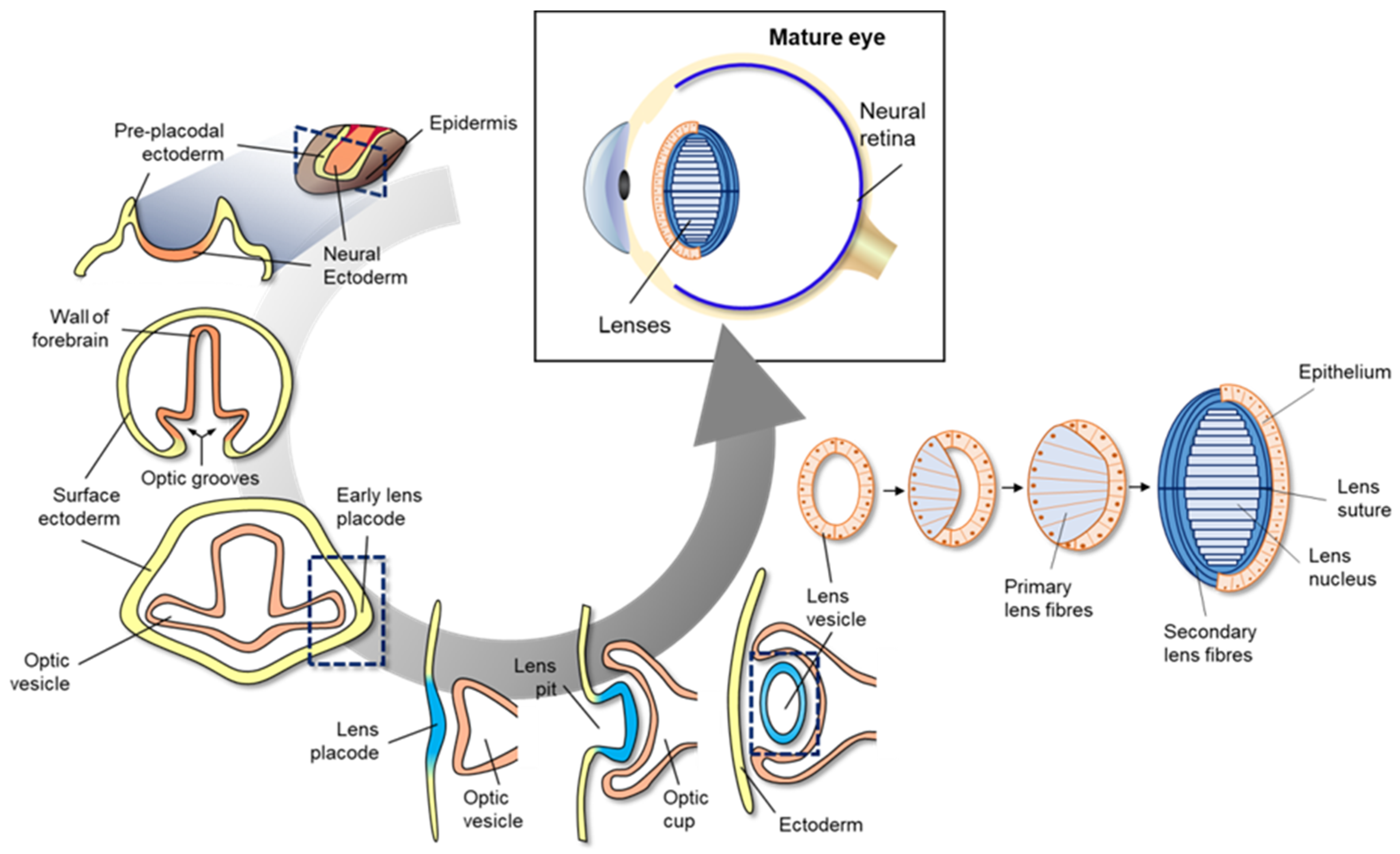
Figure 1. Schematic diagram of mammalian eye development. Eye development in vertebrates occurs in the neural ectoderm origin, surface ectoderm, and neural crest-derived periocular mesenchyme of embryonic cell. Mammalian eye development starts with formation of the wall of the forebrain on either side of the optic grooves. The optic grooves gradually deepen as the neural folds become elevated, and the neural tube is closed by surface ectoderm. The optic grooves extend to the near ectoderm to form early lens placode by thickening of the adjacent area. During the interaction of optic vesicles with the ectoderm, the generated lens placode invaginates to form the lens pit, and optic vesicles invaginate to form the double-layered optic cup. Lens vesicles are generated from the lens pit and are surrounded by the double-layered optic cup; finally, the generated lens vesicle becomes the fetal lens and mature lens after the development of primary lens fibers, secondary lens fibers, lens suture, and lens nucleus.
Toxins 13 00739 g004
Figure 2. Schematic representation of potential retinoic acid (RA) inhibition-related adverse outcome pathway (AOP). Inhibition of retinaldehyde dehydrogenase (RALDH) by known or unknown inhibitors potentially induces decreased cytosolic retinoic acid (RA) levels, which in turn, changes target gene expression, leads to abnormal embryonic eye development at individual level, and finally results in population decline of teleosts.
Many previous studies have reported that RA signaling plays a vital role in vertebrate eye development [23][24][35]. RA synthesis is mediated by three types of RALDHs, including RALDH1, RALDH2, and RALDH3. These enzyme-coding genes contribute to RA synthesis in non-overlapping locations during eye development [35]. For instance, when the optic cup forms, the RALDH2-coding gene is first expressed in the perioptic mesenchyme. Subsequently, RALDH1- and RALDH3-coding genes are expressed in the dorsal and ventral retina, respectively [35]. The synthesized RA then participates in eye development to form the optic cup, ventral retina, cornea, and eyelids [35]. Mouse knockout model-mediated genetic studies demonstrated the role of RA in embryonic eye development [35][42]. Raldh1-knockout mice had previously presented no noticeable adverse outcome and only showed slight RA activity in the dorsal retina owing to the compensation of Raldh3 during eye development [35][42]. However, Raldh1 and Raldh3 double knock out mutants induced overgrowth of mesenchyme in the cornea and eyelids due to defective apoptosis [35][43][44]. In another study, the Raldh3 knock out embryo exhibited eye and nasal defects, and thus could not survive due to blockage of the nasal passages [45]. Raldh3 knock out mutant showed abnormal embryonic development by shortening of the ventral retina. Furthermore, the study of Raldh2 knock out mutant and Raldh1/Raldh2 double knock out mutants showed the stoppage of optic cup development, which was subsequently rescued by RA addition [46]. These findings suggested that RALDH is an important molecule for embryonic eye development. A previous study had shown that citral competitively inhibits ventral RALDH of zebrafish embryo, thereby leading to reduced RA synthesis [25]. Citral administration under neurula-stage of the zebrafish embryo led to the deformity of a ventral retina in zebrafish eyes. Conversely, exogenous addition of RA induced a duplication of the zebrafish retina in a concentration-dependent manner during the initial formation of the optic primordia [24][47]. The results together indicated that the RALDH inhibition-associated undermined RA synthesis potentially causes abnormal eye development in chordates, including teleosts.
The potential AO can be sequentially explained from the molecular level to population level via the AOP framework. As shown in Figure 2, we schematized the potential AO by RALDH inhibition. At both cellular and molecular levels, RALDH inhibitors decreased the conversion of retinal to RA, thereby inhibiting the activation of target gene transcription. At the organ level, inhibited RA synthesis led to abnormal eye development, caused by inhibited optical elements. Finally, the abnormal eye development led to visual impairment at the individual level. Previous studies had reported that ocular coloboma, caused by incomplete closure of the choroid fissure, was potentially related to the ventral RA levels due to several genes; for example, Lrp6 (a bottleneck coreceptor in the canonical Wnt signaling pathway) knock out mutation resulted in ocular coloboma in mice [35][48]. The Lrp6 null mutant showed a dramatically inhibited level of RALDH in mouse eye [48]. The visual impairment potentially affected reproduction and survival of teleosts, with lowered fecundity and hence decline in the population level (Figure 2). However, the population decline caused by RALDH inhibition-mediated abnormal eye development still remains poorly understood. Therefore, the weak weight of evidence needs to be redeemed to completely construct the AOP framework.

3. Conclusions

RALDH is a key enzymatic component in cellular RA synthesis from retinaldehyde. The synthesized RA systematically regulates gene transcription via conversion of RAR or RXR receptors, thereby conducting morphogenesis and other functions during the embryonic development. In the current review, we comprehensively presented the importance of RA-mediated eye development, and potential AO derived from RALDH inhibition alongside the AOP (AOP297) framework, based on the existing knowledge.
RALDH-relevant gene deletion and RALDH-inhibitor administration caused abnormal eye development, and could potentially lead to population decline. Despite the weak weight of evidence regarding the relationship between RA inhibition and population decline, the AOP suggested the need for regulation of RALDH inhibitors to prevent potential risk of reproductive toxicity. However, further studies are required to support WoEs to construct a more complete AOP.

References

  1. Farres, J.; Wang, T.T.Y.; Cunningham, S.J.; Weiner, H. Investigation of the active site cysteine residue of rat liver mitochondrial aldehyde dehydrogenase by site-directed mutagenesis. Biochemistry 1995, 34, 2592–2598.
  2. Hempel, J.; Lindahl, R. Class III aldehyde dehydrogenase from rat liver: Super-family relationship to classes I and II and functional interpretations. Prog. Clin. Biol. Res. 1989, 290, 3–17.
  3. Lamb, A.L.; Newcomer, M.E. The structure of retinal dehydrogenase type II at 2.7 Å resolution: Implications for retinal specificity. Biochemistry 1999, 38, 6003–6011.
  4. Graham, C.E.; Brocklehurst, K.; Pickersgill, R.W.; Warren, M.J. Characterization of retinaldehyde dehydrogenase 3. Biochem. J. 2006, 394, 67–75.
  5. Moore, S.A.; Baker, H.M.; Blythe, T.J.; Kitson, K.E.; Kitson, T.M.; Baker, E.N. Sheep liver cytosolic aldehyde dehydrogenase: The structure reveals the basis for the retinal specificity of class 1 aldehyde dehydrogenases. Structure 1998, 6, 1541–1551.
  6. Duester, G. Retinoic acid synthesis and signaling during early organogenesis. Cell 2008, 134, 921–931.
  7. Pontén, F.; Jirström, K.; Uhlen, M. The Human Protein Atlas—A tool for pathology. J. Pathol. 2008, 216, 387–393.
  8. Niederreither, K.; Fraulob, V.; Garnier, J.M.; Chambon, P.; Dollé, P. Differential expression of retinoic acid-synthesizing (RALDH) enzymes during fetal development and organ differentiation in the mouse. Mech. Dev. 2002, 110, 165–171.
  9. Mey, J.; Babiuk, R.P.; Clugston, R.; Zhang, W.; Greer, J.J. Retinal dehydrogenase-2 is inhibited by compounds that induce congenital diaphragmatic hernias in rodents. Am. J. Pathol. 2003, 162, 673–679.
  10. Kot-Leibovich, H.; Fainsod, A. Ethanol induces embryonic malformations by competing for retinaldehyde dehydrogenase activity during vertebrate gastrulation. Dis. Models Mech. 2009, 2, 295–305.
  11. Lee, J.Y.; Moon, Y.J.; Lee, H.O.; Park, A.K.; Choi, S.A.; Wang, K.C.; Han, J.W.; Joung, J.G.; Kang, H.S.; Kim, J.E.; et al. Deregulation of retinaldehyde dehydrogenase 2 leads to defective angiogenic function of endothelial colony–Forming cells in pediatric moyamoya disease. Arterioscler. Thromb. Vasc. Biol. 2015, 35, 1670–1677.
  12. Cunningham, T.J.; Duester, G. Mechanisms of retinoic acid signalling and its roles in organ and limb development. Nat. Rev. Mol. 2015, 16, 110–123.
  13. Niederreither, K.; Dollé, P. Retinoic acid in development: Towards an integrated view. Nat. Rev. Genet. 2008, 9, 541–553.
  14. Påhlman, S.; Ruusala, A.I.; Abrahamsson, L.; Mattsson, M.E.K.; Esscher, T. Retinoic acid-induced differentiation of cultured human neuroblastoma cells: A comparison with phorbolester-induced differentiation. Cell Differ. 1984, 14, 135–144.
  15. Rhinn, M.; Dollé, P. Retinoic acid signalling during development. Development 2012, 139, 843–858.
  16. Rohwedel, J.; Guan, K.; Wobus, A.M. Induction of cellular differentiation by retinoic acid in vitro. Cells Tissues Organs 1999, 165, 190–202.
  17. Bayha, E.; Jørgensen, M.C.; Serup, P.; Grapin-Botton, A. Retinoic acid signaling organizes endodermal organ specification along the entire antero-posterior axis. PLoS ONE 2009, 4, e5845.
  18. Sandell, L.L.; Sanderson, B.W.; Moiseyev, G.; Johnson, T.; Mushegian, A.; Young, K.; Rey, J.P.; Ma, J.X.; Staehling-Hampton, K.; Trainor, P.A. RDH10 is essential for synthesis of embryonic retinoic acid and is required for limb, craniofacial, and organ development. Genes Dev. 2007, 21, 1113–1124.
  19. Wilson, L.; Gale, E.; Maden, M. The role of retinoic acid in the morphogenesis of the neural tube. J. Anat. 2003, 203, 357–368.
  20. Wang, Z.; Dollé, P.; Cardoso, W.V.; Niederreither, K. Retinoic acid regulates morphogenesis and patterning of posterior foregut derivatives. Dev. Biol. 2006, 297, 433–445.
  21. Osumi-Yamashita, N. Retinoic acid and mammalian craniofacial morphogenesis. J. Biosci. 1996, 21, 313–327.
  22. Asselineau, D.; Bernard, B.A.; Bailly, C.; Darmon, M. Retinoic acid improves epidermal morphogenesis. Dev. Biol. 1989, 133, 322–335.
  23. Cvekl, A.; Wang, W.L. Retinoic acid signaling in mammalian eye development. Exp. Eye Res. 2009, 89, 280–291.
  24. Hyatt, G.A.; Dowling, J.E. Retinoic acid. A key molecule for eye and photoreceptor development. Investig. Ophthalmol. Vis. Sci. 1997, 38, 1471–1475.
  25. Marsh-Armstrong, N.; McCaffery, P.; Gilbert, W.; Dowling, J.E.; Dräger, U.C. Retinoic acid is necessary for development of the ventral retina in zebrafish. Proc. Natl. Acad. Sci. USA 1994, 91, 7286–7290.
  26. Dupé, V.; Lumsden, A. Hindbrain patterning involves graded responses to retinoic acid signalling. Development 2001, 128, 2199–2208.
  27. Hack, M.A.; Sugimori, M.; Lundberg, C.; Nakafuku, M.; Götz, M. Regionalization and fate specification in neurospheres: The role of Olig2 and Pax6. Mol. Cell. Neurosci. 2004, 25, 664–678.
  28. Holland, L.Z. A chordate with a difference. Nature 2007, 447, 153–154.
  29. Niederreither, K.; Vermot, J.; Schuhbaur, B.; Chambon, P.; Dollé, P. Retinoic acid synthesis and hindbrain patterning in the mouse embryo. Development 2000, 127, 75–85.
  30. Novitch, B.G.; Wichterle, H.; Jessell, T.M.; Sockanathan, S. A requirement for retinoic acid-mediated transcriptional activation in ventral neural patterning and motor neuron specification. Neuron 2003, 40, 81–95.
  31. Zhao, X.; Duester, G. Effect of retinoic acid signaling on Wnt/β-catenin and FGF signaling during body axis extension. Gene Expr. Patterns 2009, 9, 430–435.
  32. Griffin, S.V.; Shankland, S.J. Renal hyperplasia and hypertrophy: Role of cell cycle regulatory proteins. In Seldin and Giebisch’s the Kidney; Academic Press: Cambridge, MA, USA, 2008; pp. 723–742.
  33. Mucida, D.; Park, Y.; Kim, G.; Turovskaya, O.; Scott, I.; Kronenberg, M.; Cheroutre, H. Reciprocal TH17 and regulatory T cell differentiation mediated by retinoic acid. Science 2007, 317, 256–260.
  34. Breitman, T.R.; Selonick, S.E.; Collins, S.J. Induction of differentiation of the human promyelocytic leukemia cell line (HL-60) by retinoic acid. Proc. Natl. Acad. Sci. USA 1980, 77, 2936–2940.
  35. Duester, G. Keeping an eye on retinoic acid signaling during eye development. Chem. Biol. Interact. 2009, 178, 178–181.
  36. Kumar, S.; Dollé, P.; Ghyselinck, N.B.; Duester, G. Endogenous retinoic acid signaling is required for maintenance and regeneration of cornea. Exp. Eye Res. 2017, 154, 190–195.
  37. Matt, N.; Ghyselinck, N.B.; Pellerin, I.; Dupé, V. Impairing retinoic acid signalling in the neural crest cells is sufficient to alter entire eye morphogenesis. Dev. Biol. 2008, 320, 140–148.
  38. Nedelec, B.; Rozet, J.M.; Taie, L.F. Genetic architecture of retinoic-acid signaling-associated ocular developmental defects. Hum. Genet. 2019, 138, 937–955.
  39. Smith, J.N.; Walker, H.M.; Thompson, H.; Collinson, J.M.; Vargesson, N.; Erskine, L. Lens-regulated retinoic acid signalling controls expansion of the developing eye. Development 2018, 145, dev167171.
  40. Dash, S.; Siddam, A.D.; Barnum, C.E.; Janga, S.C.; Lachke, S.A. RNA-binding proteins in eye development and disease: Implication of conserved RNA granule components. Wiley Interdiscip. Rev. RNA 2016, 7, 527–557.
  41. Heavner, W.; Pevny, L. Eye development and retinogenesis. Cold Spring Harb. Perspect. Biol. 2012, 4, a008391.
  42. Fan, X.; Molotkov, A.; Manabe, S.I.; Donmoyer, C.M.; Deltour, L.; Foglio, M.H.; Cuenca, A.E.; Blaner, W.S.; Lipton, S.A.; Duester, G. Targeted disruption of Aldh1a1 (Raldh1) provides evidence for a complex mechanism of retinoic acid synthesis in the developing retina. Mol. Cell. Biol. 2003, 23, 4637–4648.
  43. Matt, N.; Dupé, V.; Garnier, J.M.; Dennefeld, C.; Chambon, P.; Mark, M.; Ghyselinck, N.B. Retinoic acid-dependent eye morphogenesis is orchestrated by neural crest cells. Development 2005, 132, 4789–4800.
  44. Molotkov, A.; Molotkova, N.; Duester, G. Retinoic acid guides eye morphogenetic movements via paracrine signaling but is unnecessary for retinal dorsoventral patterning. Development 2006, 133, 1901–1910.
  45. Dupé, V.; Matt, N.; Garnier, J.M.; Chambon, P.; Mark, M.; Ghyselinck, N.B. A newborn lethal defect due to inactivation of retinaldehyde dehydrogenase type 3 is prevented by maternal retinoic acid treatment. Proc. Natl. Acad. Sci. USA 2003, 100, 14036–14041.
  46. Mic, F.A.; Molotkov, A.; Molotkova, N.; Duester, G. Raldh2 expression in optic vesicle generates a retinoic acid signal needed for invagination of retina during optic cup formation. Dev. Dyn. 2004, 231, 270–277.
  47. Hyatt, G.A.; Schmitt, E.A.; Marsh-Armstrong, N.R.; Dowling, J.E. Retinoic acid-induced duplication of the zebrafish retina. Proc. Natl. Acad. Sci. USA 1992, 89, 8293–8297.
  48. Zhou, C.J.; Molotkov, A.; Song, L.; Li, Y.; Pleasure, D.E.; Pleasure, S.J.; Wang, Y.Z. Ocular coloboma and dorsoventral neuroretinal patterning defects in Lrp6 mutant eyes. Dev. Dyn. 2008, 237, 3681–3689.
More
Information
Contributor MDPI registered users' name will be linked to their SciProfiles pages. To register with us, please refer to https://encyclopedia.pub/register :
View Times: 577
Entry Collection: Environmental Sciences
Revisions: 2 times (View History)
Update Date: 27 Oct 2021
1000/1000
Video Production Service