Submitted Successfully!
To reward your contribution, here is a gift for you: A free trial for our video production service.
Thank you for your contribution! You can also upload a video entry or images related to this topic.
Version Summary Created by Modification Content Size Created at Operation
1 + 4102 word(s) 4102 2021-09-07 10:18:32

Video Upload Options

We provide professional Video Production Services to translate complex research into visually appealing presentations. Would you like to try it?

Confirm

Are you sure to Delete?
Cite
If you have any further questions, please contact Encyclopedia Editorial Office.
Aboul-Soud, M.A.M. Induced Pluripotent Stem Cells. Encyclopedia. Available online: https://encyclopedia.pub/entry/14025 (accessed on 04 December 2024).
Aboul-Soud MAM. Induced Pluripotent Stem Cells. Encyclopedia. Available at: https://encyclopedia.pub/entry/14025. Accessed December 04, 2024.
Aboul-Soud, Mourad A. M.. "Induced Pluripotent Stem Cells" Encyclopedia, https://encyclopedia.pub/entry/14025 (accessed December 04, 2024).
Aboul-Soud, M.A.M. (2021, September 09). Induced Pluripotent Stem Cells. In Encyclopedia. https://encyclopedia.pub/entry/14025
Aboul-Soud, Mourad A. M.. "Induced Pluripotent Stem Cells." Encyclopedia. Web. 09 September, 2021.
Induced Pluripotent Stem Cells
Edit

The discovery of induced pluripotent stem cells (iPSCs) has made an invaluable contribution to the field of regenerative medicine, paving way for identifying the true potential of human embryonic stem cells (ESCs). iPSCs have been widely used in cardiac disease modelling, studying inherited arrhythmias, neural disorders including Alzheimer’s disease, liver disease, and spinal cord injury. Extensive research around identifying factors that are involved in maintaining the identity of ESCs during induction of pluripotency in somatic cells is undertaken. 

drug screening disease embryo induced pluripotent stem cells modelling

1. Introduction

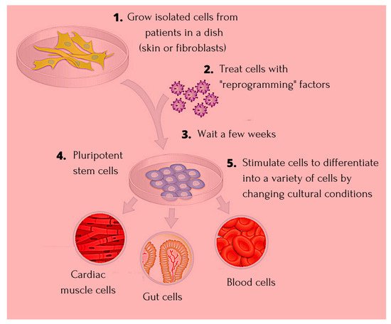
The science around terminal inactivation and deletion of genetic codes of heredity in somatic cells was postulated by the Weismann barrier theory [1]. The somatic cell nuclear transfer (SCNT) demonstration asserted the fact that the genetic code in somatic cells is not discarded, and that reactivation of the same is a possibility through careful manipulations [2]. Developmental biology entered a new dimension of achievement when the discovery of embryonic stem cells (ESCs) and their pluripotency was exhibited, and further research identified that on fusion of somatic cells like fibroblasts, and T-lymphocytes with ESCs, reprogramming of the former through expression of genes associated with pluripotency becomes a possibility [3][4]. The findings around SCNT and ESC fusion identified the possibility of reversion in somatic cells indicating the presence of reprogramming factors that bear the potential to act as epigenetic memory erasing factors [5]. The earliest study around generation of pluripotent stem cells from fibroblasts was linked to introduction of four crucial transcription factors including octamer binding transcription factor 3/4 (Oct3/4), sex determining region Y—box 2 (SRY-Sox2), Krüppel-like factor 4 (Klf4), and cellular-Myelocytomatosis (c-Myc) (OSKM) [6]. The allogenic trait of ESCs, risk of immune rejection in the recipient along with need for lifetime immunosuppression, and the ethicality around using the same, makes human induced pluripotent stem cells (iPSCs) an established candidate for regenerative therapies as they were found to not impact the host immune system [7]. The introduction of the iPSCs technology happened in the year 2006, and since then multiple observational studies have recounted its impact on cardiac diseases, ophthalmic conditions, as well as neurological disorders [8][9][10]. Figure 1 highlights the process of generating iPS cells.
Figure 1. Showing the process of progression and generating iPSC cells. Detailed description of creating iPSCs with reprogramming factors and differentiating them into a variety of cell types.
The nuclear reprogramming strategies, without compromising on safety and quality for therapeutic applications, include the integrative or nonintegrative transfer systems using viral or nonviral vectors. The first iPSCs were generated by integrating viral vectors, more popularly the retrovirus wherein the resultant iPSCs exhibited failure in complete expression of endogenous genes of pluripotency [11]. The more efficient viral vector has been documented to be the lentiviral vector (LV), which has recorded a reprogramming efficiency of between 0.1–1% [12][13][14]. To ensure increased safety for therapeutics, nonviral integrative systems have also been worked upon involving use of two plasmids; once encoding for c-Myc, and the other for the four reprogramming factors [15]. However, this system was also shown to have risk of integration, and low reprogramming efficiency. In case of nonintegrative nonviral systems for reprogramming, delivery of pluripotency marker genes has been done using self-replicating vectors, and cytoplasmic RNA. Though easy to work with, the reprogramming efficiency has been found to be lower than LV [16]. Today, research has identified possibility of successful reprogramming using microRNAs (miRNAs) which exhibit improved efficiency, wherein use of c-Myc has been replaced with miR-291-3p, miR-294, and miR-295 to generate homogenous colonies of human iPSCs [17]. The reprogramming methods have been highlighted in Table 1.
Table 1. Reprogramming strategies for iPSCs in human species. Various programming strategies with ensuring safety and quality for therapeutic applications include the integrative or nonintegrative transfer systems using viral or nonviral.

Vector

Cell Type

Genes

Efficiency

Reference

Retrovirus

Skin fibroblasts

OCT4, SOX2, KLF4

0.001%

[18]

Fibroblasts

OCT4, SOX2 and Valproic acid

0.001%

[19]

Skin cancer cell line

miR-302

Unknown

[20]

Lentivirus

Embryonic fibroblasts

OCT4, SOX2, NANOG, LIN28

0.01%

[21]

Fibroblasts

OCT4, SOX2, KLF4, c-MYC

0.01%

[22]

Adenovirus

Embryonic fibroblasts

OCT4, SOX2, KLF4, c-MYC

0.0002%

[23]

Sendai virus

Cord blood CD34+ cells

OCT4, SOX2, KLF4, c-MYC

0.2%

[24]

Recombinant protein

Fibroblasts

OCT4, SOX2, KLF4, c-MYC

0.001%

[25]

mRNA

Fibroblasts

OCT4, SOX2, NANOG, LIN28

0.05%

[26]

Note: OCT4, Octamer-binding Transcription Factor 4; SOX2, Sex-determining Region Y box 2; KLF4, Kruppel-like factor 4.
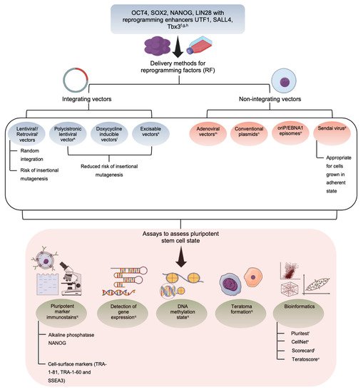
There are many assays, including molecular and functional, to evaluate the developmental efficiency of iPSCs. These include alkaline phosphatase staining of pluripotency markers, DNA demethylation, retroviral silencing, and factor independence involving assessment of self-renewal in the absence of dox-inducible trans genes. The functional assays include teratoma formation, chimera development, tetraploid complementation, germline transmission, and in vitro differentiation [14]. Considering the low reprogramming efficiency in iPSCs, many studies have identified blocks in lineage conversion. Reprogramming pathway studies in fibroblasts have identified the repel factor to be involved in mesenchymal-to-epithelial transition (MET) and BMP receptor signaling [27][28]. Further studies on the refractory fibroblasts indicate negative iPSC generation in spite of prolonged culturing and presence of homogeneous factor expression indicating loss of somatic program, and activation of endogenous pluripotency genes to be the main roadblocks in formation of iPSCs [14]. The other limiting factor has been linked to expression levels of Nanog locus which are activated late in the reprogramming process and thus limit efficiency of conversion [29]. Gene silencing by DNA methylation, involving the pluripotency genes nanog and Oct4 which causes blockage in binding of transcription factors, has also been linked to causing interference in reprogramming [30]. Though the four most popular reprogramming factors have been Oct4, Sox2, Klf4, and c-Myc, human iPSCs have also been derived using expression of Oct4, Sox2, Nanog, and Lin28, indicating that pluripotent ground state becomes achievable through activation of different transcription factors [21]. The detailed derivation of iPSC along with the assay has been highlighted in Figure 2.
Figure 2. Schematic representation on derivation and assay for human iPSCs. Detailed schematic representation of derivation of iPSC with the various assays to evaluate the developmental efficiency.
The therapeutic potential of iPSC towards personalized cell therapy and disease modelling, has extended the functionality beyond laboratory tables as a research tool in murine and human models. Animal studies have identified promising potential of iPSC around treatment of genetic disorders, including sickle cell anemia; disease modelling of complex degenerative conditions like diabetes, Alzheimer’s disease, and the feasibility to be used in organ transplantation without risk of rejection and need of immunosuppression [14][31].

2. Induced Pluripotent Stem Cells—The Niche Favoring Unique Aspects

Pluripotency and self-renewal are unique characteristics of iPSC that make them ideal for disease modelling and regenerative medicine. Their ability to indefinitely differentiate into cells of all the three germ layers makes them an important source for treating injuries as well as diseases. The availability of generating patient-specific iPSC with high efficiency and safety through protocols involving biochemical and epigenetic aspects expands the therapeutic potential of this tool. This can be assessed from the fact that a clinical trial involving iPSC-derived dopaminergic neurons have been initiated for Parkinson’s disease after successful in vivo studies involving immunodeficient mice highlighted no risk of tumorigenicity [32]. Further, tissue resident macrophages, which are critical for immunity and derived from human-iPSCs, have been found to be immunologically different and better than the traditional monocyte-derived macrophages. Studies have shown human iPSC macrophages to restrict Mycobacterium tuberculosis growth in vitro by >75%, and were found to be capable of mounting antibacterial response when challenged with pathogens [33]. The greatest niche for iPSCs is the ability to generate the same from different donor categories including the diseased, and healthy making its application in the clinical setting at any stage a feasibility without the ethical issues around the ESCs.
The fundamental use of iPSC in regenerative medicine remains undisputed, but the tumorigenic potential of residual undifferentiated stem cells necessitates the need to devise strategies to remove the same from differentiated cells. Different study reports multiple treatment methodologies for eliminating undifferentiated iPSCs and one such recent publication identified undifferentiated hiPSCs to be sensitive to treatment involving medium supplemented with high concentration of L-alanine [34]. Another study assessed the efficacy of plasma-activated medium (PAM) in eliminating undifferentiated hiSPCs through inducing oxidative stress. This study found PAM to selectively eliminate undifferentiated hiPSCs cocultured with normal human dermal fibroblasts, which were the differentiated cells. Lower expression of oxidative-stress related genes in the undifferentiated hiPSCs were found to be the underlying cause for PAM-selective cell death [35]. A recent study report describes the use of salicylic diamines to remove residual undifferentiated cells from iPSC-derived cardiomyocytes. Salicylic diamines were found to exert their specific cytotoxic activity in the pluripotent stem cells by inhibiting the oxygen consumption rate. Teratoma formation was also found to be abolished in comparison to untreated cells [36].

3. Application of iPSC in Cardiac Disease

Non-communicable diseases, including cardiovascular conditions, have emerged to be one of the leading causes for mortality in developed as well as developing nations. The trigger for myriad heart conditions exists both in genetics and the environment, which makes studying disease etiology in animal models complicated and inefficient. Animal model studies indicate up to 90% failure in new drug clinical trials, highlighting the limitation around prediction of safety and efficacy among humans. The iPSCs-based disease models have been studied for cardiac channelopathies including hereditary long QT syndrome (LQTS), dilated cardiomyopathy (DCM), hypertrophic cardiomyopathy (HCM), and arrhythmogenic right ventricular cardiomyopathy (ARVC); the endothelial cell disease including familial pulmonary arterial hypertension (FPAH); the smooth muscle cell condition including Williams-Beuren syndrome (WBS), and Marfan syndrome (MFS) [8].
LQTS is an inherited fatal arrhythmia syndrome and around 17 genes have been associated with congenital LQTS, including the three main genes; KCNQ1 (LQT1), KCNH2 (LQT2), and SCN5A (LQT3), together which account for ~75% of clinically definite cases. The current therapeutic intervention includes β-blockers and a surgical procedure named left cardiac sympathetic denervation. Though genetic markers have been defined, the occurrence of variance of unknown significance (VUS) in 1 of 3 patients adds to the dilemma of inconclusive diagnosis. The need for better diagnostic platforms to assess outcome of genetic variants as well as different therapeutics led to the introduction of iPSCs. Many studies have worked to improve the differentiation efficiency, cellular maturation, and lineage specificity, develop new high-throughput assays for cellular phenotyping, and promote clinical implementation of patient-specific genetic models. A study by Wu J.C. et al. [37], utilized patient iPSC-derived cardiomyocytes (iPSC-CMs) and devised various strategies to reduce heterogeneity. These include derivation of chamber-specific cardiomyocytes, cultivation for extended period, 3-dimensional and mechanical conditioning, rapid electric stimulation, and hormonal stimulation; use of multicellular preparations to reduce intercellular variability; and development of high-throughput cellular phenotyping using optogenetic sensors including genetically coded voltage and calcium indicators. Further, this study also established the utility of iPSC-CMs to distinguish between pathogenic and benign variants to improve diagnosis and management of LQTS using CRISPR genome editing. This study, using iPSC-CMs, also identified factors causative for prolonged QT including upregulation of genes; DLG2, KCNE4, PTRF, and HTR2C and downregulation of CAMKV gene. Thus iPSC-based model platforms aid in developing a better understanding around intractable clinical problems associated with diseases like LQTS.

4. Application of iPSC in Degenerative Diseases

Theoretically iPSC has the potential to be programmed to form any cell in the human body, and coupled with improvements in reprogramming techniques, this technology has advanced our knowledge on disease pathology, developing precise therapeutics, as well as fuel advances in regenerative medicine [38]. In case of neurodegenerative conditions, and psychiatric disorders, the genetic predisposition and its relation to the disease pathophysiology is complex, and often there is alteration at structural as well as functional levels. In case of schizophrenia, which is aptly termed the “disease of the synapses”, studies have generated iPSC from family members positive for a frameshift mutation in schizophrenia 1 (DISC1) and used gene editing to generate isogenic iPS cell lines. This study found depletion of DISC1 protein among the mutation carriers, along with dysregulation of genes associated with synapses and psychiatric disorders in the forebrain. This mutation causes deficit of synaptic vesicles among the iPS-cell derived forebrain neurons. This identification of transcriptional dysregulation in human neurons, highlights a new facet involving synaptic dysregulation in mental disorders [39]. The technology of stem cell therapy has also been used to restore the functionality in many degenerative conditions including that of the retina that leads to loss of vision. Studies have evaluated the use iPSC to overcome challenges posed by use of stem cell therapy. The proposed strategy revolves around transplantation of photoreceptors with or without the retinal pigment epithelium cells for treating retinal degradation, with minimal risk using iPSC [40].
Degenerative disease generally progresses through multiple differentiation stages, and using iPSC models, these pathways of transition can be easily identified to assess cause as well as etiopathology better. Amyotrophic lateral sclerosis (ALS) involves loss of neurons from the spinal cord and motor cortex causing paralysis and death. The research around advancement of therapeutics, requires supply of human motor neurons positive for the causative genetic mutations that will also aid in understanding the root cause of motor neuron death. One study documented the production of iPS from ALS patient specific-skin fibroblasts from two sisters. Both were identified to be positive for the L144F (Leu144 → Phe) mutation of the superoxide dismutase (SOD1) gene that is associated with a slowly progressing form of ALS. This study found successful reprogramming to be possible with only four factors; KLF4, SOX2, OCT4, and c-MYC. Further, the severe disability state of the patients used for harvesting in this case did not seem to block the transformation process or efficiency [41]. Fanconi anemia (FA) is an inherited bone marrow failure syndrome and is a chromosomal instability disorder needing transplantation of hematopoietic grafts from HLA-identical sibling donors. The reduced quality of the hematopoietic stem cells from the bone marrow of the affected limits the benefit of gene therapy trials. Studies have worked upon formation of genetically corrected FA-specific iPSCs through non-hematopoietic somatic cells reprogramming to generate large number of genetically-stable autologous hematopoietic stem cells for treating bone marrow failure in FA. The reprogramming was done on dermal fibroblasts involving two rounds of infection with mouse-stem-cell-virus-based retrovirus encoding amino-terminal flag-tagged version of the four transcription factors; OCT4, SOX2, KLF4, c-MYC. A batch of genetically corrected somatic cells using lentiviral vectors encoding FANCA or FANCD2 was also used for reprogramming to overcome the predisposition to apoptosis found in FA cells. The FANCA involved fibroblasts also underwent successful transformation to generate iPSCs. This study also found restoration of the FA pathway as a necessity to generate iPS from somatic cells of FA patients. The persistent FANCA expression in the FA-iPS cells indicated successful generation of genetically corrected FA-iPSCs with functional FA pathway, and disease-free status [42].
Parkinson’s disease (PD) is a common chronic progressive disorder due to loss of nigrostriatal dopaminergic neurons. The pathophysiology of the disease is complex and research till date lacks complete understanding. Further, sporadic cases are not linked to any genetic variation. Development of patient-specific invitro iPSC models have been attempted to understand disease etiology better. Studies have worked upon generating iPSCs from sporadic cases of PD, which have been successfully reprogrammed to form dopaminergic neurons free of the reprogramming factors. This study utilized doxycycline-inducible lentiviral vectors that were excised with Cre-/lox-recombinase, resulting in generation of iPSC free of programming factors, and which retained all the pluripotent characteristics after removal of transgenes. This removal of promoter and transgene sequences from the vector reduced risk of oncogenic transformation and re-expression of the transduced transcription factors. This study highlighted the possibility of generating stable iPS-cell line in PD for better disease modelling [43]. Another study worked on improving the safety of human and non-human primate iPSC derived dopaminergic neurons for cell transplantation treatment in PD. This study found the protocol of NCAM(+)/CD29(low) sorting to result in enriching ventral midbrain dopaminergic neurons from the pluripotent stem cell-derived neural cell populations. Further, these neurons also exhibited increased expression of FOXA2, LMX1A, TH, GIRK2, PITX3, EN1, and NURR1 mRNA. These neurons were also found to bear the potential to restore motor function among the 6-hydroxydopamine lesioned rats, 16 weeks after transplantation. Further, the primate iPSC-derived neural cell was found to have survived without any immunosuppression after one year of autologous transplant, highlighting the proof-of-concept around feasibility and safety of iPSC-derived transplantation for PD [10].
Type 1 diabetes is an autoimmune condition involving destruction of the β-cells of the pancreas wherein transplantation with β-cells as islet tissues or the entire pancreas is suggested as an alternative over the traditional exogenous insulin supplementation. However, these come with risk of rejection, need of immunosuppression, apart from difficulty in the physiological control on blood glucose levels. To circumvent this block, generation of β-cells or islet tissues from human pluripotent stem cells like iPSCs has been attempted. Many studies have generated pancreatic β-like cells which secrete insulin in response to stimuli like potassium chloride [44]. However, co-excretion of glucagon, and somatostatin, apart from releasing unsuitable amounts of insulin; make these clinically inferior. iPSC-derived pancreatic endoderm cells have been shown to retain the potential to differentiate and are functionally comparable with adult β-cells. Further, the shortage of donor islet has been overcome using iPSCs, as pancreatic cells generated from these have been evaluated in clinical trials as a new source for transplantation therapy. The differentiation of iPSCs through mimicking the natural in vivo process was facilitated using a combination of growth factors including Nodal-activin, Wnt, retinoic acid, hedgehog, epidermal and fibroblast growth factor, bone morphogenetic protein, and Notch to activate as well as inhibit the key signaling pathway. This study thus highlighted the possibility of generating patient-specific fully functional pancreatic tissue for transplantation over donor islet for diabetes treatment [45].

5. Application of iPSC in Blood Disorders

The treatment for blood disorders involves need for mature red blood cells/erythrocytes from the bone marrow or umbilical cord blood, for blood transfusion, and is limited due to incompatibility in blood group and Rh antigens, and risk of infections [46]. Erythropoiesis is a complex process for generation of mature erythrocytes from the precursor erythroblasts that are difficult to culture in vitro, as the entire process occurs in the bone marrow mediated by complex interaction between cellular and extracellular environment involving hormones, cytokines, and growth factors [47]. Further, the fully differentiated red blood cells (RBCs) are not proliferative, and setting up a system for erythropoiesis-like maturation in precursor cells is a challenge. Further, recruitment of donors, need for rare blood group types, as well as safety in sensitive population groups, add to the roadblock [48]. Studies have investigated human pluripotent stem cells, including iPSCs as an alternative source for unlimited supply of functional erythrocytes. Studies have discussed different methods devised for RBC production, including using PSCs by repeating the developmental haematopoiesis; reprogramming somatic cells through transcription factors including OCT4, SOX2, c-MYC, KLF4, NANOG, LIN28; and stimulating the maturation of hematopoietic stem cells isolated from peripheral or umbilical cord blood [48][49]. The advantage of using iPSCs is their ability to differentiate into any cell type, and can be maintained indefinitely, thus becoming a potential source for cell replacement therapies. The potential of iPSc becomes highlighted by the fact that the French National Registry of People with a Rare Blood Phenotype/Genotype claims a single iPSc clone from their database could meet 73% of the needs of sickle cell disease patients [50]. This highlights that a limited number or RBC clones have the potential to supply to the majority needs of alloimmunized patients with rare blood groups.

6. Application of iPSC in Organ Dysfunctions
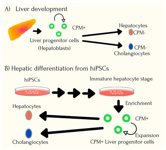
Organ donations are a key clinical need to treat end-stage organ failure conditions, and in often cases, patients are left to fight the acute shortage for the same. This apart, from identifying HLA-matched donors, handling risk of infections and rejection, as well as life-long immunosuppression, to a great extent damages quality of life for the affected as well as leads to loss of crucial time. Human iPSCs are being evaluated as a potential source for generating organs that can overcome roadblocks of shortage as well as risk of rejection. Studies have explored the possibility of generating a three-dimensional vascularized and functional liver organ from human iPSCs [51][52][53]. Generation of hepatocyte-like cells using iPSC technology has been reviewed to be fundamentally beneficial for treatment of severe liver disease, screening for drug toxicities, in liver transplantation, as well as to facilitate basic research [21]. Liver organogenesis involves delamination of specific hepatic cells from the foregut endodermal sheet to form a liver bud, which is then vascularized. One study prepared hepatic endoderm cells from human iPSCs through direct differentiation, wherein 80% of the treated cells were found to be positive for the cell fate determining hepatic marker; HNF4A. Further, to stimulate early organogenesis, the iPSCs were cocultured with stromal cells, human umbilical vein endothelial cells, and human mesenchymal stem cells, and after 48h of seeding, the human iPSCs were found to be self-organized into three-dimensional cell clusters visible macroscopically. This iPSC-derived liver bud, when further assessed by quantitative polymerase chain reaction (PCR) and microarray assay for expression analysis, highlighted the pattern to be similar to human fetal liver cell-derived liver buds. Hemodynamic stimulation to form organ was done by cranial window model, and the iPSC-derived tissue was found to perform liver-specific functions including protein synthesis and human-drug specific metabolism actions. This proof-of-concept study highlights the potential to use organ-bud transplantation for organ regeneration [54]. Figure 3 highlights the process of liver development and hepatic differentiation from hiPSCs.
Figure 3. Process of liver development and hepatic differentiation from hiPSCs. The process of isolated cells from patients can be cultured and reprogrammed into patient-specific hiPSCs and quick comparison from natural liver development.

7. Application of iPSCs in Cancer Syndromes

The iPSCs have been generated for modelling pathogenesis of many diseases, and one of the most notable additions to the same is cancer, including models for familial cancer syndromes. One such study reports on the successful establishment of Li-Fraumeni Syndrome (LFS) patient-derived iPSC to study role of p53 in development of osteosarcoma. LFS being a heterogenous cancer condition, osteosarcoma is one of the types wherein relevance of germline p53 mutations have been highly reported. The pre-existing murine LFS models have been insufficient in charting the entire tumor landscape and patient-derived iPSCs in this regard have demonstrated the feasibility to effectively study human cancer syndromes. Studies have found the LFS-derived mesenchymal stem cells to exhibit low expression of targets of p53 including p21 and MDM2; highlighting their ability to retain the defective p53 function from the parental fibroblasts. Further, p53 knockdown was found to cause upregulation of osteogenic markers in LFS osteoblasts, and the possibility to attain osteosarcoma-related phenotypes in LFS iPSC-derived osteoblasts was found. Further, gene expression analysis in LFS-derived osteoblasts was found to correlate with poor patient survival, and decreased time for recurrence. The impaired H19 restoration was also found to repress tumorigenic potential [55]. Another study involving modelling of osteosarcoma from LFS derived-iPSC identified the LFS osteoblasts to recapitulate oncogenic properties of osteosarcoma proving to be an excellent model to study disease pathogenesis [56]. In case of Noonan syndrome (NS) characterized by germline PTPN11 mutations, studies which have derived hiPSCs from hematopoietic cells and which harbor the PTPN11 mutations were found to successfully recapitulate features of NS. The iPSC-derived NS myeloid cells were found to exhibit increased STAT5 signaling and enhanced expression of micro-RNAs viz. miR-223 and miR-15a. Further, reducing miR-223 function was found to normalize myelogenesis, highlighting the role of micro-RNA dysregulation in early oncogenesis [57]. Human iPSC-derived hereditary cancer models have also aided in identifying BRCA1-deleted tumor niche to be the cause for disease progression [58].
The iPSC models around cancer aid in overcoming the hurdles posed by traditional cancer cell line systems, which may lose the characteristics of the original tumor with time, and further harnessing primary cancer cells at different stages of carcinogenesis is not feasible. The established iPSC reprogramming strategies can aid in differentiation of cancer cells to target cell lineages which can aid in studying each of the different stages in cancer progression [59]. The iPSCs developed from primary tumors, as well as cancer cell lines are invaluable tools to study genetic alterations early-on in familial cancer syndromes which is crucial in understand disease pathogenesis. Apart from cancer cell lines, patient-derived xenograft models have also been proven to be efficacious for understanding tumor heterogeneity, genetic alterations, and testing efficacy of cytotoxic drugs. However, the need for successful engraftment, technical challenges, and variable growth rates, are the key limitations. Even in case of animal models, high rate of mortality, and absence of metastasis are the limitations [60][61][62]. Advancements in iPSC models have also led researchers to be able to design autologous iPSC-based vaccine which presents a broad spectrum of tumor antigens to the immune system of the mice, and also found success in eliciting a prophylactic reaction against multiple cancer types. These studies highlight the great promise iPSC-based autologous vaccines present towards cancer prevention as well as therapy [63].

References

  1. Weismann, A. The Germ-Plasm: A Theory of Heredity; Charles Scribner’s Sons: New York, NY, USA, 1893.
  2. Gurdon, J.B.; Byrne, J.A. The first half-century of nuclear transplantation. Proc. Natl. Acad. Sci. USA 2003, 100, 8048–8052.
  3. Bradley, A.; Evans, M.; Kaufman, M.H.; Robertson, E. Formation of germ-line chimaeras from embryo-derived teratocarcinoma cell lines. Nature 1984, 309, 255–256.
  4. Cowan, C.A.; Atienza, J.; Melton, D.A.; Eggan, K. Nuclear reprogramming of somatic cells after fusion with human embryonic stem cells. Science 2005, 309, 1369–1373.
  5. Karagiannis, P.; Takahashi, K.; Saito, M.; Yoshida, Y.; Okita, K.; Watanabe, A.; Inoue, H.; Yamashita, J.K.; Todani, M.; Nakagawa, M.; et al. Induced pluripotent stem cells and their use in human models of disease and development. Physiol. Rev. 2018, 99, 79–114.
  6. Abbar, A.A.; Ngai, S.C.; Nograles, N.; Alhaji, S.Y.; Abdullah, S. Induced pluripotent stem cells: Reprogramming platforms and applications in cell replacement therapy. BioRes. Open Access 2020, 9, 121–136.
  7. Nishikawa, S.; Goldstein, R.A.; Nierras, C.R. The promise of human induced pluripotent stem cells for research and therapy. Nat. Rev. Mol. Cell Biol. 2008, 9, 725–729.
  8. Ye, L.; Ni, X.; Zhao, Z.-A.; Lei, W.; Hu, S. The application of induced pluripotent stem cells in cardiac disease modelling and drug testing. J. Cardiovasc. Transl. Res. 2018, 11, 366–374.
  9. Mandai, M.; Watanabe, A.; Kurimoto, Y.; Hirami, Y.; Morinaga, C.; Daimon, T.; Fujihara, M.; Akimaru, H.; Sakai, N.; Shibata, Y.; et al. Autologous induced stem-cell-derived retinal cells for macular degeneration. N. Engl. J. Med. 2017, 376, 1038–1046.
  10. Sundberg, M.; Bogetofte, H.; Lawson, T.; Jansson, J.; Smith, G.; Astradsson, A.; Moore, M.; Osborn, T.; Cooper, O.; Spealman, R.; et al. Improved cell therapy protocols for Parkinson’s disease based on differentiation efficiency and safety of hESC-, hiPSC-, and non-human primate iPSC-derived dopaminergic neurons. Stem Cells 2013, 31, 1548–1562.
  11. Sridharan, R.; Tchieu, J.; Mason, M.J.; Yachechko, R.; Kuoy, E.; Horvath, S.; Zhou, Q.; Plath, K. Role of the murine reprogramming factors in the induction of pluripotency. Cell 2009, 136, 364–377.
  12. Winkler, T.; Cantilena, A.; Metais, J.-Y.; Xu, X.; Nguyen, A.-D.; Borate, B.; Antosiewicz-Bourget, J.E.; Wolfsberg, T.G.; Thomson, J.A.; Dunbar, C.E. No evidence for clonal selection due to lentiviral integration sites in human induced pluripotent stem cells. Stem Cells 2010, 28, 687–694.
  13. Somers, A.; Jean, J.-C.; Sommer, C.A.; Omari, A.; Ford, C.C.; Mills, J.A.; Ying, L.; Sommer, A.G.; Jean, J.M.; Smith, B.W.; et al. Generation of transgene-free lung disease-specific human induced pluripotent stem cells using a single excisable lentiviral stem cell cassette. Stem Cells 2010, 28, 1728–1740.
  14. Stadtfeld, M.; Hochedlinger, K. Induced pluripotency: History, mechanisms, and applications. Genes Dev. 2010, 24, 2239–2263.
  15. Okita, K.; Nakagawa, M.; Hyenjong, H.; Ichisaka, T.; Yamanaka, S. Generation of mouse induced pluripotent stem cells without viral vectors. Science 2008, 322, 949–953.
  16. Lee, C.S.; Bishop, E.S.; Zhang, R.; Yu, X.; Farina, E.M.; Yan, S.; Zhao, C.; Zeng, Z.; Shu, Y.; Wu, X.; et al. Adenovirus-mediated gene delivery: Potential applications for gene and cell-based therapies in the new era of personalized medicine. Genes Dis. 2017, 4, 43–63.
  17. Ong, S.-G.; Lee, W.H.; Kodo, K.; Wu, J.C. MicroRNA-mediated regulation of differentiation and trans-differentiation in stem cells. Adv. Drug Deliv. Rev. 2015, 88, 3–15.
  18. Nakagawa, M.; Koyanagi, M.; Tanabe, K.; Takahashi, K.; Ichisaka, T.; Aoi, T.; Okita, K.; Mochiduki, Y.; Takizawa, N.; Yamanaka, S. Generation of induced pluripotent stem cells without Myc from mouse and human fibroblasts. Nat. Biotechnol. 2008, 26, 101–106.
  19. Huangfu, D.; Osafune, K.; Maehr, R.; Guo, W.; Eijkelenboom, A.; Chen, S.; Muhlestein, W.; Melton, D.A. Induction of pluripotent stem cells from primary human fibroblasts with only Oct4 and Sox2. Nat. Biotechnol. 2008, 26, 1269–1275.
  20. Lin, S.-L.; Chang, D.C.; Chang-Lin, S.; Lin, C.-H.; Wu, D.T.S.; Chen, D.T.; Ying, S.-Y. Mir-302 reprograms human skin cancer cells into a pluripotent ES-cell-like state. RNA 2008, 14, 2115–2124.
  21. Yu, Y.; Wang, X.; Nyberg, S.L. Application of induced pluripotent stem cells in liver diseases. Cell Med. 2014, 7, 1–13.
  22. Takahashi, K.; Tanabe, K.; Ohnuki, M.; Narita, M.; Ichisaka, T.; Tomoda, K.; Yamanaka, S. Induction of pluripotent stem cells from adult human fibroblasts by defined factors. Cell 2007, 131, 861–872.
  23. Zhou, W.; Freed, C.R. Adenoviral gene delivery can reprogram human fibroblasts to induced pluripotent stem cells. Stem Cells 2009, 27, 2667–2674.
  24. Nishishita, N.; Takenaka, C.; Fusaki, N.; Kawamata, S. Generation of human induced pluripotent stem cells from cord blood cells. J. Stem Cells 2011, 6, 101–108.
  25. Kim, D.; Kim, C.-H.; Moon, J.-I.; Chung, Y.-G.; Chang, M.-Y.; Han, B.-S.; Ko, S.; Yang, E.; Cha, K.Y.; Lanza, R.; et al. Generation of human induced pluripotent stem cells by direct delivery of reprogramming factors. Cell Stem Cell 2009, 4, 472–476.
  26. Yakubov, E.; Rechavi, G.; Rozenblatt, S.; Givol, D. Reprogramming of human fibroblasts to pluripotent stem cells using mRNA of four transcription factors. Biochem. Biophys. Res. Commun. 2010, 394, 189–193.
  27. Li, R.; Liang, J.; Ni, S.; Zhou, T.; Qing, X.; Li, H.; He, W.; Chen, J.; Li, F.; Zhuang, Q.; et al. A mesenchymal-to-epithelial transition initiates and is required for the nuclear reprogramming of mouse fibroblasts. Cell Stem Cell 2010, 7, 51–63.
  28. Samavarchi-Tehrani, P.; Golipour, A.; David, L.; Sung, H.-K.; Beyer, T.A.; Datti, A.; Woltjen, K.; Nagy, A.; Wrana, J.L. Functional genomics reveals a BMP-driven mesenchymal-to-epithelial transition in the initiation of somatic cell reprogramming. Cell Stem Cell 2010, 7, 64–77.
  29. Silva, J.; Nichols, J.; Theunissen, T.W.; Guo, G.; van Oosten, A.L.; Barrandon, O.; Wray, J.; Yamanaka, S.; Chambers, I.; Smith, A. Nanog is the gateway to the pluripotent ground state. Cell 2009, 138, 722–737.
  30. Gidekel, S.; Bergman, Y. A unique developmental pattern of Oct-3/4 DNA methylation is controlled by a cis-demodification element. J. Biol. Chem. 2002, 277, 34521–34530.
  31. Hanna, J.; Wernig, M.; Markoulaki, S.; Sun, C.-W.; Meissner, A.; Cassady, J.P.; Beard, C.; Brambrink, T.; Wu, L.-C.; Townes, T.M.; et al. Treatment of sickle cell anemia mouse model with iPS cells generated from autologous skin. Science 2007, 318, 1920–1923.
  32. Doi, D.; Magotani, H.; Kikuchi, T.; Ikeda, M.; Hiramatsu, S.; Yoshida, K.; Amano, N.; Nomura, M.; Umekage, M.; Morizane, A.; et al. Pre-clinical study of induced pluripotent stem cell-derived dopaminergic progenitor cells for Parkinson’s disease. Nat. Commun. 2020, 11, 3369.
  33. Nenasheva, T.; Gerasimova, T.; Serdyuk, Y.; Grigoreva, E.; Kosmiadi, G.; Nikolaev, A.; Dashinimaev, E.; Lyadova, I. Macrophages derived from human induced pluripotent stem cells are low-activated “naïve-like” cells capable of restricting Mycobacteria growth. Front. Immunol. 2020, 11, 1016.
  34. Nagashima, T.; Shimizu, K.; Matsumoto, R.; Honda, H. Selective elimination of human induced pluripotent stem cells using medium with high concentration of L-alanine. Sci. Rep. 2018, 8, 12427.
  35. Matsumoto, R.; Shimizu, K.; Nagashima, T.; Tanaka, H.; Mizuno, M.; Kikkawa, F.; Hori, M.; Honda, H. Plasma-activated medium selectively eliminates undifferentiated human induced pluripotent stem cells. Regen. Ther. 2016, 5, 55–63.
  36. Burkert, K.; Taheri, H.; Hamad, S.; Oliverio, M.; Peinkofer, G.; Kornfeld, J.-W.; Harnying, W.; Pfannkuche, K.; Hescheler, J.; Berkessel, A.; et al. Salicylic diamines selectively eliminate residual undifferentiated cells from pluripotent stem cell-derived cardiomyocyte preparations. Sci. Rep. 2021, 11, 2391.
  37. Wu, J.C.; Garg, P.; Yoshida, Y.; Yamanaka, S.; Gepstein, L.; Hulot, J.-S.; Knollmann, B.C.; Schwartz, P.J. Towards precision medicine with human iPSCs for cardiac channelopathies. Circ. Res. 2019, 125, 653–658.
  38. Kumar, S.; Blangero, J.; Curran, J.E. Induced pluripotent stem cells in disease modelling and gene identification. Methods Mol. Biol. 2018, 1706, 17–38.
  39. Wen, Z.; Nguyen, H.N.; Guo, Z.; Lalli, M.A.; Wang, X.; Su, Y.; Kim, N.-S.; Yoon, K.-J.; Shin, J.; Zhang, C.; et al. Synaptic dysregulation in a human iPS cell model of mental disorders. Nature 2014, 515, 414–418.
  40. Wong, I.Y.; Poon, M.-W.; Pang, R.T.; Lian, Q.; Wong, D. Promises of stem cell therapy for retinal degenerative diseases. Graefe’s Arch. Clin. Exp. Ophthalmol. 2011, 249, 1439–1448.
  41. Dimos, J.T.; Rodolfa, K.T.; Niakan, K.K.; Weisenthal, L.M.; Mitsumoto, H.; Chung, W.; Croft, G.F.; Saphier, G.; Leibel, R.; Goland, R.; et al. Induced pluripotent stem cells generated from patients with ALS can be differentiated inti motor neurons. Science 2008, 321, 1218–1221.
  42. Raya, A.; Rodriguez-Piza, I.; Guenechea, G.; Vassena, R.; Navarro, S.; Barrero, M.J.; Consiglio, A.; Castella, M.; Rio, P.; Sleep, E.; et al. Disease-corrected haematopoietic progenitors from Fanconi anaemia induced pluripotent stem cells. Nature 2009, 460, 53–59.
  43. Soldner, F.; Hockemeyer, D.; Beard, C.; Gao, Q.; Bell, G.W.; Cook, E.G.; Hargus, G.; Blak, A.; Cooper, O.; Mitalipova, M.; et al. Parkinson’s disease patient-derived induced pluripotent stem cells free of viral reprogramming factors. Cell 2009, 136, 964–977.
  44. D’Amour, K.A.; Bang, A.G.; Eliazer, S.; Kelly, O.G.; Agulnick, A.D.; Smart, N.G.; Moorman, M.A.; Kroon, E.; Carpenter, M.K.; Baetge, E.E. Production of pancreatic hormone-expressing endocrine cells from human embryonic stem cells. Nat. Biotechnol. 2006, 24, 1392–1401.
  45. Kondo, Y.; Toyoda, T.; Inagaki, N.; Osafune, K. iPSC technology-based regenerative therapy for diabetes. J. Diabetes Investig. 2018, 9, 234–243.
  46. Sahu, S.; Hemlata; Verma, A. Adverse events related to blood transfusion. Indian J. Anaesth. 2014, 58, 543–551.
  47. Seo, Y.; Shin, K.-H.; Kim, H.H.; Kim, H.-S. Current advances in red blood cell generation using stem cells from diverse sources. Stem Cells Int. 2019, 2019, 9281329.
  48. Chang, K.-H.; Bonig, H.; Papayannopoulou, T. Generation and characterization of erythroid cells from human embryonic stem cells and induced pluripotent stem cells: An overview. Stem Cells Int. 2011, 2011, 791604.
  49. Ebrahimi, M.; Forouzesh, M.; Raoufi, S.; Ramazii, M.; Ghaedrahmati, F.; Farzaneh, M. Differentiation of human induced pluripotent stem cells into erythroid cells. Stem Cell Res. Ther. 2020, 11, 483.
  50. Peyrard, T.; Bardiaux, L.; Krause, C.; Kobari, L.; Lapillonne, H.; Andreu, G.; Douay, L. Banking of pluripotent adult stem cells as an unlimited source for red blood cell production: Potential applications for alloimmunized patients and rare blood challenges. Transfus. Med. Rev. 2011, 25, 206–216.
  51. Ding, Q.; Cowan, C.A. Liver in a dish. Cell Res. 2013, 23, 1242–1243.
  52. Fox, I.J.; Duncan, S.A. Engineering liver tissue from induced pluripotent stem cells: A first step in generating new organs for transplantation? Hepatology 2013, 58, 2198–2201.
  53. Willenbring H and Soto-Gutierrez, A. Transplantable liver organoids made from only three ingredients. Cell Stem Cell 2013, 13, 139–140.
  54. Takebe, T.; Sekine, K.; Enomura, M.; Koike, H.; Kimura, M.; Ogaeri, T.; Zhang, R.-R.; Ueno, Y.; Zheng, Y.-W.; Koike, N.; et al. Vascularized and functional human liver from an iPSC-derived organ bud transplant. Nature 2013, 499, 481–484.
  55. Chang, C.-J.; Bouhassira, E.E. Zinc-finger nuclease-mediated correction of alpha-thalassemia in iPS cells. Blood 2012, 120, 3906–3914.
  56. Zhou, R.; Xu, A.; Tu, J.; Liu, M.; Gingold, J.A.; Zhao, R.; Lee, D.F. Modeling osteosarcoma using Li-Fraumeni syndrome patient-derived induced pluripotent stem cells. JOVE 2018, 136, 57664.
  57. Mulero-Navarro, S.; Sevilla, A.; Roman, A.C.; Lee, D.-F.; D’Souza, S.L.; Pardo, S.; Riess, I.; Su, J.; Cohen, N.; Schaniel, C.; et al. Myeloid dysregulation in a human induced pluripotent stem cell model of PTPN11–associated juvenile myelomonocytic leukemia. Cell Rep. 2015, 13, 504–515.
  58. Portier, L.; Desterke, C.; Chaker, D.; Oudrhiri, N.; Asgarova, A.; Dkhissi, F.; Turhan, A.G.; Bennaceur-Griscelli, A.; Griscelli, F. iPSC-derived hereditary breast cancer model reveals the BRCA1-deleted tumor niche as a new culprit in disease progression. Int. J. Mol. Sci. 2021, 22, 1227.
  59. Chao, H.-M.; Chern, E. Patient-derived induced pluripotent stem cells for models of cancer and cancer stem cell research. J. Formos. Med. Assoc. 2018, 117, 1046–1057.
  60. Reyal, F.; Guyader, C.; Decraene, C.; Lucchesi, C.; Auger, N.; Assayag, F.; De Plater, L.; Gentien, D.; Poupon, M.-F.; Cottu, P.; et al. Molecular profiling of patient-derived breast cancer xenografts. Breast Cancer Res. 2012, 14, R11.
  61. Krumbach, R.; Schuler, J.; Hofmann, M.; Giesemann, T.; Fiebig, H.-H.; Beckers, T. Primary resistance to cetuximab in a panel of patient-derived tumour xenograft models: Activation of MET as one mechanism for drug resistance. Eur. J. Cancer 2011, 47, 1231–1243.
  62. Chen, Z.; Trotman, L.C.; Shaffer, D.; Lin, H.-K.; Dotan, Z.A.; Niki, M.; Koutcher, J.A.; Scher, H.I.; Ludwig, T.; Gerald, W.; et al. Crucial role of p53-dependent cellular senescence in suppression of Pten-deficient tumorigenesis. Nature 2005, 436, 725–730.
  63. Wang, L.; Pegram, M.D.; Wu, J.C. Induced pluripotent stem cells as a novel cancer vaccine. Expert Opin. Biol. Ther. 2019, 19, 1191–1197.
More
Information
Subjects: Cell Biology
Contributor MDPI registered users' name will be linked to their SciProfiles pages. To register with us, please refer to https://encyclopedia.pub/register :
View Times: 826
Revision: 1 time (View History)
Update Date: 09 Sep 2021
1000/1000
ScholarVision Creations