Submitted Successfully!
To reward your contribution, here is a gift for you: A free trial for our video production service.
Thank you for your contribution! You can also upload a video entry or images related to this topic.
Version Summary Created by Modification Content Size Created at Operation
1 + 2388 word(s) 2388 2021-08-26 10:22:32 |
2 The format is correct Meta information modification 2388 2021-08-27 10:16:25 | |
3 The format is correct Meta information modification 2388 2021-08-30 08:02:13 |

Video Upload Options

We provide professional Video Production Services to translate complex research into visually appealing presentations. Would you like to try it?

Confirm

Are you sure to Delete?
Cite
If you have any further questions, please contact Encyclopedia Editorial Office.
Cremers, N. Recurrent Vulvovaginal Candidiasis and Honey. Encyclopedia. Available online: https://encyclopedia.pub/entry/13623 (accessed on 26 December 2024).
Cremers N. Recurrent Vulvovaginal Candidiasis and Honey. Encyclopedia. Available at: https://encyclopedia.pub/entry/13623. Accessed December 26, 2024.
Cremers, Niels. "Recurrent Vulvovaginal Candidiasis and Honey" Encyclopedia, https://encyclopedia.pub/entry/13623 (accessed December 26, 2024).
Cremers, N. (2021, August 27). Recurrent Vulvovaginal Candidiasis and Honey. In Encyclopedia. https://encyclopedia.pub/entry/13623
Cremers, Niels. "Recurrent Vulvovaginal Candidiasis and Honey." Encyclopedia. Web. 27 August, 2021.
Recurrent Vulvovaginal Candidiasis and Honey
Edit

Recurrent vulvovaginal candidiasis (RVVC) is a relapsing vaginal fungal infection caused by Candida species. In 57% of the cases, relapses occur within six months after fluconazole maintenance therapy, which is the current standard of care. The pathogenesis of RVVC is multifactorial, and recent studies have demonstrated that the vaginal microenvironment and activity of the immune system have a strong influence on the disease. 

recurrent vulvovaginal candidiasis medical-grade honey fluconazole alternative treatment micro-environment modulation

1. Introduction

Vulvovaginal candidiasis (VVC) is a vaginal fungal infection confirmed to be caused by Candida species, in most cases Candida albicans [1]. A total of 75% of women develop vulvovaginal candidiasis at least once in their life [2]. The symptoms related to vulvovaginal candidiasis are pruritus, soreness, irritation, dyspareunia, vaginal discharge, and discomfort. Clinical signs are exemplified by vulva erythema, edema, excoriation, and fissure formation, together with introital and vaginal erythema [1][3]. A non-malodorous clumpy white discharge is suggestive of VVC but is extremely nonspecific [1]. Women also report loss of confidence and self-esteem, inability to carry on with their normal physical activities, and difficulties with their sexual life and intimate relationships [2]. It also has a profound effect on the quality of life of affected women with additional systemic symptoms including depression and anxiety [1]. The definition of recurrent vulvovaginal candidiasis (RVVC) is at least three symptomatic episodes in the last 12 months [1]. RVVC affects about 138 million women per year worldwide (range of 103–172 million), with a global annual prevalence of 3871 per 100,000 women [2]. The highest prevalence (9%) is seen in women of an age between 25 and 34 years old. It is estimated that the population of women with recurrent vulvovaginal candidiasis will increase to almost 158 million in 2030 [2].
RVVC is a multifactorial disease whose symptoms are governed by the interaction between Candida (species and virulence factors), the Lactobacilli population, the micro-environment (inflammatory status, oxidative stress, estrogen), and the host (immune status, behavioral factors, genetic factors). A disbalance in any of these factors may induce RVVC [4].
VVC is according to the Clinical Practice Guidelines treated with topical or oral antifungals, of which azoles (miconazole, clotrimazole, and fluconazole) are the most commonly prescribed [5]. Notably, there is an increase in resistance of Candida species towards antifungal agents, causing multidrug resistance to emerge, while the long-term efficacy of antifungal agents is limited [3][6][7]. Therefore, an urgent need for alternative or complementary therapies to effectively treat RVVC and prevent it from recurrence is needed. It is important to know more about the etiology of RVVC, the different treatment options, and their efficacy to understand how novel therapies could improve the clinical outcome and quality of life.

2. Diagnosis of RVVC

RVVC is often overdiagnosed or misdiagnosed when the diagnosis is only based on clinical symptoms, which are non-specific. Laboratory testing is necessary to confirm the diagnosis of VVC, because self-diagnosis based on symptoms has an accuracy rate of only 28% for Candida albicans in self-treating women, making over-the-counter (OTC) antifungals often ineffective [5][8][9]. The golden standard for the diagnosis of VVC is by culturing the cells. It is also possible to use microscopy to identify yeast cells and hyphae. Gram staining of vaginal discharge mixed with potassium hydroxide (KOH) is used to distinguish Candida yeast cells and hyphae, which is relevant to the stage of the pathogenesis. The pH of the vaginal discharge is also an important indicator and normally stays within a range of 4.0–4.5. A pH above 4.7 is indicative of other infections such as bacterial vaginosis, trichomoniasis, or mixed infections [1][10][11]. For further differentiation between Candida species, additional culturing is needed, e.g., with chromogenic agar or Sabourad’s dextrose agar. However, culturing is not the most selective procedure, and molecular methods such as sequencing of the internal transcribed spacer (ITS sequencing) or matrix-assisted laser desorption ionization time-of-flight mass spectrometry (MALDI-ToF MS) are needed to identify the specific species [12][13]. This is especially relevant in the case of RVVC, in which non-albicans species (NAC) are becoming more prevalent. Similarly, susceptibility testing may be of adjuvant need in RVVC, as these infections are more resistant to antifungal agents [11].

3. Pathogenesis of RVVC

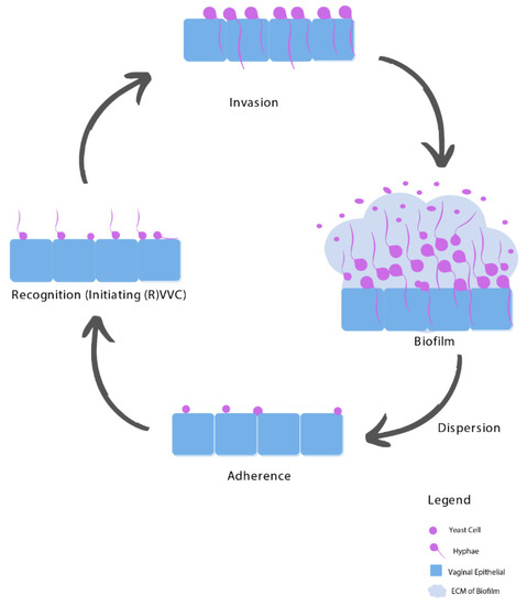
The pathogenesis of VVC is a gradual process that goes through several stages, from adherence to the vaginal epithelium, recognition (caused by a burden threshold of hyphae), becoming invasive, the possibility of biofilm formation, and dispersion of planktonic cells that reinitiates the complete process (see Figure 1).
Figure 1. Biofilm formation of Candida species during the pathogenesis of (R)VVC. Yeast cells adhere to the vaginal epithelium and subsequently form hyphae, which together with secretion of Candidalysin determine recognition by the immune system and drive damage and vulvovaginal immunopathogenesis. Invasion of the Candida follows through hyphal extension, relying on both the strain and host factors. Next, biofilms can start to form, from which Candida cells can be released (dispersion) as planktonic cells and reinitiate the complete process.

3.1. Adhesion

The most important phase of the initial contact between potential pathogen and host is the adherence of yeast cells to epithelial cells. Consequently, an interaction follows between epithelial receptors and Candida adhesins. These Candida adhesins vary depending on the morphological status of the Candida, i.e., yeast or hyphae [14]. After this initial contact, hyphae will grow out of the yeast cells, which is the first step in the pathogenesis [15]. The progress of the infection is determined by the hyphal-expressed adhesins that play a key role in the pathogenesis [14].

3.2. Recognition

The fact that Candida species have a dual lifestyle as both a vaginal commensal and opportunistic pathogen requires epithelial cells to have a mechanism to discriminate between the colonizing yeast and the invasive hyphal form [16]. Candida species can be recognized via two different pathways. The first phase is represented by an early transient response that occurs in a morphologically independent manner. The second phase starts when a burden threshold of hyphae is reached; this is a stronger response to hyphae, which in turn leads to activation of epithelial cells and the production of cytokines, chemokines, and other inflammatory mediators [14]. The hyphal burden plays a significant role in affecting epithelial activation because the presence of hyphae goes undetected below a certain threshold level. This is supported by in vivo experiments in a murine model of vaginitis, where the hyphal form was needed to cause damage to the epithelium, releasing pro-inflammatory cytokines and neutrophil recruitment [14].

3.3. Invasion

Invasion of Candida species can occur with the help of invasins after a switch from the yeast to the hyphal form [15][17]. Invasion into host cells can be achieved by endocytosis or active penetration [17]. This consequently leads to damage to the epithelial cells via necrosis and apoptosis, hence loss of epithelium. An in vivo study demonstrated a significantly lower cell damage caused by non-hyphal mutants compared to hyphal mutants [14][18].

3.4. Biofilms

Biofilm formation may be the main etiological factor contributing to antifungal resistance and is a likely contributor to treatment failure in RVVC [15][19][20][21]. It is suggested that the ability to form biofilms is a major virulence trait of Candida species in the pathogenesis of VVC [20][22]. After the invasion stage, an extracellular matrix (ECM) may be developed, which results in a ‘biofilm’ that encapsulates the Candida cells. This ECM consists of exopolymeric macromolecules, including polysaccharides, proteins, lipids, and nucleic acids, which are secreted by sessile cells within the biofilm. A mature biofilm is characterized by a structured mixture of yeast-form and hyphal cells surrounded by ECM, and it provides protection for the yeast against environmental challenges. In the final stage, the biofilm slowly disperses yeast-form cells into the surrounding, which may be able to colonize other surfaces [16][19].

4. Risk Factors of RVVC

4.1. Imbalanced Vaginal Microbiota Composition

Alteration in the mucosal ecosystem leading to fungal dysbiosis can lead to (R)VVC and its symptoms [4]. A healthy vaginal microflora consists of different microorganisms, mainly Lactobacilli, but also accommodating fungi such as Candida albicans and Candida glabrata, living in symbiosis. Lactobacilli species play an important role in maintaining a healthy vaginal microbiome [4]. Through their presence, Lactobacilli species decrease opportunism of potentially pathogenic microorganisms by microbial competition, which reduces the adherence of Candida species to the vaginal epithelium [23][24].
When the healthy microbial balance is disturbed, Lactobacilli may lose their ascendancy, and other microorganisms, such as Candida albicans, can foster and cause overgrowth. Multiple factors can alter the vaginal microbiota and disturb the balance between tolerance and invasion of Candida species. Important drivers for the pathogenesis of VVC are changes in the Lactobacilli community, elevated estrogen levels (i.e., due to oral contraceptives, hormone replacement therapy (HRT) used in post-menopause, being in the luteal phase of the menstruation cycle or pregnancy), an elevated pH, and the presence of glucose and eicosanoids (such as prostaglandin E2 and thromboxane B2). Other determinants have an inhibitory effect on VVC such as lactate and the presence of short-chain fatty acids such as acetate, butyrate, and propionate B [4][6].

4.2. Host-Related Predisposing Factors

A broad spectrum of host-related predisposing factors such as genetic background, (uncontrolled) diabetes mellitus, altered immune status, use of steroids, and antibiotics therapy, as well as behavioral factors such as sexual activity, hormone replacement therapy, and use of contraceptives including intrauterine devices, have been associated to promote VVC pathology [1][2][4].

4.3. Idiopathic RVVC

There are no predisposing factors in 20–30% of the RVVC patients. It is suggested that the Candida strain, its virulence, and inter-individual differences play a key role in idiopathic RVVC pathogenesis [1][4]. Several epidemiologic and cohort studies demonstrated that genetic mutations and polymorphisms and ethnicity play a role [4][19]. Moreover, NAC species are also associated with recurrent infections in VVC patients, likely because of their natural resistance towards azole-based antifungal agents [4].

5. Treatment of RVVC

5.1. Resistance towards Fluconazole

Since fluconazole is fungistatic rather than fungicidal, there is an increased opportunity to develop acquired resistance in the presence of this antifungal [25]. There are several challenges in fluconazole treatment such as an increase in antifungal resistance and VVC caused by NAC species, as well as the existence of biofilms [26]. Epidemiologic studies confirm that mostly all women diagnosed with fluconazole-resistant Candida albicans were previously exposed to fluconazole [26].

5.2. Unnecessary and Inappropriate Use of Fluconazole

Fluconazole is a commonly used antifungal agent and is easily accessible, which increases the risk of developing resistance. For example, the over-the-counter availability of antifungal agents combined with the frequent empiric prescription of fluconazole for sporadic VVC and the frequent use of a low weekly dose of fluconazole as a maintenance regimen facilitates fluconazole-resistance by Candida species, subsequently leading to RVVC [26]. Long-term maintenance therapy should be based on diagnostic confirmation to avoid unnecessary and inappropriate use [26].

5.3. Non-Albicans Species

VVC caused by NAC species is increasingly common due to overuse and misuse of antifungal agents [6][27][28]. Furthermore, a major concern regarding the increased incidence of VVC caused by NAC species is that such infections are often more difficult to treat because they are less susceptible to azoles and are more frequently resistant [6][27][23].

5.4. Biofilms Complicate RVVC Treatment

The biofilm formation in the pathogenesis of VVC provides elevated virulence and resistance towards antifungal therapy such as fluconazole [15][20][21]. A 1000-fold higher resistance profile of biofilms compared to their planktonic counterparts has been described [16][29][30]. Biofilms are also less sensitive to eradicate by the host immune system. A study of clinical isolates obtained from women with at least two episodes of VVC confirmed a lower antifungal susceptibility for biofilms in comparison with the planktonic antifungal susceptibility [20].

6. Medical-Grade Honey as an Alternative Treatment Option

The high recurrence rate of complaints after fluconazole treatment may be attributed to the fact that fluconazole only interacts with the yeast, hyphae, and invasive Candida stages (Figure 2). In contrast, when an established biofilm is present, the ECM prevents the fluconazole from reaching the Candida cells, and therefore it will not have an effect on biofilms [31]. Moreover, fluconazole does not affect the vaginal mucosal response [1][3][4]. Since ancient times, honey has been used for wound treatment and care because of its antimicrobial and wound healing activities. Acquired azole resistance, the epidemiological shift from Candida albicans to NAC species, and the existence of biofilms demand better treatment options. Medical-grade honey (MGH) could be an accessible, effective, and affordable option [23]. To assure the safety and efficacy of honey for clinical application, strict guidelines are followed to establish MGH [32]. MGH is effective in acute and chronic wounds and provides rapid epithelization and wound contraction, has anti-inflammatory activity, stimulates debridement, decreases pain, resolves infections, decreases wound healing time, and is cost-effective [33]. The use of honey for reducing biofilm formation on indwelling plastic devices such as urinary catheters are also considered, but more research is needed [34][35].
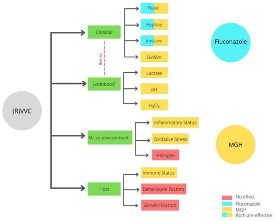
Figure 2. Differential antipathogenic activity of fluconazole and MGH on the pathogenesis of RVVC.
As mentioned before, four key factors determine the progression and development of RVVC: the presence of Candida, the population of Lactobacilli, the microenvironment, and host-related factors. In contrast to fluconazole, MGH may affect all these factors via multiple mechanisms (Figure 2, Table 1).
Table 1. Overview antipathogenic activity of fluconazole and MGH.
Characteristic Fluconazole MGH
Candida albicans + +
(Increased raise in VVC caused by) NAC species +
Biofilms +
Increased resistance +
Microenvironment/vaginal mucosal response +
Lactobacilli +−
pH +
Osmotic effect +
Antimicrobial + +
Anti-inflammatory +
Antioxidative +
−: no effect; +: positive effect; +−: possible effect.

Detailed mechanisms of how MGH affects the indicated pathways are described in the original manuscript. Extensive pre-clinical and clinical literature is discussed that supports that MGH is a promising treatment for RVVC. In addition, a new randomized-controlled trial (clinicaltrials.gov NCT04626258) is presented that intents to investigate the mycological and clinical cure and the prophylactic efficacy of the MGH-formulation L-Mesitran Soft in relation to the standard of care (fluconazole). L-Mesitran Soft is CE- and FDA approved and contains 40% MGH and different supplements that consistently have demonstrated to enhance the antimicrobial and wound healing activities. 

7. Conclusions

The population of women with RVVC is estimated to increase over the coming years. The most commonly used treatment for RVVC is fluconazole, which it seems will become inadequate in the future due to increasing resistance to fluconazole, the rise of NAC species, and the existence of biofilms. MGH might be a promising treatment in VVC and RVVC. In contrast to fluconazole, MGH is expected to have multiple beneficial mechanisms. In addition to the antifungal activity on Candida, MGH can likely also eliminate antifungal-resistant Candida species, including NAC species, and eradicate biofilms. Moreover, MGH can modulate the microenvironment by its anti-inflammatory and antioxidative activity. In vitro studies and clinical studies demonstrated honey as a promising alternative therapy for RVVC. More research is needed to investigate the exact clinical efficacy and the long-term cure rate of MGH; as proposed, this will be further investigated in a randomized controlled trial.

References

  1. Sobel, J.D. Recurrent vulvovaginal candidiasis. Am. J. Obstet. Gynecol. 2016, 214, 15–21.
  2. Denning, D.W.; Kneale, M.; Sobel, J.D.; Rautemaa-Richardson, R. Global burden of recurrent vulvovaginal candidiasis: A systematic review. Lancet Infect. Dis. 2018, 18, e339–e347.
  3. Sobel, J.D.; Wiesenfeld, H.C.; Martens, M.; Danna, P.; Hooton, T.M.; Rompalo, A.; Sperling, M.; Livengood, C., 3rd; Horowitz, B.; Von Thron, J.; et al. Maintenance fluconazole therapy for recurrent vulvovaginal candidiasis. N. Engl. J. Med. 2004, 351, 876–883.
  4. Rosati, D.; Bruno, M.; Jaeger, M.; Ten Oever, J.; Netea, M.G. Recurrent Vulvovaginal Candidiasis: An Immunological Perspective. Microorganisms 2020, 8, 144.
  5. Pappas, P.G.; Kauffman, C.A.; Andes, D.R.; Clancy, C.J.; Marr, K.A.; Ostrosky-Zeichner, L.; Reboli, A.C.; Schuster, M.G.; Vazquez, J.A.; Walsh, T.J.; et al. Clinical Practice Guideline for the Management of Candidiasis: 2016 Update by the Infectious Diseases Society of America. Clin. Infect. Dis. 2016, 62, e1–e50.
  6. Bauters, T.G.; Dhont, M.A.; Temmerman, M.I.; Nelis, H.J. Prevalence of vulvovaginal candidiasis and susceptibility to fluconazole in women. Am. J. Obstet. Gynecol. 2002, 187, 569–574.
  7. Banaeian-Borujeni, S.; Mobini, G.R.; Pourgheysari, B.; Validi, M. Comparison of the effect of honey and miconazole against Candida albicans in vitro. Adv. Biomed. Res. 2013, 2, 57.
  8. Gardella, C.E.L.; Lentz, G.M. Comprehensive Gynecology; Lentz, G.L.R., Gershenson, D., Valea, F.A., Eds.; Elsevier: Philadelphia, PA, USA, 2012; Volume 7, pp. 524–565.
  9. Workowski, K.A.; Bolan, G.A. Sexually transmitted diseases treatment guidelines, 2015. MMWR Recomm. Rep. Morb. Mortal. Wkly. Rep. Recomm. Rep. 2015, 64, 1–137.
  10. Sobel, J.D.; Faro, S.; Force, R.W.; Foxman, B.; Ledger, W.J.; Nyirjesy, P.R.; Reed, B.D.; Summers, P.R. Vulvovaginal candidiasis: Epidemiologic, diagnostic, and therapeutic considerations. Am. J. Obstet. Gynecol. 1998, 178, 203–211.
  11. Lema, V.M. Recurrent Vulvo-Vaginal Candidiasis: Diagnostic and Management Challenges in a Developing Country Context. Obstet. Gynecol. Int. J. 2017, 7, 260.
  12. Schoch, C.L.; Seifert, K.A.; Huhndorf, S.; Robert, V.; Spouge, J.L.; Levesque, C.A.; Chen, W.; Fungal Barcoding, C.; Fungal Barcoding Consortium. Nuclear ribosomal internal transcribed spacer (ITS) region as a universal DNA barcode marker for Fungi. Proc. Natl. Acad. Sci. USA 2012, 109, 6241–6246.
  13. Patel, R. A Moldy Application of MALDI: MALDI-ToF Mass Spectrometry for Fungal Identification. J. Fungi 2019, 5, 4.
  14. Moyes, D.L.; Richardson, J.P.; Naglik, J.R. Candida albicans-epithelial interactions and pathogenicity mechanisms: Scratching the surface. Virulence 2015, 6, 338–346.
  15. Kalia, N.; Singh, J.; Kaur, M. Microbiota in vaginal health and pathogenesis of recurrent vulvovaginal infections: A critical review. Ann. Clin. Microbiol. Antimicrob. 2020, 19, 5.
  16. Cauchie, M.; Desmet, S.; Lagrou, K. Candida and its dual lifestyle as a commensal and a pathogen. Res. Microbiol. 2017, 168, 802–810.
  17. Wachtler, B.; Citiulo, F.; Jablonowski, N.; Forster, S.; Dalle, F.; Schaller, M.; Wilson, D.; Hube, B. Candida albicans-epithelial interactions: Dissecting the roles of active penetration, induced endocytosis and host factors on the infection process. PLoS ONE 2012, 7, e36952.
  18. Peters, B.M.; Palmer, G.E.; Nash, A.K.; Lilly, E.A.; Fidel, P.L., Jr.; Noverr, M.C. Fungal morphogenetic pathways are required for the hallmark inflammatory response during Candida albicans vaginitis. Infect. Immun. 2014, 82, 532–543.
  19. Re, A.C.S.; Martins, J.F.; Cunha-Filho, M.; Gelfuso, G.M.; Aires, C.P.; Gratieri, T. New perspectives on the topical management of recurrent candidiasis. Drug Deliv. Transl. Res. 2021, 11, 1568–1585.
  20. Sherry, L.; Kean, R.; McKloud, E.; O’Donnell, L.E.; Metcalfe, R.; Jones, B.L.; Ramage, G. Biofilms Formed by Isolates from Recurrent Vulvovaginal Candidiasis Patients Are Heterogeneous and Insensitive to Fluconazole. Antimicrob. Agents Chemother. 2017, 61, e01065-17.
  21. Muzny, C.A.; Schwebke, J.R. Biofilms: An Underappreciated Mechanism of Treatment Failure and Recurrence in Vaginal Infections. Clin. Infect. Dis. 2015, 61, 601–606.
  22. Gulati, M.; Nobile, C.J. Candida albicans biofilms: Development, regulation, and molecular mechanisms. Microbes Infect. 2016, 18, 310–321.
  23. Zangl, I.; Pap, I.J.; Aspock, C.; Schuller, C. The role of Lactobacillus species in the control of Candida via biotrophic interactions. Microb. Cell 2019, 7, 1–14.
  24. Valenti, P.; Rosa, L.; Capobianco, D.; Lepanto, M.S.; Schiavi, E.; Cutone, A.; Paesano, R.; Mastromarino, P. Role of Lactobacilli and Lactoferrin in the Mucosal Cervicovaginal Defense. Front. Immunol. 2018, 9, 376.
  25. Berkow, E.L.; Lockhart, S.R. Fluconazole resistance in Candida species: A current perspective. Infect. Drug Resist. 2017, 10, 237–245.
  26. Marchaim, D.; Lemanek, L.; Bheemreddy, S.; Kaye, K.S.; Sobel, J.D. Fluconazole-resistant Candida albicans vulvovaginitis. Obstet. Gynecol. 2012, 120, 1407–1414.
  27. Makanjuola, O.; Bongomin, F.; Fayemiwo, S.A. An Update on the Roles of Non-albicans Candida Species in Vulvovaginitis. J. Fungi 2018, 4, 121.
  28. Jackson, S.T.; Mullings, A.M.; Rainford, L.; Miller, A. The epidemiology of mycotic vulvovaginitis and the use of antifungal agents in suspected mycotic vulvovaginitis and its implications for clinical practice. West. Indian Med. J. 2005, 54, 192–195.
  29. Rodriguez-Cerdeira, C.; Martinez-Herrera, E.; Carnero-Gregorio, M.; Lopez-Barcenas, A.; Fabbrocini, G.; Fida, M.; El-Samahy, M.; Gonzalez-Cespon, J.L. Pathogenesis and Clinical Relevance of Candida Biofilms in Vulvovaginal Candidiasis. Front. Microbiol. 2020, 11, 544480.
  30. Taff, H.T.; Mitchell, K.F.; Edward, J.A.; Andes, D.R. Mechanisms of Candida biofilm drug resistance. Future Microbiol. 2013, 8, 1325–1337.
  31. Vediyappan, G.; Rossignol, T.; d’Enfert, C. Interaction of Candida albicans biofilms with antifungals: Transcriptional response and binding of antifungals to beta-glucans. Antimicrob. Agents Chemother. 2010, 54, 2096–2111.
  32. Hermanns, R.; Mateescu, C.; Thrasyvoulou, A.; Tananaki, C.; Wagener, F.A.D.T.G.; Cremers, N.A.J. Defining the standards for medical grade honey. J. Apic. Res. 2020, 59, 125–135.
  33. Yilmaz, A.C.; Aygin, D. Honey Dressing in Wound Treatment: A Systematic Review. Complement. Ther. Med. 2020, 51, 102388.
  34. Emineke, S.; Cooper, A.J.; Fouch, S.; Birch, B.R.; Lwaleed, B.A. Diluted honey inhibits biofilm formation: Potential application in urinary catheter management? J. Clin. Pathol. 2017, 70, 140–144.
  35. Aissat, S.; Ahmed, M.; Djebli, N. Propolis-Sahara honeys preparation exhibits antibacterial and anti-biofilm activity against bacterial biofilms formed on urinary catheters. Asian Pac. J. Trop. Dis. 2016, 6, 873–877.
More
Information
Contributor MDPI registered users' name will be linked to their SciProfiles pages. To register with us, please refer to https://encyclopedia.pub/register :
View Times: 770
Revisions: 3 times (View History)
Update Date: 30 Aug 2021
1000/1000
Video Production Service