Submitted Successfully!
To reward your contribution, here is a gift for you: A free trial for our video production service.
Thank you for your contribution! You can also upload a video entry or images related to this topic.
Version Summary Created by Modification Content Size Created at Operation
1 + 5793 word(s) 5793 2021-07-20 04:31:28 |
2 format correct -21 word(s) 5772 2021-07-23 02:54:27 |

Video Upload Options

We provide professional Video Production Services to translate complex research into visually appealing presentations. Would you like to try it?

Confirm

Are you sure to Delete?
Cite
If you have any further questions, please contact Encyclopedia Editorial Office.
Peng, F. MicroRNAs in EMT of Cancer. Encyclopedia. Available online: https://encyclopedia.pub/entry/12292 (accessed on 19 November 2024).
Peng F. MicroRNAs in EMT of Cancer. Encyclopedia. Available at: https://encyclopedia.pub/entry/12292. Accessed November 19, 2024.
Peng, Fu. "MicroRNAs in EMT of Cancer" Encyclopedia, https://encyclopedia.pub/entry/12292 (accessed November 19, 2024).
Peng, F. (2021, July 22). MicroRNAs in EMT of Cancer. In Encyclopedia. https://encyclopedia.pub/entry/12292
Peng, Fu. "MicroRNAs in EMT of Cancer." Encyclopedia. Web. 22 July, 2021.
MicroRNAs in EMT of Cancer
Edit

In the last decades, a kind of small non-coding RNA molecules, called as microRNAs, has been applied as negative regulators in various types of cancer treatment through down-regulation of their targets. More recent studies exert that microRNAs play a critical role in the EMT process of cancer, promoting or inhibiting EMT progression. Interestingly, accumulating evidence suggests that pure compounds from natural plants could modulate deregulated microRNAs to inhibit EMT, resulting in the inhibition of cancer development. This small essay is on the purpose of demonstrating the significance and function of microRNAs in the EMT process as oncogenes and tumor suppressor genes according to studies mainly conducted in the last four years, providing evidence of efficient target therapy.

miRNA cancer epithelial–mesenchymal transition drug candidates

1. Introduction

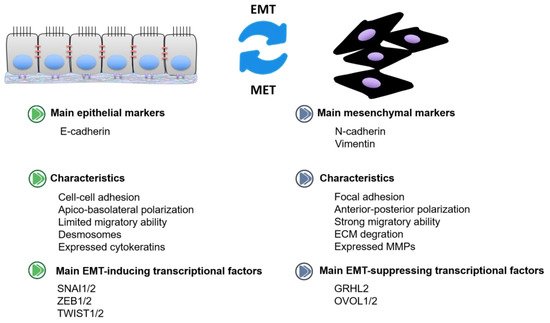
Epithelial–mesenchymal transition (EMT) refers to a process by which epithelial cells change into mesenchymal stem cells with an increase in migratory and invasive capacities [1]. EMT has been categorized into three different subtypes based on the distinguished biological settings as well as functional consequences. Among them, the “type 3” is associated with cancer development, also called as oncogenic EMT (Figure 1). Epithelial-like cells are non-mobile with adherent junctions, while mesenchymal-like cells, devoid of cell polarization, could promote the invasion and migration of primary tumor cells from one site to a distant site [2]. E-cadherin is considered as the epithelial marker, modulating cell–cell adhesion [3]. N-cadherin and vimentin are identified as mesenchymal markers. E-cadherin to N-cadherin switch in cancer leads to a more aggressive migration, invasion, and metastasis [4]. Snail and Slug transcription factors could promote epithelial cells migrating and translating into mesenchymal cells via inducing an E-cadherin to N-cadherin switch [5][6]. The mesenchymal-to-epithelial transition (MET) is the opposite process of EMT, namely, converting mesenchymal cells to epithelial cells [7]. MET allows cancer cells to colonize and form a potentially deadly metastatic lesion at the secondary tumor site [8]. EMT transcription factors (EMT-TFs) coordinate the EMT process. Among them, EMT inducers (SNAI1/2, ZEB1/2, TWIST1/2, etc.) are the most widely studied, with the capacity to suppress E-cadherin and promote mesenchymal transition [9]. In addition, some transcription factors identified as EMT suppressors (GRHL2, OVOL1/2, etc.) could drive the MET process [10]. According to the bioinformation analysis, GRHL2 is associated with epithelial characteristics across various cancers, inducing mesenchymal phenotypes [11]. Meanwhile, OVOL proteins function as the suppressors of EMT-inducing transcription factors and deletion of OVOL1/2 will result in EMT progression [12].
Figure 1. The characteristics of epithelial-like cells and mesenchymal-like cells in cancer.
MicroRNAs, also called miRNAs, are a kind of small non-coding RNA molecules of about 22 nucleotides which function as negative regulators to inhibit the expression of target genes [13]. With the base-pairing capability, miRNAs could majorly bind to the 3′ untranslated region (UTR) of its target genes, resulting in the degradation of target mRNAs or translational inhibition. Specifically, the perfect complementarity of miRNA and its targets tends to result in degradation of the targeted mRNA, while the imperfect complementary binding indicates the translational inhibition of the targets [14][15]. One miRNA could even interact with numerous mRNAs, and multiple miRNAs could regulate target genes cooperatively, which enables miRNAs to be the most substantial regulators in cells [16]. Generally speaking, miRNAs in cancers could affect cell proliferation, migration, invasion, stemness, drug resistance, angiogenesis, tumor growth, even metastasis, etc. [17]. Since a great number of studies gradually started to identify functions of miRNAs on the EMT process of cancer, miRNAs were found to play critical roles as both oncogenic genes (oncomiRs) and tumor suppressive genes (TsmiRs) in the development of EMT [18][19][20].
In the last decade, the cancer treatment efforts in the field of miRNAs have increased continuously. Although miRNA-based cancer diagnostics and prognostics have been gradually applied in cancer management, miRNA-based therapeutics are still considered as emerging challenges. The main approaches focus on inactivating or abating oncomiRs, activating or promoting tsmiRs, and modulating deregulated miRNAs [21]. Besides single strand interfering RNAs, such as miR MRX34 in a phase I study (NCT01829971), natural compounds have gained attention due to their possible applications in the regulation of miRNAs. Interestingly, these related findings even point out that single compounds are promising drug candidates in that they can regulate multiple miRNAs at the same time [22]. Thus, this review is aimed at illustrating the function and importance of miRNAs in the EMT process as promising targets in cancer treatment and the collection of compounds with the capacity to suppress EMT via regulating microRNA in cancer.

2. OncomiRs in EMT

In most conditions, it was found that oncomiRs were overexpressed or upregulated, promoting the development of EMT and giving rise to the metastatic potential. OncomiRs could promote the EMT process through activating TGFβ, FGF, and Notch signaling pathways, suppressing EMT inhibitors, and enhancing invasion and motility [23].

2.1. Breast Cancer

Breast cancer is ranked as the most common cause of cancer-death among women worldwide. The expression of miR-27a was higher in breast cancer tissues and cell lines than the normal counterparts. Overexpressed miR-27a could induce the EMT process and promote migration. FBXW7 was expressed at a low level in breast cancer tissues and cell lines and rescuing FBXW7 would at least partly reverse the oncogenic effect of miR-27a [24]. MiR-122-5p was highly expressed in triple-negative breast cancer (TNBC) while CHMP3 expression was low, which was associated with poor survival in patients with TNBC. At the cellular level, miR-122-5p promoted TNBC cell viability, proliferation, and invasion but inhibited apoptosis by targeting CHMP3 mRNA. Furthermore, miR-122-5p compelled the EMT process and MAPK signaling in TNBC [25]. MiR-155 was expressed at a high level in breast cancer tissues compared with paired normal tissues, while TGFBR2 was expressed at a low level. High expression of miR-155 could promote tumor growth and metastasis. Transfecting miR-155 inhibitors could inhibit cell proliferation and even EMT process, and increase TGFBR2 expression. Luciferase assay exerted that TGFBR2 was the direct target of miR-155, suggesting that miR-155 might promote breast cancer progression through the down-regulation of TGFBR2 [26]. High expression of miR-615-3p was determined in invasive breast cancer cells and metastatic breast cancer tissues. Overexpressed miR-615-3p could critically promote cell motility in vitro and pulmonary metastasis in vivo. Interestingly, the study also exerted that PICK1 was the direct target of miR-615-3p, and PICK1 inhibited the processing of pre-miR-615-3p to mature miR-615-3p, resulting in a negative feedback loop [27].

2.2. Colorectal Cancer

In recent years, the occurrence of colorectal cancer has been gradually increasing with poor overall survival rate [28]. The miR-410-3p expression levels were significantly up-regulated in colorectal cancer (CRC) cells and tissues, while its target, namely, ZCCHC10 was markedly down-regulated. In the functional assay, miR-410-3p could promote EMT with the enhanced ability of migration and invasion of HT29 and SW480 cells. Interestingly, the increase in ZCCHC10 could reverse the effect of miR-410-3p [29]. MiR-425-5p promoted CRC tumor growth and metastasis in vivo. Transfection of miR-425-5p mimic in SW480 cells exerted a promotive effect on cell viability, cell cycle entry, migration, invasion, and EMT process. Moreover, high expression of miR-425-5p promoted β-catenin translocating to the nucleus. Transfection of miR-425-5p inhibitor in LOVO cells demonstrated opposite effects. Luciferase reporter assay confirmed that CTNND1 was the direct target of miR-425-5p. Silencing of CTNND1 would mimic the effect of miR-425-5p overexpression [30]. RASSF6 hypermethylation has been shown to be frequent in a variety of solid cancers, according to extensive research. A recent study showed that miR-496 was aberrantly up-regulated in CRC and RASSF6 was a direct target of miR-496. Further research revealed that the stimulation of Wnt signaling by miR-496 targeting RASSF6 might have facilitated cell migration and EMT, yet had little effect on cell survival. In summary, it was confirmed that miR-496/RASSF6 axis was engaged in Wnt pathway-mediated tumor metastasis which may be a therapeutic target for CRC [31].

2.3. Cervical Cancer

Cervical cancer is one of the common types of cancer occurring among women in developing countries. MiR-150-5p has been found to up-regulate in cervical carcinoma. The research suggested that the levels of miR-150-5p were obviously different in C-33A and HeLa cells. MiR-150-5p was strongly associated with cell growth. Furthermore, SRCIN1 acted as a direct target of miR-150-5p. When transducing miR-150-5p mimics, the expression of SRCIN1 increased, the proliferation and EMT of cervical cancer cells were suppressed significantly, and apoptosis was accelerated by SRCIN1 [32]. Recently, a study revealed that the expression of miR-199a-5p was high in cervical carcinoma tissues and cell lines, and thus promoted cell proliferation and metastasis. Meanwhile, the vimentin and N-cadherin expressions were enhanced while E-cadherin was reduced by high levels of miR-199a-5p, which were the significant markers of EMT. Further research indicated that PIAS3 was the direct targets of miR-199a-5p and was negatively correlated with miR-199a-5p. In contrast, the effects of miR-199a-5p were reversed by up-regulation of PIAS3 in cervical carcinoma [33].

2.4. Gastric Cancer

Evidence showed that the aberrantly high expression of miR-21 contributed to early metastasis in gastric cancer. In this study, miR-21 was higher in gastric cancer cell lines compared with a gastric mucosal epithelial cell line. Furthermore, miR-21 mimic could increase N-cadherin and vimentin, and decrease E-cadherin to promote EMT in gastric cancer cells whereas miR-21 inhibitor showed the opposite effects. As a result, up-regulation of miR-21 facilitated cell metastasis and invasion of MGC-803 cells. The decreased expression of miR-21 suppressed cell metastasis and invasion in gastric cancer [34]. Overexpressed miR-130b-3p was detected in gastric cancer tissues, and miR-130b-3p promoted proliferation, migration, and EMT of gastric cancer cells as well as tumor formation and metastasis in vivo. Additionally, miR-130b-3p could directly target MLL3, and MLL3 could promote the expression and function of GRHL2. Decreasing miR-130b-3p or upregulating of GRHL2 inhibited tumor growth and angiogenesis in gastric cancer [35]. The overall survival rates of advanced gastric cancer are still poor despite the progression of pharmacotherapy [36]. MiR-616-3p was expressed at a high level in cancer tissues, and high expression of miR-616-3p was positively correlated with poor prognosis. Functional assay exerted that overexpressed miR-616-3p enhanced angiogenesis and EMT in gastric cancer cells through the down-regulation of PTEN. Furthermore, rescuing PTEN could at least partly inhibit the effect of miR-616-3p on gastric cancer cells [37].

2.5. Nasopharyngeal Carcinoma

Nasopharyngeal carcinoma is a kind of cancer developed from nasopharyngeal epithelium [38]. A study showed that increased expression of miRNA-10b promoted the growth of nasopharyngeal carcinoma cells CNE1 in vitro by regulating EMT, proliferation, and migration [39]. The study demonstrated that miR-205-5p was significantly higher in the cisplatin-resistant nasopharyngeal carcinoma cell line HNE1/DDP than in its parental cell HNE1. According to the findings, low expression of miR-205-5p inhibited EMT development in HNE1/DDP cells. Further research suggested that down-regulation of the expression of PTEN, a downstream candidate gene of miR-205-5p, could counterbalance the impact of miR-205-5p inhibitors. Therefore, miR-205-5p was shown to regulate EMT through the PI3K/AKT pathway by targeting PTEN on HNE1/DDP cells [40].

2.6. Lung Cancer

Lung cancer is considered as one of the most common cancers with extremely poor prognosis over the years. MiR-21 was overexpressed in human non-small cell lung cancer (NSCLC) cells, and overexpressed miRNA-21 induced cyclin D1 and cyclin E1 expression and promoted proliferation of cancer cells. In addition, high expression of miRNA-21 increased the EMT process through PTEN/Akt/GSK3β signaling [41]. MiR-942 was detected expressed at a high level in human NSCLC tissues and cells. Overexpressed miR-942 could promote cell migration, invasion, and angiogenesis. Furthermore, miR-942 was attributed to NSCLC metastasis in tail vein xenograft models. Three bioinformatics software predicted BARX2 as a downstream target of miR-942, and luciferase assay validated that miR-942 could directly bind to the 3′UTR region of BARX2. Western blot results exerted that miR-942 increased EMT-associated proteins by down-regulation of BARX2 [42].

2.7. Osteosarcoma

Osteosarcoma is the most common malignant bone tumor occurring in youth [43]. In osteosarcoma tissues and cell lines, there was obviously increasing expression of miR-17-5p, which played a role in suppressing cell proliferation, EMT, and accelerating apoptosis in osteosarcoma. Further study revealed that SRCIN1, an anti-oncogene, was served as a target of miR-17-5p. When silencing miR-17-5p, the EMT-related proteins expression changed, and thus regulated EMT progression of osteosarcoma [44].

2.8. Other Types of Cancer

Intrahepatic cholangiocarcinoma is ranked as the second most common malignant hepatic tumor with a relatively high postoperative recurrence rate [45]. High expression of miR-19b-3p was detected in intrahepatic cholangiocarcinoma tissues compared with adjacent tissues, and overexpressed miR-19b-3p could promote cell proliferation and EMT, and inhibit apoptosis. Further study exerted that CCDC6 was a predicted target of miR-19b-3p, and in vivo study displayed that miR-19b-3p/CCDC6 axis modulated EMT to promote intrahepatic cholangiocarcinoma development [46]. Ovarian cancer is related to gynecological tumors with an extremely poor prognosis [47]. MiR-27a was expressed at a high level in ovarian cancer tissues and cells. Functional assays exerted that overexpressed miR-27a promoted migration and invasion of HO8910 and OV90 cells, while decreasing miR-27a displayed the opposite effect. Furthermore, high expression of miR-27a induced EMT markers and processes. FOXO1 was found as the direct target of miR-27a, and miR-27a could significantly inhibit FOXO1 expression as well as the Wnt pathway [48]. Prostate cancer is one of the most common cancers among men, resulting in over 11,000 deaths per year [49]. Real-time PCR results determined the overexpression of miR-19a in prostate cancer. Clinical data suggested a negative correlation between miR-19a expression and poor outcome of patients. Luciferase reporter assay exerted that miR-19a could directly target the mRNA 3′-UTR of CUL5 in PC3 cells. In addition, decreased CUL5 expression would predict a worse outcome for prostate cancer patients. Functional assay found that miR-19a promoted cell migration, invasion, EMT, and metastasis in prostate cancer through down-regulation of CUL5, while rescuing CUL5 would partially reverse the roles of miR-19a on prostate cancer [50]. Renal cancer belongs to the most common type of kidney carcinoma, accounting for about 3% of tumors in adults [51]. MiR-155-5p was expressed at a high level in renal cancer, and overexpressed miR-155-5p could promote cell proliferation, colony formation, migration, and invasiveness of renal cancer cells. In contrast, decreasing miR-155-5p could inhibit cancer development. Meanwhile, miR-155-5p could increase N-cadherin and Snail, and promote the EMT process [52].

3. TsmiRs in EMT

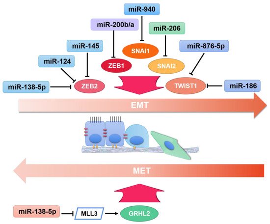
TsmiRs, as tumor suppressors, show the opposite function of oncomiRs in EMT pathways. TsmiRs were found to be dramatically downregulated or completely eliminated in cancer cells, and the rescue of tsmiRs could inhibit EMT development, with the increase in E-cadherin and decrease in N-cadherin. Some tsmiRs could directly target SNAI, Twist, and ZEB families, inhibiting their expression (Figure 2).
Figure 2. The regulation of microRNAs on transcription regulators impacting EMT, namely EMT-inducers and EMT-suppressors in cancer.

3.1. Breast Cancer

Bioinformatics analysis indicated that Gli1 was the direct target of miR-34b. Further study determined that miR-34b was markedly decreased in MCF-7 and MDA-MB-231 cells than in normal mammary gland epithelial MCF-10A cells, while the expression of Gli1 was the opposite. Reducing expression of miR-34b would enhance Gli1, promote the EMT process as well as cell invasion, while increasing its expression could reverse the effects [53]. Real-time PCR results determined that miR-124 was expressed at a low level in TNBC tissues and cells. Overexpressed miR-124 could inhibit proliferation, migration, invasion, and the EMT process of TNBC cells. ZEB2 was a direct target of miR-124 according to the results of luciferase reporter assay. Rescue assays manifested that miR-124 inhibited EMT and metastasis through the inhibition of ZEB2 [54]. In vitro and vivo experiments revealed that the ectopic expression of miR-135 obviously suppressed cell proliferation, migration, invasion, and EMT in breast cancer cell lines MDA-MB-468 and MCF-7 cells, and reduced tumor volume. Moreover, mechanistic studies suggested that the miR-135 mimic could down-regulate the N-cadherin and vimentin at mRNA and protein levels, and enhance the expression of E-cadherin. Meanwhile, miR-135 could increase p-GSK3β expression and decrease Wnt and β-catenin expression which suggested that miR-135 partly inactivated the Wnt/β-catenin pathway to suppress breast cancer [55]. It has been observed that DUSP4 expression was significantly up-regulated in breast cancer cell lines, and the miR-137 level was dramatically down-regulated. Moreover, the introduction of miR-137 significantly inhibited resistance to doxorubicin of breast cancer cells by decreasing EMT. Besides, up-regulation of DUSP4 in breast cancer cells partially abolished the chemo-resistance of miR-137 mimic. Moreover, the results of miR-137 enhanced the sensitivity of breast cancer cells to doxorubicin in vivo and were consistent with that in vitro [56]. The low expression of miR-186 in breast cancer tissues and cells is related to tumor metastasis and a poor overall survival in breast cancer patients. MiR-186 with high expression suppressed breast cancer cells proliferation, migration, and EMT progression, while miR-186 inhibition showed the opposite results. Moreover, it was confirmed that miR-186 directly targeted Twist1 in breast cancer cells. The activities of miR-186 were attenuated by restoring Twist1 [57]. In this study, researchers found up-regulation of miR-214 inhibited cell proliferation and invasion, while inhibition of miR-214 expression could have the opposite result. In addition, the survival rate of breast cancer patients was favorably connected with miR-214 levels and negatively connected with RNF8 levels which is a recently discovered regulator that promotes EMT. Further study firstly suggested that miR-214 could inhibit RNF8 to regulate EMT and thus suppress breast cancer [58]. MiR-516a-3p expression was expressed at a low level in human breast cancer tissues and cells, while the expression of Pygo2 was high. Additionally, the expression of miR-516a-3p was negatively associated with poor prognosis for patients with breast cancer, while the expression of Pygo2 exerted a positive correlation to poor prognosis. Luciferase reporter assay validated Pygo2 as the direct target of miR-516a-3p. Functional study exerted that increasing miR-516a-3p expression could suppress cell growth, migration, invasion, and EMT process, and reducing miR-516a-3p had a reverse effect [59]. MiR-574-5p was down-regulated in breast cancer tissues and cells. MiR-574-5p inhibited proliferation, migration, and EMT in TNBC cells. Furthermore, miR-574-5p had an inhibitory effect on tumor size and metastasis in vivo. Luciferase assay exerted that BCL11A and SOX2 were the direct target of miR-574-5p. The suppressive effect of miR-574-5p in TNBC cells was at least partly due to SOX2 and BCL11A [60]. The up-regulation of miR-6838-5p in TNBC could inhibit cell migration, invasion, and Wnt signaling pathway, while down-regulating miR-6838-5p had a reverse effect. Luciferase reporter and RNA immunoprecipitation assay confirmed that WNT3A was the direct target of miR-6838-5p. Interestingly, rescuing WNT3A would at least partly reverse the suppressive effect of miR-6838-5p on the TNBC EMT process [61].

3.2. Colorectal Cancer

Bioinformatic analysis displayed that miR-1-3p was expressed at a low level in CRC tissues, and real-time PCR results validated the expression in CRC cells. Overexpressed miR-1-3p could inhibit CRC cell proliferation and invasion. Advanced study exerted that miR-1-3p could directly bind to YWHAZ, resulting in inhibition of the EMT process [62]. Research suggested that the expression of miR-9-5p was decreased and the expression of FOXP2 was increased in CRC tissues and cell lines. Moreover, FOXP2 had oncogenic roles in CRC and miR-9-5p could inhibit cell metastasis and EMT in CRC by targeting FOXP2. The low miR-9-5p levels and the high FOXP2 levels were associated with poor prognosis of CRC patients. Therefore, miR-9-5p/FOXP2 axis is key to diagnose and treat CRC [63]. Real-time PCR showed that the expression of miR-205 was negatively correlated with MDM4 in CRC tissues and cells. Luciferase assay displayed a direct binding between miR-205 and MDM4. Advanced study demonstrated that overexpressed miR-25 inhibited proliferation, migration, and invasion of HCT116 cells. Additionally, the decreased N-cadherin, vimentin, MMP2, and MMP9 with the increase of E-cadherin were observed in miR-205-overexpressed cells [64]. MiR-145-5p were down-regulated in CRC samples and cell lines. Elevated miR-145-5p expression inhibited cell viability, migration, invasion and EMT of LoVo and SW480 cells, while reducing miR-145-5p displayed the opposite effect. CDCA3 was confirmed as a direct target of miR-145-5p according to the luciferase reporter assay. In addition, rescuing CDCA3 in SW480 cells could at least partly reverse the influence induced by miR-145-5p [65]. It was interesting to find that the expression of miR-330 was negatively correlated with HMGA2. The high expression of HMGA2 and low expression of miR-330 were detected in CRCs with poor long-term patient survival. Evaluated miR-330 expression in HCT116 and SW480 cells suppressed the oncogenic effect of HMGA2. Further study exerted that miR-330 could inhibit cell migration and viability, the Akt pathway, as well as the essential regulators in the EMT process through the down-regulation of HMGA2 [66]. MiR-370-3p played a key role in repressing CRC proliferation and EMT in vitro. The underlying mechanism proved that miR-370-3p could reduce tumor-related inflammatory factors, such as TNF-α, IL-1 β, and IL-6, thereby inhibiting proteins such as P53, β-catenin, and ki67, which are closely related to tumor growth [67]. MiR-873-5p overexpression dramatically inhibited CRC cell proliferation and EMT. Mechanistically, miR-873-5p directly targeted JMJD8 which was significantly up-regulated in CRC tissues and cell lines to impact the proliferation and EMT process. Finally, the research also found that miR-873-5p could suppress the NF-кB pathway in CRC. Taken together, miR-873-5p could inhibit proliferation and EMT in CRC by targeting JMJD8 and suppressing the NF-кB signaling pathway, which may provide a new antitumor strategy to carcinogenesis [68]. MiR-3622a-3p was remarkably down-regulated in CRC according to the TCGA database. Functional assays exerted that miR-3622a-3p could inhibit proliferation, apoptosis, cell cycle, migration, and invasion of CRC cells. The results from dual luciferase assay, RNA immunoprecipitation assay, and pull-down assay showed that SALL4 was the direct target of miR-3622a-3p. Further studies showed that miR-3622a-3p inhibited stemness and the EMT process of CRC cells through SALL4 mRNA decrease. Additionally, miR-3622a-3p also manifested the anti-cancer effect in a tumor xenograft model and an in vivo metastasis model [69].

3.3. Gastric Cancer

Research indicated that the level of miR-105 was low in gastric cancer tissues and cells, and SOX9 was expressed highly in gastric cancer cells. Further study suggested that miR-105 was negatively correlated with SOX9, and directly targeted it to suppress cell migration, invasion, and EMT in gastric cancer. The partial effects of miR-105 on cell migration and invasion could be reversed by SOX9. SOX9 knockdown could inhibit gastric cancer cell migration, invasion, and EMT [70]. In this study, miR-129-5p was expressed lower in gastric cancer cells than in GES-1. In addition, the high levels of miR-129-5p could suppress EMT and proliferation through directly targeting HMGB1 which was up-regulated in gastric cancer cells [71]. Low expression of miR-203 was detected in gastric cancer tissues and cell lines. Overexpressed miR-203 could suppress invasion and the EMT process. Bioinformatics analysis indicated annexin A4 as the direct target of miR-203, and the binding was validated by luciferase reporter assay. Additionally, rescuing annexin A4 in GC cells at least partly reversed the inhibitory effect of miR-203 [72]. A recent study indicated that the expression of FOXQ1 was so high in gastric cancer cells that it greatly promoted proliferation, migration, invasion, and EMT. Further study suggested that FOXQ1 was directly regulated by miR-519 which was observed weakly expressed in both gastric cancer tissues and cells. In other words, miRNA-519 functioned as a suppressor in regulating gastric cancer EMT by targeting FOXQ1 [73]. Low expression of miR-630 was detected in gastric cancer cell lines. MiR-630 mimic transfection could inhibit migration and invasion in SGC-7901 and BGC-823 cells. Furthermore, EMT phenotype was suppressed after the increase in miR-630 [74]. The decrease in miRNA-665 expression is associated with gastric cancer occurrence. In vitro, miRNA endogenous mimics could repress gastric cancer cell lines growth by targeting the 3′-UTR of the CRIM1 gene which was found to promote the EMT process in GC cell lines. Interestingly, up-regulating the expression of CRIM1 would eliminate the inhibitory effect of miRNA-665 on gastric cancer [75].

3.4. Glioma

Glioblastoma multiforme (GBM) is considered as the most common malignant brain tumor, identified as a Grade IV astrocytoma [76]. Functional assay displayed that high expression of miR-96 could inhibit migration, invasion, and proliferation in GBM cells. Luciferase assay indicated AEG-1 as the direct target of miR-96. Further study exerted that miR-96 could regulate EMT markers and suppress EMT process through the downregulation of AEG-1 in GBM cells. Additionally, the down-regulation of AEG-1 would result in a similar effect as overexpressed miR-96 in GBM [77]. MiR-205 was found to express lowly in glioma tissues and human glioma cell lines U87 and U251. In vitro, the overexpression of miR-205 increased E-cadherin, decreased mesenchymal markers, and inhibited cell proliferation, migration, and invasion. Furthermore, miR-205 was reported to directly bind to HOXD9 and decreased its expression so that EMT progression was reversed [78]. When glioma tissues from patients were compared with normal cells, the expression of miR-218-5p was shown to be lower. After transfecting miR-218-5p into U251 and U87 cells, the findings showed that miR-218-5p could suppress cell activity, proliferation, and invasive ability as well as EMT. Further research revealed LHFPL3 was highly expressed in glioma and a direct target of miR-218-5p. Namely, miR-218-5p could bind to LHFPL3 to inhibit the invasion of glioma cells [79]. The miR-802 level was dramatically down-regulated in GBM tissues, and the SIX4 expression was significantly up-regulated. Moreover, high SIX4 expression was strongly associated with a low miR-802 level in GBM tissues. In addition, the introduction of miR-802 significantly inhibited proliferation, invasion, and EMT of GBM cells. Furthermore, knockdown of SIX4 had similar effects with miR-802 overexpression on GBM cells [80]. In vitro, the expression of miR-876-5p was observed to decrease in all of the GBM cell lines while TWIST1 expression levels showed the opposite trend. This finding indicated that miR-876-5p inhibited EMT in GBM accompanied with downregulation of gene TWIST1, and thus is a promising target for the treatment of GBM [81].

3.5. Lung Cancer

The down-regulation of miR-138-5p was detected in lung adenocarcinoma tissues and cell lines. The high levels of miR-138-5p inhibited EMT, proliferation, and metastasis in A549 and H1299 cells. Additionally, miR-138-5p was identified to bind directly to ZEB2, and decreased ZEB2 to suppress the EMT process, proliferation, and metastasis of lung adenocarcinoma cells [82]. In this study, it was verified that miR-145 was associated well with invasive ability as a result of low expression in NSCLC cell lines H1299, PC7, and SPCA-1 cells. When compared with adjacent normal tissue, the expression of miR-145 was lower in cancer tissue. The important markers of EMT, namely, N-cadherin, vimentin, and E-cadherin were down-regulated by overexpression of miR-145. Further research indicated that the oncogene ZEB2 was bound by miR-145, and miR-145 could decrease ZEB2 to inhibit EMT progression in NSCLC as well [83]. MiR-146b expression was down-regulated in cisplatin resistant human lung adenocarcinoma cells (A549/DDP and H1299/DDP), which indicated that miR-146b could function as a tumor suppressor in cisplatin resistant human lung adenocarcinoma cells. Furthermore, PTP1B was a target of miR-146b. Then, miR-146b suppressed the EMT of H1299/DDP and A549/DDP cells and improved cisplatin sensitivity. Additionally, to detect the impact of miR-146b up-regulation on tumor growth in vivo, xenograft model mice were used, and these findings were consistent with that of in vitro [84]. MiR-335-5p exerted a significant effect on repressing TGF-β1-mediated NSCLC migration and invasion. More importantly, over-expressed miR-335-5p could specifically bind to ROCK1, thus inhibiting the EMT, migration, and invasion in NSCLC. Consistently, deceased ROCK1 efficiently impaired TGF-β1-mediated EMT and migratory and invasive capabilities of NSCLC cells. In other words, overexpression of miR-335-5p inhibited cell proliferation and EMT of NSCLC cells by directly down-regulating ROCK1 expression [85]. In a recent study, findings showed that the overexpression of miR-363-3p could inhibit EMT to suppress cell migration and invasion in NSCLC while knocking down miR-363-3p had the opposite results. Moreover, it was identified that miR-363-3p bound to 3′UTR of NEDD9 and SOX4, negatively regulating their expression. Intriguingly, NEDD9 or SOX4 knockdown could reverse the metastasis-promoting function of antagomiR-363-3p and the inhibitory effects of agomiR-363-3p. Collectively, miR-363-3p may act as an anti-oncogene to be a potential therapeutic target for NSCLC [86]. The lower expression of miR-379 expression in NSCLC tissues and cell lines compared with normal was associated with a poor survival rate in NSCLC patients. The research demonstrated that miR-379 could combine with the 3′UTR of CHUK and obviously down-regulate its expression in NSCLC cells. MiR-379 played an important role in suppressing cell invasion of NSCLC through regulating EMT. Furthermore, CHUK was an oncogene and the overexpression of miR-379 could inhibit CHUK and NF-кB pathway to prevent NSCLC [87]. The low expression of miR-874 was detected in NSCLC tissues while its expression was relatively higher in corresponding adjacent nontumor tissues. The results from functional assays exerted that miR-874 could inhibit cell proliferation and mobility, while decreasing miR-874 in A549 and H1299 cells showed an opposite effect. Luciferase assay displayed that miR-874 down-regulated the expression of AQP3 through a direct binding to the 3 ‘UTR of AQP3 mRNA. MiR-874 inhibited AQP3 and the PI3K/AKT signaling pathway, resulting in the inhibition of EMT in A549 cells. Additionally, miR-874 had a suppressive effect on NSCLC growth in vivo [88]. Recently, a study found miR-940 could inhibit the proliferation of NSCLC by targeting SNAI1 and inhibiting TGF-β-induced EMT and invasion in vitro. Namely, this finding indicated that knocking down SNAI1, which is regulated by miRNA-940, could repress TGF-β-induced EMT and migration and invasion of NSCLC [89].

3.6. Pancreatic Cancer

Screening of transcription profiles revealed that 113 microRNAs and 1749 messenger RNAs expressed differentially in pancreatic cancer tissues. In particular, the expression of miR-203a-3p was markedly down-regulated in both pancreatic cancer tissues and cells, while the expression of SLUG was opposite. Luciferase reporter assay and RNA immunoprecipitation confirmed that miR-203a-3p could directly target SLUG. Further study displayed that miR-203a-3p mimic transfection could suppress migration, invasion, and EMT process through sponging SLUG [90]. A recent study showed that miR-338-5p was lowly expressed in pancreatic cancer tissues and the expression was negatively correlated with lymph node metastasis. Additionally, miR-338-5p overexpression could inhibit EMT and the metastasis process of pancreatic cancer cells. Furthermore, miR-338-5p could target EGFR and suppress EGFR/ERK pathways. These findings suggested miR-338-5p as a negative regulator of the EMT process in pancreatic cancer [91].

3.7. Papillary Thyroid Cancer

Papillary thyroid carcinoma is the most common type of thyroid cancer, with an increasing morbidity rate in recent years [92]. Mechanically, miR-451a played an important role in suppressing proliferation, EMT, and apoptosis in papillary thyroid cancer cells in vitro. Furthermore, luciferase reporter assays proved that miR-451a could bind to 3′UTR of PSMB8 directly. In addition, in vitro, functional experiments showed miR-451a could suppress the ontogenetic capabilities of PTC cells by targeting PSMB8 as well, which was consistent with that of in vivo [93]. Research revealed that the expression of miR-630 was decreased in papillary thyroid carcinoma cell lines TIPC-1 and SW1736 cells. Up-regulation of miR-630 could suppress the cell proliferation and promote cell apoptosis in PTC by inhibiting the expression of caspase-3 and caspase-6, combined with inhibiting PTC cell migration and invasion through preventing EMT progress. Moreover, enhanced miR-630 expression reduced protein phosphorylation levels in the JAK2/STAT3 signaling pathway [94].

3.8. Other Types of Cancer

It has been observed that the expression level of miR-499a-5p was decreased in cervical cancer tissues and cell lines. The research showed increased expression of miR-499a-5p inhibited cervical cancer cell proliferation, invasion, migration, and EMT by targeting the gene eIF4E [95]. MiR-373-3p could inhibit EMT progress in choriocarcinoma JEG-3 and JAR cells through the inhibition of the TGF-β signaling pathway. MiR-373-3p impede the migratory and invasive ability of choriocarcinoma cells. Microarray analysis indicted an interaction between miR-373-3p and TGF-β R2, and luciferase assay validated that miR-373-3p could directly bind to TGF-β R2 [96]. MiR-4429 was found to express lowly in clear cell renal cell carcinoma tissues and cells and could inhibit cell proliferation, migration, and invasion combined with the EMT process. Further study indicated that miR-4429 targeted CDK6 and had a negative correlation with it [97]. MiR-34a-5p played an anti-esophageal squamous cell carcinoma (ESCC) role by targeting the Hippo-YAP1/TAZ signaling pathway while the expression of LEF1 was high in ESCC tissue and cell lines. Inexplicably, high expression of miR-34a-5p promoted cancer cell viability, while up-regulation of LEFA1 could impair the inhibitory effect of miR-34a-5p on ESCC cell lines [98]. Real-time PCR results showed that miR-22 was expressed at a low level in ovarian cancer tissues and cells, and the expression of miR-22 had a negative correlation with poor prognosis. NLRP3 was up-regulated in ovarian cancer, and the expression of NLRP3 was negatively correlated with miR-22. Further study showed that miR-22 inhibited cell viability and EMT through direct targeting of NLRP3 mRNA, and the NLRP3 overexpression partially reversed the effects of miR-22 [99]. GRHL2 could maintain the epithelial phenotype epithelial status of ovarian cancer. ChIP-seq pointed that GRHL2 could bind to the promoter site of miR-200b/a, and ZEB1 was the direct target of miR-200b/a. Then, GRHL2 and ZEB1 formed a negative feedback to suppress EMT process through the regulation of miR-200b/a [100]. The expression of miR-490-5p in primary pharyngolaryngeal cancer tissues and cell lines including BICR 18 and FaDu cells was obviously decreased. Research demonstrated that the high levels of MAP3K9 could facilitate cell viability, migration and invasion rates, the EMT process, and ability of cloning. Furthermore, miR-490-5p was found to target MAP3K9 and then regulate cell proliferation, migration, invasion, and EMT in pharyngolaryngeal cancer [101]. IL-8 acted as a tongue squamous cell carcinoma (TSCC) growth factor through promoting invasion and EMT of TSCC cells, which could be suppressed by miR-940 up-regulation. Interestingly, if gene CXCR2 was silenced or NF-кB inhibitor existed, the positive effect of miR-940 disappeared as well, suggesting that miR-940 regulated the growth of TSCC cells through targeting CXCR2/NF-кB system [102].

References

  1. Demirkan, B. The Roles of Epithelial-to-Mesenchymal Transition (EMT) and Mesenchymal-to-Epithelial Transition (MET) in Breast Cancer Bone Metastasis: Potential Targets for Prevention and Treatment. J. Clin. Med. 2013, 2, 264–282.
  2. Gupta, S.; Maitra, A. EMT: Matter of Life or Death? Cell 2016, 164, 840–842.
  3. Biswas, K.H. Molecular Mobility-Mediated Regulation of E-Cadherin Adhesion. Trends Biochem. Sci. 2020, 45, 163–173.
  4. Nakagawa, M.; Bando, Y.; Nagao, T.; Morimoto, M.; Takai, C.; Ohnishi, T.; Honda, J.; Moriya, T.; Izumi, K.; Takahashi, M.; et al. Expression of p53, Ki-67, E-cadherin, N-cadherin and TOP2A in triple-negative breast cancer. Anticancer Res. 2011, 31, 2389–2393.
  5. Wang, Y.; Shi, J.; Chai, K.; Ying, X.; Zhou, B.P. The Role of Snail in EMT and Tumorigenesis. Curr. Cancer Drug Targets 2013, 13, 963–972.
  6. Alves, C.C.; Carneiro, F.; Hoefler, H.; Becker, K.F. Role of the epithelial-mesenchymal transition regulator Slug in primary human cancers. Front. Biosci. 2009, 14, 3041–3050.
  7. Cho, E.S.; Kang, H.E.; Kim, N.H.; Yook, J.I. Therapeutic implications of cancer epithelial-mesenchymal transition (EMT). Arch. Pharmacal Res. 2019, 42, 14–24.
  8. Singh, M.; Yelle, N.; Venugopal, C.; Singh, S.K. EMT: Mechanisms and therapeutic implications. Pharmacol. Ther. 2018, 182, 80–94.
  9. Georgakopoulos-Soares, I.; Chartoumpekis, D.V.; Kyriazopoulou, V.; Zaravinos, A. EMT Factors and Metabolic Pathways in Cancer. Front. Oncol. 2020, 10, 499.
  10. Chung, V.Y.; Tan, T.Z.; Ye, J.R.; Huang, R.L.; Lai, H.C.; Kappei, D.; Wollmann, H.; Guccione, E.; Huang, R.Y.J. The role of GRHL2 and epigenetic remodeling in epithelial-mesenchymal plasticity in ovarian cancer cells. Commun. Biol. 2019, 2, 15.
  11. Mooney, S.M.; Talebian, V.; Jolly, M.K.; Jia, D.; Gromala, M.; Levine, H.; McConkey, B.J. The GRHL2/ZEB Feedback Loop-A Key Axis in the Regulation of EMT in Breast Cancer. J. Cell. Biochem. 2017, 118, 2559–2570.
  12. Li, S.; Yang, J. Ovol Proteins: Guardians against EMT during Epithelial Differentiation. Dev. Cell 2014, 29, 1–2.
  13. Khoshnaw, S.M.; Green, A.; Powe, D.G.; Ellis, I. MicroRNA involvement in the pathogenesis and management of breast cancer. J. Clin. Pathol. 2009, 62, 422–428.
  14. Lee, Y.; Kim, M.; Han, J.; Yeom, K.-H.; Lee, S.; Baek, S.H.; Kim, V.N. MicroRNA genes are transcribed by RNA polymerase II. EMBO J. 2004, 23, 4051–4060.
  15. Lee, Y.; Ahn, C.; Han, J.; Choi, H.; Kim, J.; Yim, J.; Lee, J.; Provost, P.; Radmark, O.; Kim, S.; et al. The nuclear RNase III Drosha initiates microRNA processing. Nature 2003, 425, 415–419.
  16. Grimaldi, A.M.; Salvatore, M.; Incoronato, M. miRNA-Based Therapeutics in Breast Cancer: A Systematic Review. Front. Oncol. 2021, 11, 14.
  17. Hill, M.; Tran, N. miRNA interplay: Mechanisms and consequences in cancer. Dis. Model. Mech. 2021, 14.
  18. Berindan-Neagoe, I.; Calin, G.A. Molecular Pathways: MicroRNAs, Cancer Cells, and Microenvironment. Clin. Cancer Res. 2014, 20, 6247–6253.
  19. D’Amato, N.C.; Howe, E.N.; Richer, J.K. MicroRNA regulation of epithelial plasticity in cancer. Cancer Lett. 2013, 341, 46–55.
  20. Tang, J.; Li, Y.; Wang, J.; Wen, Z.; Lai, M.; Zhang, H. Molecular mechanisms of microRNAs in regulating epithelial–mesenchymal transitions in human cancers. Cancer Lett. 2016, 371, 301–313.
  21. Peng, F.; Xie, X.; Peng, C. Chinese Herbal Medicine-Based Cancer Therapy: Novel Anticancer Agents Targeting MicroRNAs to Regulate Tumor Growth and Metastasis. Am. J. Chin. Med. 2019, 47, 1711–1735.
  22. Ahmed, F.; Ijaz, B.; Ahmad, Z.; Farooq, N.; Sarwar, M.B.; Husnain, T. Modification of miRNA Expression through plant extracts and compounds against breast cancer: Mechanism and translational significance. Phytomedicine 2020, 68, 153168.
  23. Sreekumar, R.; Sayan, B.S.; Mirnezami, A.H.; Sayan, A.E. MicroRNA Control of Invasion and Metastasis Pathways. Front. Genet. 2011, 2, 58.
  24. Jiang, G.B.; Shi, W.W.; Fang, H.Y.; Zhang, X.H. miR-27a promotes human breast cancer cell migration by inducing EMT in a FBXW7-dependent manner. Mol. Med. Rep. 2018, 18, 5417–5426.
  25. Wang, Z.; Wang, X.Y. miR-122-5p promotes aggression and epithelial-mesenchymal transition in triple-negative breast cancer by suppressing charged multivesicular body protein 3 through mitogen-activated protein kinase signaling. J. Cell. Physiol. 2020, 235, 2825–2835.
  26. Liu, X.Y.; Li, Y.J.; Li, Z.; Hou, T. miR-155 promotes proliferation and epithelial-mesenchymal transition of MCF-7 cells. Exp. Ther. Med. 2021, 21, 7.
  27. Lei, B.; Wang, D.D.; Zhang, M.; Deng, Y.W.; Jiang, H.J.; Li, Y.W. miR-615-3p promotes the epithelial-mesenchymal transition and metastasis of breast cancer by targeting PICK1/TGFBRI axis. J. Exp. Clin. Cancer Res. 2020, 39, 14.
  28. Hu, Y.; Su, Y.; He, Y.; Liu, W.; Xiao, B. Arginine methyltransferase PRMT3 promote tumorigenesis through regulating c-MYC stabilization in colorectal cancer. Gene 2021, 791, 145718.
  29. Ma, Z.H.; Shi, P.D.; Wan, B.S. MiR-410-3p activates the NF-kappa B pathway by targeting ZCCHC10 to promote migration, invasion and EMT of colorectal cancer. Cytokine 2021, 140, 10.
  30. Liu, D.S.; Zhang, H.; Cui, M.M.; Chen, C.S.; Feng, Y. Hsa-miR-425-5p promotes tumor growth and metastasis by activating the CTNND1-mediated beta-catenin pathway and EMT in colorectal cancer. Cell Cycle 2020, 19, 1917–1927.
  31. Wang, H.; Yan, B.; Zhang, P.; Liu, S.; Li, Q.; Yang, J.; Yang, F.; Chen, E. MiR-496 promotes migration and epithelial-mesenchymal transition by targeting RASSF6 in colorectal cancer. J. Cell. Physiol. 2020, 235, 1469–1479.
  32. Zhu, J.; Han, S. miR-150-5p promotes the proliferation and epithelial-mesenchymal transition of cervical carcinoma cells via targeting SRCIN1. Pathol. Res. Pr. 2019, 215, 738–747.
  33. Qu, D.; Yang, Y.; Huang, X. miR-199a-5p promotes proliferation and metastasis and epithelial-mesenchymal transition through targeting PIAS3 in cervical carcinoma. J. Cell. Biochem. 2019, 120, 13562–13572.
  34. Xiao, T.; Jie, Z. MiR-21 Promotes the Invasion and Metastasis of Gastric Cancer Cells by Activating Epithelial-Mesenchymal Transition. Eur. Surg. Res. 2019, 60, 208–218.
  35. Zhang, Y.; Meng, W.; Yue, P.; Li, X. M2 macrophage-derived extracellular vesicles promote gastric cancer progression via a microRNA-130b-3p/MLL3/GRHL2 signaling cascade. J. Exp. Clin. Cancer Res. 2020, 39, 1–20.
  36. Ma, R.; Shimura, T.; Yin, C.; Okugawa, Y.; Kitajima, T.; Koike, Y.; Okita, Y.; Ohi, M.; Uchida, K.; Goel, A.; et al. Antitumor effects of Andrographis via ferroptosis-associated genes in gastric cancer. Oncol. Lett. 2021, 22, 1–8.
  37. Wu, Z.-H.; Lin, C.; Liu, C.-C.; Jiang, W.-W.; Huang, M.-Z.; Liu, X.; Guo, W.-J. MiR-616-3p promotes angiogenesis and EMT in gastric cancer via the PTEN/AKT/mTOR pathway. Biochem. Biophys. Res. Commun. 2018, 501, 1068–1073.
  38. Chen, H.; Luo, M.; Wang, X.; Liang, T.; Huang, C.; Huang, C.; Wei, L. Inhibition of PAD4 enhances radiosensitivity and inhibits aggressive phenotypes of nasopharyngeal carcinoma cells. Cell. Mol. Biol. Lett. 2021, 26, 1–12.
  39. Cheng, Z.Q.; Qiang, H.L.; Wang, B.; Ding, Z.R.; Jiang, C.Y. mir-10b modulates the epithelial-mesenchymal transition, proliferation and migration of nasopharyngeal carcinoma cells. Acta Medica Mediterr. 2020, 36, 941–945.
  40. Zhang, P.; Lu, X.; Shi, Z.; Li, X.; Zhang, Y.; Zhao, S.; Liu, H. miR-205-5p regulates epithelial-mesenchymal transition by targeting PTEN via PI3K/AKT signaling pathway in cisplatin-resistant nasopharyngeal carcinoma cells. Gene 2019, 710, 103–113.
  41. Dai, L.H.; Chen, F.H.; Zheng, Y.H.; Zhang, D.; Qian, B.; Ji, H.X.; Long, F.; Cretoiu, D. miR-21 regulates growth and EMT in lung cancer cells via PTEN/Akt/GSK3 beta signaling. Front. Biosci. 2019, 24, 1426–1439.
  42. Yang, F.M.; Shao, C.C.; Wei, K.; Jing, X.M.; Qin, Z.Q.; Shi, Y.N.; Shu, Y.Q.; Shen, H. miR-942 promotes tumor migration, invasion, and angiogenesis by regulating EMT via BARX2 in non-small-cell lung cancer. J. Cell. Physiol. 2019, 234, 23596–23607.
  43. Marina, N.; Gebhardt, M.; Teot, L.; Gorlick, R. Biology and Therapeutic Advances for Pediatric Osteosarcoma. Oncologist 2004, 9, 422–441.
  44. Zhao, X.; Xu, Y.; Sun, X.; Ma, Y.; Zhang, Y.; Wang, Y.; Guan, H.; Jia, Z.; Li, Y.; Wang, Y. miR-17-5p promotes proliferation and epithelial-mesenchymal transition in human osteosarcoma cells by targeting SRC kinase signaling inhibitor 1. J. Cell. Biochem. 2019, 120, 5495–5504.
  45. Li, F.; Chen, Q.; Yang, Y.; Li, M.; Zhang, L.; Yan, Z.; Zhang, J.; Wang, K. ESR1 as a recurrence-related gene in intrahepatic cholangiocarcinoma: A weighted gene coexpression network analysis. Cancer Cell Int. 2021, 21, 1–12.
  46. Tang, Y.; Yang, J.R.; Wang, Y.G.; Tang, Z.Y.; Liu, S.L.; Tang, Y.T. MiR-19b-3p facilitates the proliferation and epithelial-mesenchymal transition, and inhibits the apoptosis of intrahepatic cholangiocarcinoma by suppressing coiled-coil domain containing 6. Arch. Biochem. Biophys. 2020, 686, 11.
  47. Ye, Y.; Dai, Q.J.; Qi, H.B. A novel defined pyroptosis-related gene signature for predicting the prognosis of ovarian cancer. Cell Death Discov. 2021, 7, 11.
  48. Zhang, L.Y.; Chen, Y.; Jia, J.; Zhu, X.; He, Y.; Wu, L.M. MiR-27a promotes EMT in ovarian cancer through active Wnt/beta-catenin signalling by targeting FOXO1. Cancer Biomark. 2019, 24, 31–42.
  49. Coffey, K. Targeting the Hippo Pathway in Prostate Cancer: What’s New? Cancers 2021, 13, 611.
  50. Wang, Y.; Hu, J.D.; Qi, G.Y.; Wang, S.H.; Gao, J.J. miR-19a promotes the metastasis and EMT through CUL5 in prostate cancer cell line PC3. J. Buon 2020, 25, 2028–2035.
  51. Chou, S.-T.; Ho, B.-Y.; Tai, Y.-T.; Huang, C.-J.; Chao, W.-W. Bidirect effects from cisplatin combine with rosmarinic acid (RA) or hot water extracts of Glechoma hederacea (HWG) on renal cancer cells. Chin. Med. 2020, 15, 1–13.
  52. Lei, Q.-Q.; Huang, Y.; Li, B.; Han, L.; Lv, C. MiR-155-5p promotes metastasis and epithelial–mesenchymal transition of renal cell carcinoma by targeting apoptosis-inducing factor. Int. J. Biol. Markers 2021, 36, 20–27.
  53. Shen, J.L.; Lu, Y.Q.; Li, N.; Zhang, Y.; Hu, F.; Dai, H.; Cai, H.F.; Yan, J.Y. miR-34b Inhibits Breast Cancer Cell Epithelial-Mesenchymal Transition (EMT) and Invasion via Targeting Glioma-Associated Oncogene Protein 1 (Gli1). J. Biomater. Tissue Eng. 2020, 10, 1465–1470.
  54. Jia, H.; Sang, M.X.; Liu, F.; Ai, N.; Geng, C.Z. miR-124 regulates EMT based on ZEB2 target to inhibit invasion and metastasis in triple-negative breast cancer. Pathol. Res. Pract. 2019, 215, 697–704.
  55. Jiang, D.; Zhou, B.; Xiong, Y.; Cai, H. miR-135 regulated breast cancer proliferation and epithelial-mesenchymal transition acts by the Wnt/β-catenin signaling pathway. Int. J. Mol. Med. 2019, 43, 1623–1634.
  56. Du, F.; Yu, L.; Wu, Y.; Wang, S.; Yao, J.; Zheng, X.; Xie, S.; Zhang, S.; Lu, X.; Liu, Y.; et al. miR-137 alleviates doxorubicin resistance in breast cancer through inhibition of epithelial-mesenchymal transition by targeting DUSP4. Cell Death Dis. 2019, 10, 1–10.
  57. Sun, W.J.; Zhang, Y.N.; Xue, P. miR-186 inhibits proliferation, migration, and epithelial-mesenchymal transition in breast cancer cells by targeting Twist1. J. Cell. Biochem. 2019, 120, 10001–10009.
  58. Min, L.; Liu, C.; Kuang, J.; Wu, X.; Zhu, L. miR-214 inhibits epithelial–mesenchymal transition of breast cancer cells via downregulation of RNF8. Acta Biochim. Biophys. Sin. 2019, 51, 791–798.
  59. Chi, Y.Y.; Wang, F.; Zhang, T.F.; Xu, H.; Zhang, Y.N.; Shan, Z.Z.; Wu, S.X.; Fan, Q.X.; Sun, Y. miR-516a-3p inhibits breast cancer cell growth and EMT by blocking the Pygo2/Wnt signalling pathway. J. Cell Mol. Med. 2019, 23, 6295–6307.
  60. Zhang, K.; Hu, Y.; Luo, N.; Li, X.; Chen, F.; Yuan, J.; Guo, L. miR-574-5p attenuates proliferation, migration and EMT in triple-negative breast cancer cells by targeting BCL11A and SOX2 to inhibit the SKIL/TAZ/CTGF axis. Int. J. Oncol. 2020, 56, 1240–1251.
  61. Liu, G.; Wang, P.; Zhang, H. MiR-6838-5p suppresses cell metastasis and the EMT process in triple-negative breast cancer by targeting WNT3A to inhibit the Wnt pathway. J. Gene Med. 2019, 21, e3129.
  62. Du, G.; Yu, X.; Chen, Y.; Cai, W. MiR-1-3p Suppresses Colorectal Cancer Cell Proliferation and Metastasis by Inhibiting YWHAZ-Mediated Epithelial–Mesenchymal Transition. Front. Oncol. 2021, 11.
  63. Wang, W.-X.; Yu, H.-L.; Liu, X. MiR-9-5p suppresses cell metastasis and epithelial-mesenchymal transition through targeting FOXP2 and predicts prognosis of colorectal carcinoma. Eur. Rev. Med. Pharmacol. Sci. 2019, 23, 6467–6477.
  64. Fan, Y.; Wang, K. miR-205 suppresses cell migration, invasion and EMT of colon cancer by targeting mouse double minute 4. Mol. Med. Rep. 2020, 22, 633–642.
  65. Chen, Q.; Zhou, L.; Ye, X.; Tao, M.; Wu, J. miR-145-5p suppresses proliferation, metastasis and EMT of colorectal cancer by targeting CDCA3. Pathol. Res. Pr. 2020, 216, 152872.
  66. Mansoori, B.; Mohammadi, A.; Naghizadeh, S.; Gjerstorff, M.; Shanehbandi, D.; Shirjang, S.; Najafi, S.; Holmskov, U.; Khaze, V.; Duijf, P.H.; et al. miR-330 suppresses EMT and induces apoptosis by downregulating HMGA2 in human colorectal cancer. J. Cell. Physiol. 2020, 235, 920–931.
  67. Lin, L.J.; Wang, D.X.; Qu, S.X.; Zhao, H.; Lin, Y. miR-370-3p Alleviates Ulcerative Colitis-Related Colorectal Cancer in Mice Through Inhibiting the Inflammatory Response and Epitheliale-Mesenchymal Transition. Drug Des. Dev. Ther. 2020, 14, 1127–1141.
  68. Wang, L.Q.; Jiang, F.Q.; Ma, F.; Zhang, B. MiR-873-5p suppresses cell proliferation and epithelial-mesenchymal transition via directly targeting Jumonji domain-containing protein 8 through the NF-kappa B pathway in colorectal cancer. J. Cell Commun. Signal. 2019, 13, 549–560.
  69. Chang, S.; Sun, G.; Zhang, D.; Li, Q.; Qian, H. MiR-3622a-3p acts as a tumor suppressor in colorectal cancer by reducing stemness features and EMT through targeting spalt-like transcription factor 4. Cell Death Dis. 2020, 11, 1–19.
  70. Shang, J.-C.; Yu, G.-Z.; Ji, Z.-W.; Wang, X.-Q.; Xia, L. MiR-105 inhibits gastric cancer cells metastasis, epithelial-mesenchymal transition by targeting SOX9. Eur. Rev. Med. Pharmacol. Sci. 2019, 23, 6160–6169.
  71. Wang, S.; Chen, Y.; Yu, X.; Lu, Y.; Wang, H.; Wu, F.; Teng, L. miR-129-5p attenuates cell proliferation and epithelial mesenchymal transition via HMGB1 in gastric cancer. Pathol. Res. Pract. 2019, 215, 676–682.
  72. Li, J.Y.; Zhang, B.; Cui, J.Z.; Liang, Z.; Liu, K.X. miR-203 Inhibits the Invasion and EMT of Gastric Cancer Cells by Directly Targeting Annexin A4. Oncol. Res. 2019, 27, 789–799.
  73. Xu, J.P.; You, Q.; Wei, Z.R.; Fu, H.P.; Zhang, Y.; Hu, Z.Q.; Cai, Q.P. miR-519 inhibits epithelial-mesenchymal transition and biologic behavior of gastric cancer cells by down-regulating FOXQ1. Int. J. Clin. Exp. Pathol. 2020, 13, 425–436.
  74. Li, D.; Tian, B.; Jin, X.S. miR-630 Inhibits Epithelial-to-Mesenchymal Transition (EMT) by Regulating the Wnt/beta-Catenin Pathway in Gastric Cancer Cells. Oncol. Res. 2019, 27, 9–17.
  75. Wu, K.Z.; Zhang, C.D.; Zhang, C.; Pei, J.P.; Dai, D.Q. miR-665 Suppresses the Epithelial-Mesenchymal Transition and Progression of Gastric Cancer by Targeting CRIM1. Cancer Manag. Res. 2020, 12, 3489–3501.
  76. Ostrom, Q.; Gittleman, H.; Liao, P.; Rouse, C.; Chen, Y.; Dowling, J.; Wolinsky, Y.; Kruchko, C.; Barnholtz-Sloan, J. CBTRUS Statistical Report: Primary Brain and Central Nervous System Tumors Diagnosed in the United States in 2007–2011. Neuro-Oncology 2014, 16, iv1–iv63.
  77. Feng, S.Y.; Yao, J.; Zhang, Z.B.; Zhang, Y.Y.; Zhang, Z.Y.; Liu, J.L.; Tan, W.L.; Sun, C.H.; Chen, L.; Yu, X.G. miR-96 inhibits EMT by targeting AEG-1 in glioblastoma cancer cells. Mol. Med. Rep. 2018, 17, 2964–2972.
  78. Dai, B.; Zhou, G.; Hu, Z.; Zhu, G.; Mao, B.; Su, H.; Jia, Q. MiR-205 suppresses epithelial–mesenchymal transition and inhibits tumor growth of human glioma through down-regulation of HOXD9. Biosci. Rep. 2019, 39.
  79. Li, Z.; Qian, R.; Zhang, J.; Shi, X. MiR-218-5p targets LHFPL3 to regulate proliferation, migration, and epithelial–mesenchymal transitions of human glioma cells. Biosci. Rep. 2019, 39.
  80. Huang, W.; Shi, Y.; Han, B.; Wang, Q.L.; Zhang, B.; Qi, C.J.; Liu, F. miR-802 inhibits the proliferation, invasion, and epithelial-mesenchymal transition of glioblastoma multiforme cells by directly targeting SIX4. Cell Biochem. Funct. 2020, 38, 66–76.
  81. Ma, B.Y.; Xu, J.; Chen, G.; Wei, D.; Gu, P.Y.; Li, L.X.; Hu, W.X. miR-876-5p exerts tumor suppressor function by targeting TWIST1 and regulating the epithelial-mesenchymal transition in glioblastoma. Int. J. Clin. Exp. Med. 2020, 13, 1454–1463.
  82. Zhu, D.; Gu, L.; Li, Z.; Jin, W.; Lu, Q.; Ren, T. MiR-138-5p suppresses lung adenocarcinoma cell epithelial-mesenchymal transition, proliferation and metastasis by targeting ZEB2. Pathol. Res. Pr. 2019, 215, 861–872.
  83. Liu, Q.; Chen, J.; Wang, B.; Zheng, Y.; Wan, Y.; Wang, Y.; Zhou, L.; Liu, S.; Li, G.; Yan, Y. miR-145 modulates epithelial-mesenchymal transition and invasion by targeting ZEB2 in non–small cell lung cancer cell lines. J. Cell. Biochem. 2019, 120, 8409–8418.
  84. Han, Q.; Cheng, P.; Yang, H.J.; Liang, H.P.; Lin, F.C. miR-146b Reverses epithelial-mesenchymal transition via targeting PTP1B in cisplatin-resistance human lung adenocarcinoma cells. J. Cell. Biochem. 2020, 121, 3901–3912.
  85. Du, W.W.; Tang, H.C.; Lei, Z.; Zhu, J.J.; Zeng, Y.Y.; Liu, Z.Y.; Huang, J.A. miR-335-5p inhibits TGF-beta 1-induced epithelial-mesenchymal transition in non-small cell lung cancer via ROCK1. Respir. Res. 2019, 20, 11.
  86. Chang, J.; Gao, F.; Chu, H.; Lou, L.; Wang, H.; Chen, Y. miR-363-3p inhibits migration, invasion, and epithelial-mesenchymal transition by targeting NEDD9 and SOX4 in non-small-cell lung cancer. J. Cell. Physiol. 2020, 235, 1808–1820.
  87. Liu, B.; Wang, Z.; Cheng, S.; Du, L.; Yin, Y.; Yang, Z.; Zhou, J. miR-379 inhibits cell proliferation and epithelial-mesenchymal transition by targeting CHUK through the NF-κB pathway in non-small cell lung cancer. Mol. Med. Rep. 2019, 20, 1418–1428.
  88. Wang, S.H.; Wu, Y.Y.; Yang, S.H.; Liu, X.C.; Lu, Y.; Liu, F.X.; Li, G.X.; Tian, G.R. miR-874 directly targets AQP3 to inhibit cell proliferation, mobility and EMT in non-small cell lung cancer. Thorac. Cancer 2020, 11, 1550–1558.
  89. Jiang, K.Q.; Zhao, T.; Shen, M.J.; Zhang, F.Q.; Duan, S.Z.; Lei, Z.; Chen, Y.B. MiR-940 inhibits TGF-beta-induced epithelial-mesenchymal transition and cell invasion by targeting Snail in non-small cell lung cancer (vol 10, pg 2735, 2019). J. Cancer 2020, 11, 4897–4898.
  90. An, N.; Zheng, B. MiR-203a-3p Inhibits Pancreatic Cancer Cell Proliferation, EMT, and Apoptosis by Regulating SLUG. Technol. Cancer Res. Treat. 2020, 19.
  91. Sun, J.; Chen, L.; Dong, M. MiR-338-5p Inhibits EGF-Induced EMT in Pancreatic Cancer Cells by Targeting EGFR/ERK Signaling. Front. Oncol. 2021, 11, 11.
  92. Zhang, W.; Ji, W.; Li, T.; Liu, T.; Zhao, X. MiR-145 functions as a tumor suppressor in Papillary Thyroid Cancer by inhibiting RAB5C. Int. J. Med. Sci. 2020, 17, 1992–2001.
  93. Fan, X.; Zhao, Y. miR-451a inhibits cancer growth, epithelial-mesenchymal transition and induces apoptosis in papillary thyroid cancer by targeting PSMB8. J. Cell. Mol. Med. 2019, 23, 8067–8075.
  94. Pan, X.-M.; He, X.-Y.; Yang, Y.-L.; Jia, W.-J.; Yang, Z.-Q.; Yan, D.; Ma, J.-X. MiR-630 inhibits papillary thyroid carcinoma cell growth, metastasis, and epithelial-mesenchymal transition by suppressing JAK2/STAT3 signaling pathway. Eur. Rev. Med. Pharmacol. Sci. 2019, 23, 2453–2460.
  95. Gu, X.B.; Dong, M.L.; Liu, Z.Y.; Yang, J.; Shi, Y.G. MiR-499a-5p Inhibits Proliferation, Invasion, Migration, and Epithelial-Mesenchymal Transition, and Enhances Radiosensitivity of Cervical Cancer Cells via Targeting eIF4E. Oncotarget Ther. 2020, 13, 2913–2924.
  96. Lu, Y.J.; Li, X.R.; Zuo, Y.Z.; Xu, Q.; Liu, L.; Wu, H.Y.; Chen, L.; Zhang, Y.; Liu, Y.; Li, Y.H. miR-373-3p inhibits epithelial-mesenchymal transition via regulation of TGF beta R2 in choriocarcinoma. J. Obstet. Gynaecol. Res. 2021, 47, 2417–2432.
  97. Pan, H.; Hong, Y.; Yu, B.; Li, L.; Zhang, X. miR-4429 Inhibits Tumor Progression and Epithelial-Mesenchymal Transition Via Targeting CDK6 in Clear Cell Renal Cell Carcinoma. Cancer Biotherapy Radiopharm. 2019, 34, 334–341.
  98. Wang, X.; Zhao, Y.; Lu, Q.; Fei, X.; Lu, C.; Li, C.; Chen, H. MiR-34a-5p Inhibits Proliferation, Migration, Invasion and Epithelial-mesenchymal Transition in Esophageal Squamous Cell Carcinoma by Targeting LEF1 and Inactivation of the Hippo-YAP1/TAZ Signaling Pathway. J. Cancer 2020, 11, 3072–3081.
  99. Wu, H.; Liu, J.; Zhang, Y.; Li, Q.; Wang, Q.; Gu, Z. miR-22 suppresses cell viability and EMT of ovarian cancer cells via NLRP3 and inhibits PI3K/AKT signaling pathway. Clin. Transl. Oncol. 2021, 23, 257–264.
  100. Chung, V.Y.; Tan, T.Z.; Tan, M.; Wong, M.K.; Kuay, K.T.; Yang, Z.; Ye, J.R.; Muller, J.; Koh, C.M.; Guccione, E.; et al. GRHL2-miR-200-ZEB1 maintains the epithelial status of ovarian cancer through transcriptional regulation and histone modification. Sci. Rep. 2016, 6, 15.
  101. Abdeyrim, A.; Cheng, X.; Lian, M.; Tan, Y. miR-490-5p regulates the proliferation, migration, invasion and epithelial-mesenchymal transition of pharyngolaryngeal cancer cells by targeting mitogen-activated protein kinase kinasekinase 9. Int. J. Mol. Med. 2019, 44, 240–252.
  102. Ma, T.; Zhao, Z.G.; Wang, Z.M.; Wang, C.N.; Zhang, L.P. MiR-940 inhibits migration and invasion of tongue squamous cell carcinoma via regulating CXCR2/NF-kappa B system-mediated epithelial-mesenchymal transition. Naunyn-Schmiedebergs Arch. Pharmacol. 2019, 392, 1359–1369.
More
Information
Subjects: Oncology
Contributor MDPI registered users' name will be linked to their SciProfiles pages. To register with us, please refer to https://encyclopedia.pub/register :
View Times: 622
Revisions: 2 times (View History)
Update Date: 23 Jul 2021
1000/1000
ScholarVision Creations