Submitted Successfully!
To reward your contribution, here is a gift for you: A free trial for our video production service.
Thank you for your contribution! You can also upload a video entry or images related to this topic.
Version Summary Created by Modification Content Size Created at Operation
1 + 4713 word(s) 4713 2021-06-22 05:58:26 |
2 format correct Meta information modification 4713 2021-07-01 03:25:07 | |
3 format correct Meta information modification 4713 2021-07-01 03:25:34 |

Video Upload Options

Do you have a full video?

Confirm

Are you sure to Delete?
Cite
If you have any further questions, please contact Encyclopedia Editorial Office.
Cantó Santos, J. Mitochondrial Pathways Altered in NMD. Encyclopedia. Available online: https://encyclopedia.pub/entry/11539 (accessed on 29 March 2024).
Cantó Santos J. Mitochondrial Pathways Altered in NMD. Encyclopedia. Available at: https://encyclopedia.pub/entry/11539. Accessed March 29, 2024.
Cantó Santos, Judith. "Mitochondrial Pathways Altered in NMD" Encyclopedia, https://encyclopedia.pub/entry/11539 (accessed March 29, 2024).
Cantó Santos, J. (2021, June 30). Mitochondrial Pathways Altered in NMD. In Encyclopedia. https://encyclopedia.pub/entry/11539
Cantó Santos, Judith. "Mitochondrial Pathways Altered in NMD." Encyclopedia. Web. 30 June, 2021.
Mitochondrial Pathways Altered in NMD
Edit

Neuromuscular diseases (NMDs) are a heterogeneous group of acquired or inherited rare disorders caused by injury or dysfunction of the anterior horn cells of the spinal cord (lower motor neurons), peripheral nerves, neuromuscular junctions, or skeletal muscles leading to muscle weakness and waste. Unfortunately, most of them entail serious or even fatal consequences. The prevalence rates among NMDs range between 1 and 10 per 100,000 population, but their rarity and diversity pose difficulties for healthcare and research. Some molecular hallmarks are being explored to elucidate the mechanisms triggering disease, to set the path for further advances. In fact, in the present review we outline the metabolic alterations of NMDs, mainly focusing on the role of mitochondria. The aim of the review is to discuss the mechanisms underlying energy production, oxidative stress generation, cell signaling, autophagy, and inflammation triggered or conditioned by the mitochondria. Briefly, increased levels of inflammation have been linked to reactive oxygen species (ROS) accumulation, which is key in mitochondrial genomic instability and mitochondrial respiratory chain (MRC) dysfunction. ROS burst, impaired autophagy, and increased inflammation are observed in many NMDs. Increasing knowledge of the etiology of NMDs will help to develop better diagnosis and treatments, eventually reducing the health and economic burden of NMDs for patients and healthcare systems.

neuromuscular diseases (NMDs) mitophagy reactive oxygen species (ROS) damage-associated molecular patterns (DAMPs) oxidative stress mitochondrial respiratory chain (MRC)

1. Mitochondrial Genome and mtDNA Mutations

Mitochondria contain their own genome, which encodes 13 proteins, 22 tRNAs, and 2 rRNAs. The remaining proteins involved in mitochondrial structure and function are encoded by the nuclear DNA, translated in the cytosol and translocated into mitochondria [1]. Although mtDNA only encodes ±1% of the mitochondrial proteins, these elements are critical components of the MRC, essential for mitochondrial function [2]. Nuclear and mitochondrial intergenomic communication is essential to build the MRC complexes that enables cell bioenergetics and promotes healthy organ function [3].
If mtDNA mutates, these mutations can be present in all mtDNA copies in the cell (homoplasmy) or only in a fraction (heteroplasmy). Almost all humans carry low levels of heteroplasmic mtDNA point mutations, which could be pathogenic [4]. Both the subcellular distribution of mutated mtDNA and the intrinsic pathogenicity of the mutation itself may also be important in determining whether a mtDNA defect is expressed phenotypically [5]. mtDNA mutations are also related to the aging process or age-associated diseases [2].
The distribution of a mutation in different tissues may change with time depending on the mitotic activity of the tissue [1]. In tissues with ongoing cell division, there is a chance that a defect in mtDNA may be selected in or out. This may explain the predilection for mitochondrial diseases to manifest clinically in postmitotic tissues such as the central nervous system and skeletal muscle. This mechanism may also explain why some mitochondrial phenotypes change with time [5][6].
Pathogenic mtDNA mutations must be present in 60–90% of mitochondria in a cell to trigger MRC dysfunction [7]. Computational models estimate that only mtDNA mutations generated in early life could expand to reach the threshold and cause MRC dysfunction in aging human adults [8]. One of the reasons is because mtDNA is organized into mitochondrial nucleoids [9] and coupled with mitochondrial transcription factor A (TFAM), to make mtDNA less accessible to mutations than naked DNA. In fact, lower amount of TFAM is found in hypothyroid myopathy (endocrine myopathy, Table 2), triggering reduced mtDNA copy number and mitochondrial dysfunction [10].
Other NMDs associated to mtDNA mutations are metabolic myopathies, ALS, CMT2, mitochondrial myopathies, and Friedreich’s ataxia (Table 2, Table 3 and Table 4).
Apart from mtDNA point mutations, single large deletion in mtDNA at high levels cause multisystem diseases in children and NMDs associated with chronic progressive external ophthalmoplegia in adults and proximal myopathy [11][12]. Other NMDs with mtDNA deletions are sporadic inclusion body myositis (sIBM) (reported in 67% of sIBM patients) [13][14] and giant axonal neuropathy (GAN). These deletions are generally spontaneously acquired [7], probably as accidents during mtDNA replication in the oocyte, in the early embryo or later on, along lifetime. Some multiple large mtDNA deletions are inherited, as they are caused by mutations in nuclear genes involved in mtDNA replication, nucleotide synthesis or transport [1]. Spontaneous mtDNA deletions appear to accumulate with time, and may increase the likelihood that tissue function will be impaired with time [5].
Above all, mtDNA variants (mutations and deletions) have a broad range of impact, from the ones that are causal of monogenic disorders to those that are a risky allele for complex diseases, in which clinical penetrance also depend on environmental factors. For example, de novo mtDNA monogenic mutations m.3243A > G and m.8344A > G trigger MELAS and MERRF, respectively, and can arise spontaneously, because of replication errors. Another high impact variant is a large 4,977-bp deletion of mtDNA, the most common cause of Kearns–Sayre syndrome. These mutations usually occur de novo within two or three human generations and, for deletions, tend to affect only one generation [6]. In all cases, the specific mutation, the level of heteroplasmy, the threshold effect that determines the level of phenotypic penetrance and which tissues are affected, directly determine the inheritance of the disease and the onset and progression of these disorders.

2. Mitochondrial Respiratory Chain (MRC)

The MRC contains five multimeric enzymatic complexes. Complexes I–IV oxidize and transfer their electrons, in the form of hydrogen ions, to the next complex. Once in complex IV, electrons are transferred to molecular oxygen, producing water [5][15]. Complex V generates ATP from adenosine diphosphate (ADP) using the transmembrane electrical potential generated by proton translocation along the MRC [16]. This production of ATP from the reduction of oxygen, known as mitochondrial coupling, is what generates the energy needed for cellular function [17]. In parallel, MRC generates ROS as a subproduct of oxygen metabolism, especially in case of disease. Mutations in mtDNA can trigger the impairment of MRC, affecting energy production and cellular dysfunction [18], observed in some NMDs and mitochondrial diseases [19]. mtDNA mutates more than ten times faster than nuclear DNA, and this high mutation rate in mtDNA is based on the proximity of mtDNA to the inner mitochondrial membrane, where ROS are generated as by-products of MRC chain, coupled with a lack of histones and efficient DNA repair mechanisms. In addition, mtDNA has low number of noncoding regions between genes, leading to random mutations that impact coding sequences of genes responsible of MRC.
In particular, most of the muscular dystrophies (MD) have some MRC complexes with impaired or decreased function (Table 1): complex I in Becker MD (BMD), complexes III and IV in DMD, and complexes I and V in oculopharyngeal (OPMD) and in distal MD (DD). Other NMDs with MRC alterations are myofibrillar myopathies (complexes I and IV) (Table 2), ALS (CIV subunit I) (Table 3), SMA (complexes I and IV) (Table 3), GAN (complexes I and IV) (Table 4), and sIBM (complex IV) [13]. Impaired MRC function promotes mitochondrial dysfunction, eventually leading to progressive weakness and muscle wasting.
To understand the extent of the how MRC dysfunction affects the progression of a NMDs or vice versa, here we present the case of hypothyroid myopathy (endocrine myopathy, Table 2) and ALS (motor neuron diseases, Table 3).
In hypothyroid myopathy, there is an abnormal activity of the thyroid gland, leading to reduced hormone levels. Thyroid hormones affect skeletal muscle through T3 receptors, located on the mitochondrial membrane of skeletal muscle fibers [20][21]. This MRC-thyroid hormones connection is mediated by several molecular mechanisms that include both direct and indirect effects on mitochondrial structure, function, and biogenesis. One effect in hypothyroid myopathy is a reduced mitochondrial oxidative metabolism, due to decreased hormonal levels and a reduced amount and activity of complex IV (cytochrome c oxidase negative fibers (or COX-)). In addition, T3 receptors, together with the thyroid hormone receptor, increase the expression of some nuclear-encoded respiratory genes, such as cytochrome c1 and b-F1- ATPase subunit genes [10].
Regarding ALS, ALS-associated P525L mutant interacts with ATP synthase complex subunit TP5B, disrupting the formation of ATP synthase complex, thus inhibiting the production of ATP [22]. Both are examples of the complex and intricated pathways governing NMDs in which mitochondria, and herein explained, MRC, play a role.

3. Mitochondrial Quality Control

3.1. Mitochondrial Biogenesis

Mitochondrial biogenesis increases the number of mitochondria, what can lead to a higher energy supply and survival of cells; or if mutated mtDNA expands to higher levels, low energy supply, and cell death. Both outcomes depend on random clonal expansion of mitochondria [4]. Although mitochondrial biogenesis is energy-consuming, it helps to increase energy production and often restores mitochondrial function. Mitochondrial biogenesis can react to many external stimuli, such as exercise, hormones, and possibly dietary restriction, in addition to mitochondrial dysfunction [1]. In the case of mitochondrial dysfunction, coordination between mitochondrial synthesis (mitochondrial biogenesis) and degradation (mitophagy) regulates mitochondrial content, quality, and function.
To initiate mitochondrial biogenesis, synthesis of more MRC complexes is required. MRC components are both encoded in nuclear and mtDNA, although their transcripts are not synthesized simultaneously. Cytosolic translation unidirectionally controls mitochondrial translation, both orchestrated by the nuclear genome. The nuclear genes coding for the MRC are rapidly induced under nutrient shift, whereas mitochondrial genes coding for the MRC are induced more slowly. Mitochondrial MRC’s slow kinetics may underlie the lack of environment-responsive mitochondrial transcription factors. Instead of transcription factors, mitochondria contain mRNA-specific translational activators, involved in initiation, elongation, and feedback control of MRC complexes assembly [3].
If mitochondrial biogenesis is disrupted, it may result in mitochondrial dysfunction and loss of respiratory capacity [4], observed in patients with mitochondrial diseases. However, excessive mitochondrial biogenesis can also be a sign of disease. In fact, in the muscle of patients with mitochondrial disease, mitochondrial biogenesis generates the so-called ragged-red fibers (RRF), an increase in the number of mitochondria surrounding muscle fibers aimed to compensate mitochondrial dysfunction (Figure 1). RRF typically loss COX activity (partially encoded in the mitochondrial genome) and increase complex II function (succinate dehydrogenase, SDH, entirely encoded in the nuclear genome). Thus, RRF mainly accumulate abnormal proliferated mitochondria [2].
Figure 1. Schematic view of the mechanistic model of mitochondrial-related pathways in a cell. 1. MRC generates ATP in the inner mitochondrial membrane. In this process, also ROS are produced. 2. In case of excessive ROS levels, mitochondrial membrane potential decreases, allowing MPTP to open and release DAMPs (mtDNA, cardiolipin, ceramides) to the cytosol. These DAMPs can trigger an inflammatory response. In this scheme, the inflammatory cascade represented is the one activated by mtDNA. mtDNA activates cGAS in cytosol, that eventually induces the expression of interferon-related genes (IRF3/IRF7). In parallel, mtDNA activates TLR9, which activates NF-kB, and thus, NLRP3 inflammasome, expression of pro-inflammatory cytokines, and among them, IL-1 response. 3. Increased ROS levels can result from MRC dysfunction. To compensate dysfunctional mitochondria, mitochondrial biogenesis increases the number of mitochondria in a cell. Excessive mitochondria around muscle cells form RRF. 4. Excessive ROS, RRF, inflammation, and ER stress damage mitochondria, which alterations are sensed by autophagy and mitophagy. Autophagy and mitophagy remove damaged mitochondria, and consequently stop the inflammatory response, supporting repair mechanisms in the cell. If autophagy and mitophagy are altered, mitochondria cannot be recycled, leading to apoptosis. Abbreviations: MRC: mitochondrial respiratory chain; ROS: reactive oxygen species; MPTP: mitochondrial permeability transition pore; DAMPs: damage-associated molecular patterns; mtDNA: mitochondrial DNA; cGAS: cyclic GMP-AMP synthase; TLR9: toll-like receptor 9; NF-kB: nuclear factor kappa-light-chain-enhancer of activated B cells; NLRP3: NOD-like receptor protein 3; RRF: ragged-red fibers; ER: endoplasmic reticulum; MAVS: mitochondrial antiviral signaling protein. Figure created with BioRender.com.
Alterations in some pathways may trigger accumulation of mitochondria in RRF (e.g., changes in mitochondrial protein synthesis). In addition, RRF usually contain a higher amount of mutant mtDNA genomes than non-RRF. Overall, RRF are a tag of defective oxidative phosphorylation in the affected muscle fibers [5]. In fact, RRF are found in many NMDs like limb-girdle MD (LGMD), sIBM, myofibrillar myopathies, myasthenia gravis, ALS, and mitochondrial myopathies (Table 1, Table 2, Table 3 and Table 4).

3.2. Mitochondrial Dynamics

Mitochondria are highly dynamic organelles that continuously fuse, divide, and move, and mitochondrial function is controlled and maintained by these morphologic changes. Mitochondrial fission is mainly mediated by dynamin-related guanosine triphosphatase (GTPase) protein 1 (Drp1); while in mitochondrial fusion, the outer and inner mitochondrial membranes primarily fuse mediated by dynamin-related GTPases mitofusin (Mfn 1 and 2) and optic atrophy 1 (OPA1), respectively [4]. The most direct consequence of mitochondrial division and fusion is the change in size of the mitochondria [7]. Interestingly, these processes promote the exchange of genetic and structural material among participant mitochondria, thus enabling the renewal of damaged components, or its expansion throughout the mitochondrial net (Figure 2). However, mitochondria can also increase in number when there are the specific requirements.
When mitochondria are damaged, they undergo changes in mitochondrial membrane potential (MMP) causing depolarization. Altered MMP can also impact the mitochondrial fission/fusion machinery and thereby mitochondrial morphology. Mitochondrial fission generates fragmented mitochondria with increased ROS production [8], which may help in the separation of dysfunctional mitochondria from healthy mitochondria and is a prerequisite for mitochondrial degradation (in a process called mitophagy) [9]. The detrimental effect of mitochondrial fission is the release of cytochrome c from mitochondria, which could activate apoptosis signaling through activation of Bax/Bak pore [4]. Apoptotic cell death may be an important mediator of nervous system injury in the mitochondrial encephalomyopathies [5]. Other NMD with increased apoptosis are OPMD, spinal-bulbar muscular atrophy (SBMA), and ion channel diseases. Defects in ion channel diseases may directly reduce MMP and delay repolarization, affecting the modulation of cell survival and increasing cell apoptosis.
Mitochondrial dynamics is also implicated in the pathogenesis of NMDs like inherited peripheral neuropathy, GAN [11], infantile-onset encephalopathy [23], autosomal dominant optic atrophy [24], and CMT2A [25]. The last three are characterized by mutations involving mitochondrial fusion regulatory genes, OPA1 and MFN2, respectively [26]. Some mutations in mitochondrial dynamics genes can trigger severe effects or even death. An example is a lethal dominant negative allele of DRP1 that affects mitochondrial and peroxisomes fission in newborns and triggers abnormalities in brain development and optic atrophy [27].
Mitochondrial dynamics have also been thoroughly studied in ALS. As mutations in several genes are responsible of ALS, depending on the gene mutated, fusion or fission of mitochondria are enhanced. For example, TDP-43 mutations enhance Mitochondrial fission 1 protein (Fis 1) expression, which increases DRP1 recruitment, leading to higher mitochondrial fission, and a fragmented morphology of mitochondria. In ALS caused by SOD1, increased DRP1 and reduced Mfn1/Opa1 lead to higher level of mitochondrial fission and a fragmented mitochondrial network; while in ALS related to C9ORF72 mutation, higher Mfn1 levels triggered increased mitochondrial fusion and an elongated morphology [28]. Thus, the deregulation of mitochondrial dynamics is frequently associated to NMDs and its proper function protects and counteracts the progression of some NMDs.

3.3. Autophagy and Mitophagy

Lysosomes were discovered in 1955 [14], and soon was observed that cytoplasmic components could be located in lysosomes [29]. This was the basis for De Duve (1963) to coin the term autophagy [30]. Autophagy is a lysosomal degradation process characterized by the formation of double-membrane autophagosomes from cytoplasmic material and damaged organelles. Autophagosomes fuse with lysosomes to degrade their content by acidic hydrolases. Autophagic degradation generates amino acids and fatty acids for protein synthesis or oxidation in the MRC to produce ATP for cell survival under starvation conditions. Other functions of autophagy include removal of damaged organelles and protein aggregates, control of cellular biomass and elimination of intracellular pathogens [1]. There are three subtypes of autophagy: macroautophagy, microautophagy, and chaperone-mediated autophagy, divided according to the mechanism used to send intracellular material to be degraded in the lysosomes and the kind of cargo for degradation.
Briefly, mitophagy—the specific autophagy of mitochondria—initiates with the recognition of damaged mitochondria by the E3-ubiquitin ligase Parkin, which decorates the outer mitochondrial membrane proteins with poly-Ub chains [31]. p62 binds polyubiquitinated proteins and damaged organelles and target them to autophagosomal clearance via its ubiquitin association domain (UBA) and LC3 binding motif (LIR), respectively [32]. Absence of specific mitophagy proteins can block autophagosomal or lysosomal clearance leading to an accumulation of damaged proteins and dysfunctional organelles in autophagosomes [33][34].
Mitophagy can occur under several circumstances: metabolic alterations (starvation, particularly), differentiation of some cell types (requiring mitochondrial clearance), and selective targeting and removal of dysfunctional mitochondria [35]. Mitophagy of dysfunctional mitochondria is also called PTEN-induced putative kinase 1 (PINK1)-Parkin-mediated mitochondrial quality-control system [1], since both PTEN and PINK1 are among the main effectors of mitophagy.
The extent of mitophagy varies in different tissues. For instance, heart, skeletal muscle, nervous system, hepatic and renal tissue have higher mitophagy than thymus and spleen [36]. Defects in mitophagy can lead to accumulation of dysfunctional mitochondria and increased levels of mitochondrial ROS and damage-associated molecular pattern (DAMPs), which are self-molecules that resemble pathogenic ones and can trigger an inflammatory response (Figure 2). Both mitochondrial ROS and DAMPs can initiate an inflammatory response [37]. In addition, impaired mitophagy has been linked with aging, metabolic disorders, cancer, senescence, inflammation, and genomic instability [9]. Regarding NMDs, impaired autophagy has been revealed in congenital MD (CMD), endocrine myopathies, inflammatory myopathies, myofibrillar myopathy and congenital myasthenic syndromes, among others.
In CMD linked to collagen VI deficiency, impaired autophagy is responsible for spontaneous apoptosis, dysfunctional mitochondria and myofibers degeneration. Defective autophagy causes accumulation of abnormal organelles and apoptotic degeneration of muscle fibers. Accumulation of defective mitochondria increases oxidative stress and ROS production, which also contribute to increase apoptosis. The cause of defective autophagy in CMD is a reduced protein amount of beclin-1 and Bnip3, what leads to a defective activation of the autophagy process [38].
As mitophagy protects from the accumulation of defective mitochondria and ROS overproduction, this process is frequently impaired in NMDs affecting both nerve and muscle function.

4. Mitochondrial ROS and ER Stress

ROS comprises superoxide anion (O2•−), hydrogen peroxide (H2O2), hydroxyl radical (OH), peroxyl radical (ROO), and nitric oxide (NO). Although mitochondrial oxidative metabolism is a major source of ROS in many cell types, outside mitochondria there are other enzymes associated with ROS production, like NADPH oxidase, neural nitric oxide synthase, and monoamine oxidase [39]. A balance between oxidative species and antioxidant defense mechanisms is required for cell homeostasis [40]. Briefly, the antioxidant response is mediated by enzymes responsible for metabolizing or neutralizing ROS, such as catalase, glutathione peroxidase, or superoxide dismutase (SODs) [41].
The high proportion of ROS generation in MRC is due to the transfer of electrons in the inner mitochondrial membrane until they encounter oxygen, when electrons are converted in water. Under hypoxia or pathologic conditions this process is not completed, resulting in an increase of superoxide anions [41]. For example, mutation in SOD2, an antioxidant enzyme encoded in the mitochondrial genome, triggers the development of CMT, a NMD [42]. Mutations in SOD1 gene are found in ALS. SOD1 encodes the Cu-Zn superoxide dismutase, one of three proteins involved in the conversion of free superoxide radicals to molecular oxygen and hydrogen peroxide. Mutations in the SOD1 gene are found in 10–20% of familial ALS cases and 1–5% of sporadic ALS cases globally [43]; so far, more than 170 mutations of the SOD1 gene are known in ALS [28].
Other NMDs in which high ROS levels are not compensated and cells hold excessive oxidative stress are BMD, DMD, FSHD, ALS, SBMA, mitochondrial myopathies, and FA.
When overproduction of ROS cannot be compensated by antioxidant defenses or mitophagy, ER receives ROS signals through MAMs, which may eventually activate cell stress responses to compensate and overcome this adverse situation [44].
ER is the main organelle in charge of protein biosynthesis and folding and one of the main cell sensors to stress insults. Alterations of the ER redox status can negatively affect protein folding and result in ER stress. ER stress occurs when the rate at which new protein entering the ER exceeds its folding capacity, which can lead to the activation of three transmembrane proteins: inositol-requiring enzyme 1α (IRE1α), PKR-like ER kinase (PERK), and the activation transcription factor 6 (ATF6), which altogether trigger a UPR [44]. The UPR is comprised of a series of transcriptional, translational, and post-translational processes that can lower the rate of protein synthesis and increase the protein folding capacity of the ER machinery. This results in an increase of misfolded protein elimination, and escalating the size of the ER compartment thereby reducing the ER stress [45]. Usually, transient ER stress can be overcome by UPR, but if the damaging stimulus persists, inflammatory response genes are activated [41].
ER-mitochondria crosstalk is involved in several pathways including cell proliferation, apoptosis, autophagy, lipid metabolism, Ca2+ signaling, UPR, inflammation, and bioenergetics. Alterations in this ER-mitochondria crosstalk are associated with multiple diseases, like motor neuron diseases, myotonic dystrophy, OPMD, endocrine myopathies, myofibrillar myopathies, SBMA, CMT, and mitochondrial myopathies. In ALS, over expression of both wild-type and mutated TDP-43 leads to a decrease in ER-mitochondria physical and functional coupling, affecting Ca2+ regulation. Ca2+ dysregulation is considered to be a primary cause of motor neuron death in ALS [46]. Ca2+ is responsible for the link between metabolic impairment and MAMs interaction. This is seen in cancer cells, which are able to remodel their intracellular Ca2+ signaling, enhancing their survival and proliferation. Modulation of ER-mitochondrial Ca2+ crosstalk favors resistance to apoptosis [47].
In addition to triggering ER stress, if ROS damage overwhelms these repair mechanisms, MMP may decrease, thus potentially leading to the increase in the permeability of mitochondrial membranes. Higher permeability in mitochondrial membrane can activate the mitochondrial permeability transition pore (MPTP) and thus, the release of DAMPs to the cytosol (mtDNA), ceramides, formaldehyde) [48][49]. MPTP and ROS can also promote the activation of mitochondrial fission and mitophagy to eliminate damaged mitochondria (Figure 2). In mitochondrial fission, MAMs signal the ER tubules to encircle mitochondria and tag the sites for mitochondrial division [50]. Accordingly to mitochondrial implication in NMDs, dysregulation of MPTP opening and defective autophagy have been associated with muscular dystrophies, like collagen VI myopathies (CMD), BMD, DMD, LGMD, and DD [16][38] and, remarkably, most of NMDs show some level of deregulation in most of these pathways.

5. Mitochondrially Induced Inflammatory Response

Inflammatory responses recognize pathogen-associated molecular patterns (PAMPs), derived from infection, through a variety of receptors. However, because of the bacterial origin of mitochondria, their proteins are structurally similar to those in bacteria and enable their recognition by the same receptors of the immune system, reinforcing the notion of mitochondria as hubs of immunity [51]. Thus, mitochondrial DAMPs can also trigger an inflammatory response [37]. DAMPs include proteins and peptides, such as N-formyl peptides and TFAM, as well as lipids, and metabolites such as cardiolipin, succinate and ATP, and mtDNA [50].

5.1. Implication of mtDNA in Inflammation

In particular, mtDNA is recognized by multiple innate immune receptors: cytosolic cyclic GMP-AMP synthase (cGAS), endosomal Toll-like receptor 9 (TLR9), and NOD, LRR, and Pyrin domain-containing protein 3 (NLRP3) [140]. Here we briefly remark the main effects of these innate immune responses.
Cytosolic mtDNA enhances cytosolic antiviral signaling and expression of interferon-stimulated genes (IRF3/IRF7), which results in the activation of cGAS DNA sensor and STING-IRF3-dependent signaling [141]. These pathways activate transcription factors NF- kB and IRF3 through the kinases IKK and TBK1, respectively. MtDNA stress triggered by TFAM deficiency has been reported to release mtDNA to the cytosol [101].
TLR9 is a nucleic acid sensor that binds to unmethylated CpG DNA, like mtDNA. In basal conditions, TLR9 is located in the ER, but when it is stimulated, TLR9 translocates to the membrane of endosomes or lysosomes, where it binds to ligands and triggers cell inflammation [140].
TLR9 interaction with mtDNA occurs in the endolysosomal compartment and activates the myeloid differentiation primary response protein 88 (MyD88), which induces a number of kinases and transcriptional factors, namely mitogen-activated protein kinases (MAPK) [101]. MAPK leads to nuclear factor-kB (NF-B) and IRF7 activation to enhance pro-inflammatory and interferon type I (IFN-1) responses, respectively [89]. NF-kB signaling increases the expression of other pro-inflammatory cytokines such as tumor necrosis factor-a (TNF-a), interleukin (IL)-6, IL-1b [128]. Furthermore, TLR9 senses incomplete digestion in lysosomes of damaged mitochondria, because of impaired mitophagy, without the mtDNA release to cytosol.
The activation of the inflammasome NLRP3 involves two sequential signals. The inflammasome priming and assembly signal is induced by NF-B, which acts downstream of TLRs and other immune receptors [142]. NF-kB activates the expression of inflammasome components and inactive forms of the cytokines, including pro-IL-1b. When active, NLRP3 inflammasome activates caspase-1, which processes pro-IL-1  and pro-IL-18 [100, 143]. DAMPs directly activate NLRP3 inflammasome as
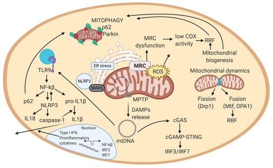
well. Mitochondria are suggested to regulate the activity of the NLRP3 inflammasome complex, by: activating mitophagy (reduces inflammation by clearing mitochondrial-bound NLRP3 complexes) and releasing mitochondrial ROS (amplifies inflammasome immunogenic signal) [140].
In normal conditions, NLRP3 protein is located on the ER. Under oxidative stress and in response to inflammation, NLRP3 can be translocated to MAMs. MAMs transmit danger signals through physical interaction, promoting coordinated responses to oxidative stress, such as triggering mitophagy to remove damaged mitochondria [131].
Altogether, mitochondria-induced inflammation usually is initiated by mitochondrial ROS, which can cause the activation of MAPKs by inhibiting MAPK phosphatase. Activated MAPK may aid in the production of IL-6 and TNF. Mitochondrial ROS accumulation can also activate NLRP3 inflammasome, which promotes the maturation of IL-1  and IL-18. mtDNA accumulation in the cytosol can interact with TLR-9 (in lysosomes) to induce inflammatory responses (e.g., in autosomal
dominant optic atrophy [101]. This pathologic feature has been described in sIBM and in LMNA-related NMDs (CMD, Emery-Dreifuss MD, LGMD, CMT, etc.)) [144]. Further, cytosolic mtDNA can activate cGAS-STING to induce IFN-1 or the NLRP3 inflammasome which can induce the maturation/secretion of pro-inflammatory cytokines [101]. Thus, mtDNA can activate major innate immune responses by acting as a DAMP from the mitochondria. Overall, mtDNA is released from mitochondria to the cytosol, where it is recognized by immune receptors, which trigger a signaling cascade that leads to the production of cytokines or transcription of inflammatory genes in the nucleus (Figure 2) [143]. This close association between mitochondria and inflammation triggered from affected cells, explains why NMDs are usually associated with inflammatory effects.

5.2. Implication of Mitochondrial ROS in Inflammation

Regarding mitochondrial ROS, they are sensed by mitochondrial antiviral signaling protein (MAVS), key in viral RNA infections. MAVS can activate pathways that regulate NF-kB and IRF3/IRF7 to induce gene expression [145]. It also can interact with the outer mitochondrial membrane, where mitochondrial ROS can trigger MAVS oligomerization, promoting IFN-1 production. In addition, MAVS can associate with NLRP3 and triggers its oligomerization, leading to caspase-1 activation [146].
NLRP3 is activated by mitochondrial ROS but also by cardiolipin, an inner mitochondrial membrane lipid. Cardiolipin translocates to the outer mitochondrial membrane after mitochondrial membrane depolarization, where it can recruit NLRP3. Cardiolipin-NLRP3 interaction suggests the role of mitochondria as a hub for the activation of innate immunity [143].
Similar to MAVS, NLRP3 also responds to mitochondrial ROS and can cause mitochondrial damage that promotes the generation of additional ROS. NLRP3 drives the production of IL-1, IL-18, and pyroptosis. mtDNA can also activate NLRP3, and is also sensed by TLR9, which leads to immune and inflammatory gene expression. Mitochondria are therefore critical for signaling by three major innate immune pathways: RIG-I/MAVS, NLRP3, and TLR9 [143]. All these pathways rely on mitochondria-inflammatory response and, consequently, are altered in numerous NMDs.

5.3. Feedback Regulation of the Mitochondrial Inflammatory Response

NF-kB has a role as a pro-inflammatory and anti-inflammatory transcription factor in mitochondrial-induced inflammation. NF-kB anti-inflammatory functions prevent premature and excessive NLRP3-inflammasome activation, avoiding chronic inflammatory disease and inflammation caused by cell and tissue stress [147]. Self-limiting inflammation is also important for maintaining homeostasis, tissue repair, and regeneration, which restores integrity of epithelial barriers and thereby
attenuates PAMP and DAMP availability [100].
Autophagy is the mechanism underlying NF-kB-mediated inhibition of NLRP3 inflammasome. After inducing the expression of pro-inflammatory cytokines and NLRP3, NF-kB is able to switch the initial pro-inflammatory to an anti-inflammatory state to avoid excessive inflammation. Thus, NF-kB induces the expression of p62, which eliminates NLRP3 by mitophagy [138]. Therefore, the “NF-kB-p62/SQSTM1-mitophagy” pathway provides an essential regulatory loop through which NF-kB orchestrates a reparative inflammatory response and prevents excessive collateral damage [100].
Dysregulation of the NLRP3-inflammasome is frequently associated with diverse inflammatory, metabolic, and malignant diseases, including gouty arthritis, Alzheimer’s disease, obesity, type II diabetes, and colorectal cancer [148]. Other NMDs with increased inflammation are ALS (with aberrant activation of NF-kB) [17], muscular dystrophies (like BMD and DMD), and inflammatory myopathies. Therefore, proper control of NLRP3-inflammasome activity and overall inflammatory cascade are
critical for preventing disease development.
The aging process itself is linked to elevated low-grade inflammation or para-inflammation, characterized by constitutive production of low amounts of IL-1b, TNF, and IL-6. The NLRP3-inflammasome activation and insucient mitophagy link accumulation of damaged mitochondria to the para-inflammatory state. Normal, healthy aging may be compromised and greatly enhanced by accumulation of somatically mutated mitochondria, which are more likely to release mitochondrial ROS and fragmented mtDNA that act as direct NLRP3-inflammasome activators [100]. This explains why most of NMDs arise hand by hand to aging processes.

References

  1. Kauppila, T.E.S.; Kauppila, J.H.K.; Ran Larsson, N.-G. Cell Metabolism Review Mammalian Mitochondria and Aging: An Update. Cell Metab. 2017, 25, 57–71.
  2. Piantadosi, C.A.; Suliman, H.B. Transcriptional control of mitochondrial biogenesis and its interface with inflammatory processes. Biochim. Biophys. Acta 2012, 1820, 532–541.
  3. Couvillion, M.T.; Soto, I.C.; Shipkovenska, G.; Stirling, L. Synchronized mitochondrial and cytosolic translation programs. Nature 2016, 533, 499–503.
  4. Tokuyama, T.; Hirai, A.; Shiiba, I.; Ito, N.; Matsuno, K.; Takeda, K.; Saito, K.; Mii, K.; Matsushita, N.; Fukuda, T.; et al. Mitochondrial Dynamics Regulation in Skin Fibroblasts from Mitochondrial Disease Patients. Biomolecules 2020, 10, 450.
  5. Nardin, R.A.; Johns, D.R. Mitochondrial dysfunction and neuromuscular disease. Muscle Nerve 2001, 24, 170–191.
  6. Stewart, J.B.; Chinnery, P.F. The dynamics of mitochondrial DNA heteroplasmy: Implications for human health and disease. Nat. Rev. Genet. 2015, 16, 530–542.
  7. Picca, A.; Lezza, A.M.S.; Leeuwenburgh, C.; Pesce, V.; Calvani, R.; Landi, F.; Bernabei, R.; Marzetti, E. Fueling inflamm-aging through mitochondrial dysfunction: Mechanisms and molecular targets. Int. J. Mol. Sci. 2017, 18.
  8. Frank, M.; Duvezin-Caubet, S.; Koob, S.; Occhipinti, A.; Jagasia, R.; Petcherski, A.; Ruonala, M.O.; Priault, M.; Salin, B.; Reichert, A.S. Mitophagy is triggered by mild oxidative stress in a mitochondrial fission dependent manner. Biochim. Biophys. Acta Mol. Cell Res. 2012, 1823, 2297–2310.
  9. Babbar, M.; Basu, S.; Yang, B.; Croteau, D.L.; Bohr, V.A. Mitophagy and DNA damage signaling in human aging. Mech. Ageing Dev. 2020, 186.
  10. Siciliano, G.; Monzani, F.; Manca, M.L.; Tessa, A.; Caraccio, N.; Tozzi, G.; Piemonte, F.; Mancuso, M.; Santorelli, F.M.; Ferrannini, E.; et al. Human Mitochondrial Transcription Factor A Reduction and Mitochondrial Dysfunction in Hashimoto’s Hypothyroid Myopathy. Mol. Med. 2002, 8, 326–333.
  11. Bomont, P.; Cavalier, L.; Blondeau, F.; Hamida, C.B.; Belal, S.; Tazir, M.; Demir, E.; Topaloglu, H.; Korinthenberg, R.; Tüysüz, B.; et al. The gene encoding gigaxonin, a new member of the cytoskeletal BTB/kelch repeat family, is mutated in giant axonal neuropathy. Nat. Lett. 2000, 26, 370–374.
  12. Vincent, A.E.; Ng, Y.S.; White, K.; Davey, T.; Mannella, C.; Falkous, G.; Feeney, C.; Schaefer, A.M.; Mcfarland, R.; Gorman, G.S.; et al. The Spectrum of Mitochondrial Ultrastructural Defects in Mitochondrial Myopathy. Sci. Rep. 2016, 6, 30610.
  13. Bhatt, P.S.; Tzoulis, C.; Balafkan, N.; Miletic, H.; Tran, G.T.T.; Sanaker, P.S.; Bindoff, L.A. Mitochondrial DNA depletion in sporadic inclusion body myositis. Neuromuscul. Disord. 2019, 29, 242–246.
  14. De Duve, C.; Pressman, B.C.; Gianetto, R.; Wattiaux, R.; Appelmans, F. Tissue fractionation studies. Biochem. J. 1955, 60, 604–617.
  15. Kepp, O.; Galluzzi, L.; Kroemer, G. Mitochondrial control of the NLRP3 inflammasome. Nat. Immunol. 2011, 12.
  16. Katsetos, C.D.; Koutzaki, S.; Melvin, J.J. Mitochondrial dysfunction in neuromuscular disorders. Semin. Pediatric Neurol. 2013, 20, 202–215.
  17. Koopman, W.J.H.; Willems, P.H.G.M.; Smeitink, J.A.M. Monogenic Mitochondrial Diseases. N. Engl. J. Med. 2012, 366, 1132–1141.
  18. Keogh, M.J.; Chinnery, P.F. Mitochondrial DNA mutations in neurodegeneration. Biochim. Biophys. Acta Bioenerg. 2015, 1847, 1401–1411.
  19. Lightowlers, R.N.; Taylor, R.W.; Turnbull, D.M. Mutations causing mitochondrial disease: What is new and what challenges remain? Science 2015, 349, 1494–1499.
  20. Argov, Z.; Renshaw, P.F.; Boden, B.; Winokur, A.; Bank, W.J. Effects of thyroid hormones on skeletal muscle bioenergetics. In vivo phosphorus-31 magnetic resonance spectroscopy study of humans and rats. J. Clin. Investig. 1988, 81, 1695–1701.
  21. Monzani, F.; Caraccio, N.; Siciliano, G.; Manca, L.; Murri, L.; Ferrannini, E. Clinical and Biochemical Features of Muscle Dysfunction in Subclinical Hypothyroidism. J. Clin. Endocrinol. Metab. 1997, 82, 3315–3318.
  22. Deng, J.; Wang, P.; Chen, X.; Cheng, H.; Liu, J.; Fushimi, K.; Zhu, L.; Wu, J.Y. FUS interacts with ATP synthase beta subunit and induces mitochondrial unfolded protein response in cellular and animal models. Proc. Natl. Acad. Sci. USA 2018, 115, E9678–E9686.
  23. Spiegel, R.; Saada, A.; Flannery, P.J.; Burté, F.; Soiferman, D.; Khayat, M.; Eisner, V.; Vladovski, E.; Taylor, R.W.; Bindoff, L.A.; et al. Fatal infantile mitochondrial encephalomyopathy, hypertrophic cardiomyopathy and optic atrophy associated with a homozygous OPA1 mutation. J. Med. Genet. 2016, 53, 127–131.
  24. Rodríguez-Nuevo, A.; Díaz-Ramos, A.; Noguera, E.; Díaz-Sáez, F.; Duran, X.; Muñoz, J.P.; Romero, M.; Plana, N.; Sebastián, D.; Tezze, C.; et al. Mitochondrial DNA and TLR9 drive muscle inflammation upon Opa1 deficiency. EMBO J. 2018, 37.
  25. Züchner, S.; Mersiyanova, I.V.; Muglia, M.; Bissar-Tadmouri, N.; Rochelle, J.; Dadali, E.L.; Zappia, M.; Nelis, E.; Patitucci, A.; Senderek, J.; et al. Mutations in the mitochondrial GTPase mitofusin 2 cause Charcot-Marie-Tooth neuropathy type 2A. Nat. Genet. 2004, 36, 449–451.
  26. Mishra, P.; Carelli, V.; Manfredi, G.; Chan, D.C. Proteolytic cleavage of Opa1 stimulates mitochondrial inner membrane fusion and couples fusion to oxidative phosphorylation. Cell Metab. 2014, 19, 630–641.
  27. Waterham, H.R.; Koster, J.; van Roermund, C.W.T.; Mooyer, P.A.W.; Wanders, R.J.A.; Leonard, J.V. A Lethal Defect of Mitochondrial and Peroxisomal Fission. N. Engl. J. Med. 2007, 356, 1736–1741.
  28. Kodavati, M.; Wang, H.; Hegde, M.L. Altered Mitochondrial Dynamics in Motor Neuron Disease: An Emerging Perspective. Cells 2020, 9, 1065.
  29. Clark, S. Newborn mice studied with the electron mircroscope. Biophys. Biochem. Cytol. 1957, 3, 349–362.
  30. Ohsumi, Y. Historical landmarks of autophagy research. Cell Res. 2014, 24, 9–23.
  31. Eiyama, A.; Okamoto, K. PINK1/Parkin-mediated mitophagy in mammalian cells. Curr. Opin. Cell Biol. 2015, 33, 95–101.
  32. Seabright, A.P.; Fine, N.H.F.; Barlow, J.P.; Lord, S.O.; Musa, I.; Gray, A.; Bryant, J.A.; Banzhaf, M.; Lavery, G.G.; Hardie, D.G.; et al. AMPK activation induces mitophagy and promotes mitochondrial fission while activating TBK1 in a PINK1-Parkin independent manner. FASEB J. 2020, 34, 6284–6301.
  33. García-Prat, L.; Martínez-Vicente, M.; Perdiguero, E.; Ortet, L.; Rodríguez-Ubreva, J.; Rebollo, E.; Ruiz-Bonilla, V.; Gutarra, S.; Ballestar, E.; Serrano, A.L.; et al. Autophagy maintains stemness by preventing senescence. Nature 2016, 529, 37–42.
  34. Lemasters, J.J. Selective mitochondrial autophagy, or mitophagy, as a targeted defense against oxidative stress, mitochondrial dysfunction, and aging. Rejuvenation Res. 2005, 8, 3–5.
  35. Youle, R.J.; Narendra, D.P. Mechanisms of mitophagy. Nat. Rev. Mol. Cell Biol. 2011, 12, 9–14.
  36. Sun, N.; Yun, J.; Liu, J.; Malide, D.; Liu, C.; Rovira, I.I.; Holmström, K.M.; Fergusson, M.M.; Yoo, Y.H.; Combs, C.A.; et al. Measuring In Vivo Mitophagy. Mol. Cell 2015, 60, 685–696.
  37. Zhang, Q.; Raoof, M.; Chen, Y.; Sumi, Y.; Sursal, T.; Junger, W.; Brohi, K.; Itagaki, K.; Hauser, C.J. Circulating mitochondrial DAMPs cause inflammatory responses to injury. Nat. Lett. 2010, 464, 104–107.
  38. Grumati, P.; Grumati, P.; Coletto, L.; Sabatelli, P.; Cescon, M.; Angelin, A.; Bertaggia, E.; Blaauw, B.; Urciuolo, A.; Tiepolo, T.; et al. Autophagy is defective in collagen VI muscular dystrophies, and its reactivation rescues myofiber degeneration. Nat. Med. 2010, 16, 1313–1320.
  39. Zhang, W.; Hu, X.; Shen, Q.; Xing, D. Mitochondria-specific drug release and reactive oxygen species burst induced by polyprodrug nanoreactors can enhance chemotherapy. Nat. Commun. 2019, 10, 1–14.
  40. Lerner, C.A.; Sundar, I.K.; Rahman, I. Mitochondrial redox system, dynamics, and dysfunction in lung inflammaging and COPD. Int. J. Biochem. Cell Biol. 2016, 81, 294–306.
  41. Domènech, B.E.; Marfany, G. The Relevance of Oxidative Stress in the Pathogenesis and Therapy of Retinal Dystrophies. Antioxidants 2020, 9, 347.
  42. Peterman, E.M.; Sullivan, C.; Goody, M.F.; Rodriguez-Nunez, I.; Yoder, J.A.; Kim, C.H. Neutralization of mitochondrial superoxide by superoxide dismutase 2 promotes bacterial clearance and regulates phagocyte numbers in zebrafish. Infect. Immun. 2015, 83, 430–440.
  43. Andersen, P.M.; Nilsson, P.; Forsgren, L.; Marklund, S.L. CuZn-Superoxide Dismutase, Extracellular Superoxide Dismutase, and Glutathione Peroxidase in Blood from Individuals Homozygous for Asp90Ala CuZn-Superoxide Dismutase Mutation. J. Neurochem. 2002, 70, 715–720.
  44. Sano, R.; Reed, J.C. ER stress-induced cell death mechanisms. Biochim. Biophys. Acta Mol. Cell Res. 2013, 1833, 3460–3470.
  45. Manevski, M.; Muthumalage, T.; Devadoss, D.; Sundar, I.K.; Wang, Q.; Singh, K.P.; Unwalla, H.J.; Chand, H.S.; Rahman, I. Cellular stress responses and dysfunctional Mitochondrial–cellular senescence, and therapeutics in chronic respiratory diseases. Redox Biol. 2020, 33, 101443.
  46. Smith, E.F.; Shaw, P.J.; De Vos, K.J. The role of mitochondria in amyotrophic lateral sclerosis. Neurosci. Lett. 2019, 710, 132933.
  47. Naon, D.; Scorrano, L. At the right distance: ER-mitochondria juxtaposition in cell life and death. Biochim. Biophys. Acta Mol. Cell Res. 2014, 1843, 2184–2194.
  48. Nadalutti, C.A.; Stefanick, D.F.; Zhao, M.-L.; Horton, J.K.; Prasad, R.; Brooks, A.M.; Griffith, J.D.; Wilson, S.H. Mitochondrial dysfunction and DNA damage accompany enhanced levels of formaldehyde in cultured primary human fibroblasts. Sci. Rep. 2020, 10, 5575.
  49. Nakahira, K.; Haspel, J.A.; Rathinam, V.A.K.; Lee, S.-J.; Dolinay, T.; Lam, H.C.; Englert, J.A.; Rabinovitch, M.; Cernadas, M.; Pyo Kim, H.; et al. Autophagy proteins regulate innate immune responses by inhibiting the release of mitochondrial DNA mediated by the NALP3 inflammasome. Nat. Immunol. 2011, 8, 222–230.
  50. Pérez-Treviño, P.; Velásquez, M.; García, N. Mechanisms of mitochondrial DNA escape and its relationship with different metabolic diseases. BBA Mol. Basis Dis. 2020, 1866, 165761.
  51. Rodríguez-Nuevo, A.; Zorzano, A. The sensing of mitochondrial DAMPs by non-immune cells. Cell Stress 2019, 3, 195–207.
More
Information
Subjects: Others
Contributor MDPI registered users' name will be linked to their SciProfiles pages. To register with us, please refer to https://encyclopedia.pub/register :
View Times: 457
Revisions: 3 times (View History)
Update Date: 01 Jul 2021
1000/1000