Jasmonate Compounds
Edit

There are four known stereoisomers of jasmonic acid: trans-(−)-(3R,7R), abbreviated as (−)-JA; trans-(+)-(3S,7S) abbreviated as (+)-JA; cis-(−)-(3S,7R) abbreviated as (−)-epi-JA; cis-(+)-(3R,7S) abbreviated as (+)-epi-JA [15]. The naturally occurring jasmonic acid in plants is (−)-JA and (+)-epi-JA. Due to the fact that the cis stereoisomers are thermodynamically less stable, they epimerize at the C-7 atom to the stable trans form, which at the same time shows a higher biological activity. The biological activity of jasmonic acid has been found to be dependent on the presence of a carboxyl group at the C-1 position, a keto or hydroxyl group at the C-6 position, and a pentenyl side chain at the C-7 position. Because of this structure, jasmonates inhibit, induce and/or stimulate changes that occur in plants at the morphological, physiological, cellular and molecular levels.

jasmonic acid anti-cancer drugs structure–activity relationship

1. Introduction

Neoplastic diseases constitute a major problem in modern medicine, mainly due to the insufficient effectiveness of currently available methods of anti-cancer therapy. Therefore, when analyzing the basic mechanisms regulating the development of neoplasms, new therapeutic solutions look for mechanisms that would prevent or extinguish the neoplastic process [1]. Particular attention is focused on compounds of natural origin that can be used both for the prevention and treatment of cancer. The National Cancer Institute has screened about 35,000 plant species that have potential anticancer activity, of which 3000 species have fully confirmed such activity [2]. Jasmonic acid and its derivatives are also among such compounds and show anti-cancer properties. However, while this action is not always effective enough for the needs of effective therapies, attempts are also made to combine jasmonic acid or its derivatives with other anti-cancer agents or radiotherapy, as well as chemically modifying the structure of these compounds in order to create derivatives with effective anti-cancer properties [3].

2. Jasmonic Acid and Its Derivatives Occurring in Plants

Jasmon compounds, called jasmonates, occur in almost all tissues of higher plants, i.e., flower plants, bryophytes and ferns, where they play the role of endogenous regulators of growth and development [4]. They are present in, among others, stems, roots, tubers, leaves, flowers, fruits and pollen. Jasmonic acid, the main representative of jasmon compounds, was first detected in the fungi Lasiodiplodia theobromae, and its methyl ester was isolated from the essential oils of the Jasminum grandiflorum olive family (now obtained commercially by synthesis) [5][6]. Jasmonians are found in over 160 plant families.
Jasmonic acid occurs in the green parts of plants in the form of conjugates with amino acids; in flowers, it is found with phenylalanine, tryptophan and tyrosine, in leaves with isoleucine or valine, and in fruits with isoleucine [7]. Moreover, jasmonic acid methyl ester is a component of essential oils and gives fragrance to many flowers (e.g., jasmine) and fruit (e.g., apple). Because of its fragrance properties, it is used by the perfume industry.
The content of jasmonates in plants is very diverse and ranges from 10 to 100 ng/g of fresh weight [5][8], depending on the type, species and age of the plant. It has been shown that there are more of these compounds in the generative parts of plants (pericarp, fruit and seeds) than in the vegetative parts (stems and leaves). Higher concentrations are also found in young plants as, with age, the jasmonates level decreases [5]. The increase in the content of jasmonates in the plant is also influenced by biological factors (insects and pathogens) and physicochemical factors (osmotic stress, drought, UV radiation, cooling and increased temperature, ozone) as well as mechanical damage (herbivores, mechanical stress) [8][9]. In plants, jasmonic acid has been found to be more frequently present than its methyl ester [2]. However, in the case of, for example, Malus sylvestris fruit, both compounds occur simultaneously. In recent years, these compounds have also been detected in lower plants (e.g., algaeChorella) and fungi (Gibberella fujikuroi and Botryodiplodia theobromae). Unfortunately, previous studies have not shown in which specific parts of the plant they are synthesized.

3. Chemical Structure of Jasmonates

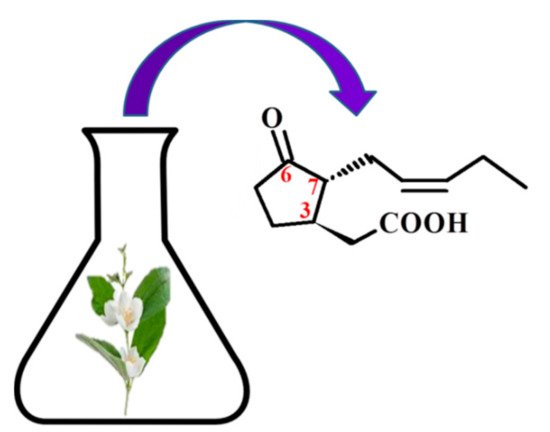
The basis of the structure of jasmonates is a cyclopentane ring with three different substituents in the C-3, C-6 and C-7 position, showing optical activity (Figure 1) [10]. The parent compound belonging to the jasmonate group is jasmonic acid 3-oxo-2-(pent-2’-enyl)cyclopentane acetic acid (IUPAC name), synthesized from linolenic acid found in the chloroplast membrane. The metabolites of jasmonic acid include various compounds obtained as a result of conjugation with isoleucine to obtain jasmonylisoleucine (JA-Ile) [9][11][12]; methylation to methyl jasmonate (MJ) (MJ) [9]; ester formation with glucose to 12-glucosyljasmonic acid [13]; decarboxylation to cis-jasmon [14] and hydroxylation to 12-hydroxyasmonic acid.
Figure 1. The structure of trans-(-)-(3R,7R)-jasmonic acid.
There are four known stereoisomers of jasmonic acid: trans-(−)-(3R,7R), abbreviated as (−)-JA; trans-(+)-(3S,7S) abbreviated as (+)-JA; cis-(−)-(3S,7R) abbreviated as (−)-epi-JA; cis-(+)-(3R,7S) abbreviated as (+)-epi-JA [15]. The naturally occurring jasmonic acid in plants is (−)-JA and (+)-epi-JA. Due to the fact that the cis stereoisomers are thermodynamically less stable, they epimerize at the C-7 atom to the stable trans form, which at the same time shows a higher biological activity. The biological activity of jasmonic acid has been found to be dependent on the presence of a carboxyl group at the C-1 position, a keto or hydroxyl group at the C-6 position, and a pentenyl side chain at the C-7 position [16][17][18]. Because of this structure, jasmonates inhibit, induce and/or stimulate changes that occur in plants at the morphological, physiological, cellular and molecular levels.

4. Pharmacological Activity of Jasmonates

The jasmonate family mainly consists of jasmonic acid, cis-jasmonate and methyl jasmonate (MJ), which are structurally similar to prostaglandins, especially those with anti-inflammatory action: prostaglandin D2 (PGD2) and 15-Deoxy-Δ12,14-prostaglandin J2 (15d -PGJ2) [19]. For this reason, many studies have been conducted to assess their effect on mammalian cells, which showed that jasmonates have a cytotoxic effect on neoplastic cells, while having no effect on healthy cells [20]. Jasmonates have been found to possess properties characteristic of anticancer drugs. Their action is characterized by high selectivity in relation to neoplastic cells, as well as effectiveness against neoplastic cells resistant to antineoplastic drugs. A cytotoxic effect of jasmonates was demonstrated in lymphoblastic leukemia cells (MOL-4) for the first time [21]. Further studies have also shown susceptibility to jasmonate in breast cancer cells (MCF-7), human melanoma cells (SK-28), androgen-responsive human prostate adenocarcinoma (LNCaP), cervix and murine lymphoma cells (EL-4) [19][22][23]. The greatest sensitivity to JA (increasing with increasing concentration, 0.5–3 mM) showed MOLT-4 cells, while SK-28, LNCaP and MCF-7 cells showed less sensitivity. MJ was more cytotoxic than jasmine acid. MJ induced 87.5% cytotoxicity in Molt-4 cells at a concentration of 0.5 mM. The same cytotoxicity was induced by JA at the highest used concentration [21]. The cytotoxic effect of MJ on cervical cancer cells was evaluated using a selected range of cervical carcinoma derived cell lines, including SiHa and CaSki (contain HPV16 DNA), HeLa (contain HPV18 DNA) and C33A (not containing HPV DNA). Cells were treated with 1–5 mM MJ for 24 h. C33A and CaSki cells were more sensitive to the MJ compared to HeLa and SiHa. The IC50 values were 1.7 mM for CaSki, 2.2 mM for C33A, 3 mM for HeLa and 3.3 mM for SiHa [24].
The antitumor activity of methyl jasmonate (MJ) was found to be higher than other jasmonates; therefore, MJ and its synthetic derivatives have recently been studied more intensively as promising compounds for the treatment of cancer [25]. MJ has been shown to prolong the survival of mice with EL-4 lymphoma and mice vaccinated with multiple myeloma cells (MM.1S) [21][26]. Survival rates were significantly higher in the group of lymphoma (EL-4) in mice treated with MJ (236 mg/kg) compared to untreated mice. Forty-five percent of the treated mice still live more than 5 months after the inoculation of the tumor cells [21]. In the case of murine multiple myeloma (MM) cell lines, MJ (0.5–2.5 mM by 24 h) significantly increased the survival rate. IC50 values for MJ were observed to be less than or equal to 1.5 mM for 15 of 16 (94%) cell lines. At the highest concentration tested, MJ caused more than a 90% reduction in the viability of all MM cell lines [26]. The cytotoxic effect of MJ (0.2–2000 µM) was also selective for MDA-MB-361 and T-47D human breast cancer cells [25]. The highest inhibition of the growth of T-47D and MDA-MB-361 cells was observed at 2 mM MJ (cell survival was 47% and 78%, respectively).
The preventive effect of MJ is primarily due to the ability to direct neoplastic cells to the path of apoptosis or necrosis. It is associated with the overproduction of reactive oxygen species (ROS) and the influence on the expression of proteins, such as p53, p21, and proteins from Bcl-2 and Bax families [24][27]. Western blot analyses indicated that MJ (2 mM, 5 and 24h incubation) induced a significant increase in p53 and decrease in p53 levels in CaSki cells. In HeLa and SiHa cells, a reduction in p53 and p21 levels was observed after 24 h of treatment [24]. In human non-small cell lung cancer cells (A549), jasmonate causes an increase in the expression of pro-apoptotic proteins from the Bcl-2 and Bax families [28][29]. MJ (2 mM) inhibits Bcl-2 protein expression in vitro in the case of prostate cancer cells (PC-3) [30]. The induction of apoptosis by methyl jasmonate is associated with decreased fluidity of tumor cell membranes and increased expression of tumor necrosis factor (TNFα) and its receptor 1 (TNFR1) in breast cancer cells (MDA-MB-435 and MCF7). The IC50 value of MJ for MDA-MB-435 cells was 1.9 mM and 2.0 mM for MCF-7 cells. The consequence of this action of jasmonate is the activation of caspase-8 in the external pro-apoptotic pathway. MDA-MB-435 and MCF-7 cells showed 35.0% and 37.2% apoptosis upon treatment MJ, respectively [31]. In contrast, MJ in human neuroblastoma cell lines caused decreased expression of XIAP protein and surwinin and MJ IC50 value for SK-N-SH and BE (2)-C cells were 1.39 and 1.35 mmol/L, respectively [32]. These results suggest that MJ may induce apoptosis through the activation of different signaling pathways in cervical cancer cells.

5. Therapeutic Benefits of Combining Jasmonates with Anti-Cancer Drugs

In addition to the fact that jasmonates induce apoptosis in tumor cells, they can also be combined with other anti-tumor agents to achieve synergistic anti-tumor effects. In fact, many modern chemotherapy procedures use multicomponent combinations of drugs that allow for lower doses to be administered, can reduce undesirable side effects and even overcome drug resistance [20]. Therefore, studies have been carried out to evaluate the combination of the effects of MJ and various other anticancer agents [33] that are routinely used in clinical practice: BCNU (carmustine), cisplatin, paclitaxel (taxol) [34] and doxorubicin (adriamycin) or 3-bromopyruvate (3-BrP) (Table 1).
Table 1. Effect of MJ combined with other anticancer agents.
Jasmonates/Drug Cancer Concentration Range Action/Effects References
MJ + BCNU
(in vitro)
MJ + taxol
(in vitro)
Pancreatic cell:
PaCa-2
BCL1
MCF-7
DA-3
D-122
MJ: 0.1 mM
BCNU: 1, 10, 25 µg/mL-PaCa-2
2.5, 5 µg/mL-BCL1
taxol: 1, 2.5, 5, 10 μg/ml
mitochondriotoxic synergic cytotoxicity
IC50
[35]
MJ + POH
or
MJ + cisplatin
or
MJ + cisplatin + POH
(in vitro)
Breast cancer cell lines:
MDA-MB-231
MDA-MB-435
MCF7
IC2O (POH)
MDA-MB-231: 0.76 mM
MDA-MB-435: 0.6 mM
MCF7: 0.8 mM
MJ + POH:
cytotoxicity ↑
apoptosis ↑
TNFR1 ↑
MJ + POH:
apoptosis ↑
[36]
MJ + 2DG
2-deoxyglucose
(in vitro)
Sarcoma:
SaOS-2
MCA-105
MJ: 0.5–3 mM
2DG: 1 and 2 mM
synergic cytotoxicity ↑
ATP glycolysis ↑
[37]
MJ + TRAIL
(in vitro)
CRC cell lines:
SW480, HT29, LS180, HCT116
MJ: 0.5 mM
TRAIL: 100–200 ng/ml
IAP (survivin) ↓
caspase activity ↑
TRAIL-induced apoptosis ↑
[38]
MJ + Smac7N
(in vitro)
prostate carcinoma cells:
DU145, PC-3
proximal tubular epithelial cells:
HK-2
MJ: 0.5–2 mM Smac7N:
MJ-induced cytotoxicity ↑
ran caspase-9 dependent and independent pathways
[39]
MJ + 3-BrP
(in vitro)
Mice breast carcinoma cell line:
4 T1
MJ: 0.5–3 mM
3-BrP: 12.5, 25, 50, 100, 200, 400 μM
ALT ↑ AST ↑
tumor growth ↓
antitumor activity ↑
[40]
MJ + cisplatin
MJ + X-rays
MJ + α-rays
Cervical cancer cells:
SiHa,
CaSki,
HeLa
C33A
MJ: 0.1–1 mM
Cisplatin: 0.1–0.5 μM
X-rays: 0 25–3 Gy
cell survival ↓
IC50 radiation dose ↓
cell viability ↓
[41]
3-BrP—3-bromopyruvate; BCNU—1.3-bis-(2-chloroethyl)-1-nitrosourea; 2DG—2-deoxy-D-glucose; IAP—inhibitors of apoptosis; MJ—methyl Jasmonate; POH—perillyl alcohol; Smac7N—a peptide that contains the N-terminal seven residues of smac; TNFR1—tumor-necrosis factor receptor-1; TRAIL—tumor necrosis factor- (TNF-)related apoptosis-inducing ligand.
The interactions of these combination drugs have been observed in many cell lines of malignant tumors such as: breast, lung, prostate and pancreatic cancer as well as leukemia [40], where MJ drastically reduced the IC50 values of the used chemotherapeutic drugs, while reducing of side effects of these drugs. The combination of MJ (0.1 nM) and BCNU (carmustine) therapy (1, 10 and 25 µg/mL—PaCa-2 cell; 2.5 and 5 µg/mL—BCL1 cell) had an adverse effect on pancreatic cancer cells (PaCa-2; BCL1) causing their apoptosis, which was not observed with BCNU alone. It follows that the influence of BCNU on mitochondria [35] makes them hypersensitive to MJ, resulting in over-additive cytotoxic effects. Another study also showed a positive effect of MJ together with perillyl alcohol (POH), which increased the cytotoxicity of cisplatin in breast cancer cells (MDA-MB-231, MDA-MB-435, MCF7) [36].
The combination of MJ (0.5–3 mM) and 2DG (2-deoxyglucose, glycolysis inhibitor) (1 and 2 mM) also resulted in a synergistic cytotoxic effect on tumor cells (sarcoma SaOS-2; MCA-105), possibly due to the interaction of both MJ-induced oxidative phosphorylation of ATP biosynthesis and 2DG-induced ATP glycolysis [22][37]. Most importantly, in vivo experiments have shown that the combination of MJ and doxorubicin has a synergistic effect on mouse leukemia (BCL1) [22]. Moreover, pre-incubation with MJ (0.5 mM), at non-cytotoxic concentrations, may sensitize colorectal cancer (CRC) cells to ligand-induced apoptosis, inducing TRAIL (tumor necrosis factor-related apoptosis-inducing ligand) (100–200 ng/mL), resulting in synergistic cell death through enhanced caspase activity [42][38].
TRAIL receptors are highly expressed in primary tumors and various tumor cell lines [43], which makes this pathway of cytotoxicity very specific for neoplastic cells while sparing most of the normal cells. Therefore, TRAIL-induced cell apoptosis is a very attractive potential target of anti-cancer therapy [42]. TRAIL, induced by MJ, mediates the reduction in survivin, a member of the Inhibitors of Apoptosis Proteins family (IAP). This study also shows that MJ, by inhibiting transcription, influenced the signal transduction pathways, resulting in a reduction in survivin mRNA [20]. Nevertheless, many tumor cells are intrinsically resistant to TRAIL-induced apoptosis. Therefore, the results of synergistic cell death achieved by the combination of MJ and TRAIL, through bypassing the TRAIL resistance barrier, have great potential in tumor therapy as both agents are highly selective for tumor cells.
Moreover, studies on prostate (PC-3) and breast cancer cell lines (MDA-MB-435) showed that treatment with jasmonates resulted in increased expression of TNFR1 and caspase-8 and caspase-3 activation [22], showing that jasmonates can act directly on the extrinsic apoptotic pathway in addition to intrinsic mitochondrial apoptotic pathway. Additionally, the IAP antagonist, an N-terminal peptide consisting of seven Smac residues (SmacN7), synergistically significantly increased MJ-induced cytotoxicity in human cancer cells, but not in normal epithelial cells, and acted simultaneously through caspase-9 dependent and independent pathways [39][44]. These findings suggest that inhibition of IAP may facilitate MJ-induced cytotoxicity and may be of advantageous value for the further development of jasmonate-based chemotherapy.
MJ (0.5–3 mM) also increased the effectiveness of therapy with the use of 3-BrP (12.5; 25; 50; 100; 200; 400 μM; IC50 value was 70 μM). This polytherapy was more effective than monotherapy with 3-BrP, MJ, and also surprisingly with cyclophosphamide as a routine treatment for breast cancer in tumor bearing mice, as observed by reducing tumor volume and increasing the percent inhibition of tumor growth. Moreover, the applied therapy had no appreciable side effects on the kidneys, liver, immune system and body weight [40].
Recently, MJ has also been shown to be effective in cooperating with cisplatin and radiotherapy in the treatment of cervical cancer cells by significantly reducing the doses of radiation and cisplatin required to inhibit these cells’ survival [22]. This study showed, for the first time, that alpha radiation selectively reduces cell viability and cervical cancer cell survival, and that alpha radiation also works with MJ in reducing cell viability, as shown in some of the cervical cancer cell lines used (SiHa, CaSki, HeLa and C33A) [41]. In addition, MJ can be administered along with conventional X-ray (0.25–3 Gy) and cisplatin therapies, increasing their cytotoxic efficacy while lowering the dose, avoiding possible side effects.

References

  1. Choudhary, D.; Goykar, H.; Karanwad, T.; Kannaujia, S.; Gadekar, V.; Misra, M. An Understanding of Mitochondria and Its Role in Targeting Nanocarriers for Diagnosis and Treatment of Cancer. Asian J. Pharm. Sci. 2020.
  2. Kuruppu, A.I.; Paranagama, P.; Goonasekara, C.L. Medicinal Plants Commonly Used against Cancer in Traditional Medicine Formulae in Sri Lanka. Saudi Pharm. J. SPJ Off. Publ. Saudi Pharm. Soc. 2019, 27, 565–573.
  3. Koklesova, L.; Liskova, A.; Samec, M.; Qaradakhi, T.; Zulli, A.; Smejkal, K.; Kajo, K.; Jakubikova, J.; Behzadi, P.; Pec, M.; et al. Genoprotective Activities of Plant Natural Substances in Cancer and Chemopreventive Strategies in the Context of 3P Medicine. EPMA J. 2020, 11, 261–287.
  4. Wilmowicz, E.; Frankowski, K.; Sidłowska, M.; Kućko, A.; Kesy, J.; Gasiorowski, A.; Glazińska, P.; Kopcewicz, J. [Jasmonate biosynthesis—The latest discoveries]. Postepy Biochem. 2012, 58, 26–33.
  5. Ghasemi Pirbalouti, A.; Sajjadi, S.E.; Parang, K. A Review (Research and Patents) on Jasmonic Acid and Its Derivatives. Arch. Pharm. 2014, 347, 229–239.
  6. Umukoro, S.; Alabi, A.O.; Eduviere, A.T.; Ajayi, A.M.; Oluwole, O.G. Anti-Inflammatory and Membrane Stabilizing Properties of Methyl Jasmonate in Rats. Chin. J. Nat. Med. 2017, 15, 202–209.
  7. Kramell, R.; Atzorn, R.; Schneider, G.; Miersch, O.; Brückner, C.; Schmidt, J.; Sembdner, G.; Parthier, B. Occurrence and Identification of Jasmonic Acid and Its Amino Acid Conjugates Induced by Osmotic Stress in Barley Leaf Tissue. J. Plant Growth Regul. 1995, 14, 29.
  8. Wasternack, C. Jasmonates: An Update on Biosynthesis, Signal Transduction and Action in Plant Stress Response, Growth and Development. Ann. Bot. 2007, 100, 681–697.
  9. Wang, J.; Song, L.; Gong, X.; Xu, J.; Li, M. Functions of Jasmonic Acid in Plant Regulation and Response to Abiotic Stress. Int. J. Mol. Sci. 2020, 21, 1446.
  10. Wasternack, C.; Kombrink, E. Jasmonates: Structural Requirements for Lipid-Derived Signals Active in Plant Stress Responses and Development. ACS Chem. Biol. 2010, 5, 63–77.
  11. Staswick, P.E.; Tiryaki, I. The Oxylipin Signal Jasmonic Acid Is Activated by an Enzyme That Conjugates It to Isoleucine in Arabidopsis. Plant Cell 2004, 16, 2117–2127.
  12. Suza, W.P.; Staswick, P.E. The Role of JAR1 in Jasmonoyl-L: -Isoleucine Production during Arabidopsis Wound Response. Planta 2008, 227, 1221–1232.
  13. Heitz, T.; Smirnova, E.; Widemann, E.; Aubert, Y.; Pinot, F.; Ménard, R. The Rise and Fall of Jasmonate Biological Activities. In Lipids in Plant and Algae Development; Subcellular Biochemistry; Nakamura, Y., Li-Beisson, Y., Eds.; Springer International Publishing: Cham, Switzerland, 2016; pp. 405–426. ISBN 978-3-319-25979-6.
  14. Swiatek, A.; Van Dongen, W.; Esmans, E.L.; Van Onckelen, H. Metabolic Fate of Jasmonates in Tobacco Bright Yellow-2 Cells. Plant Physiol. 2004, 135, 161–172.
  15. Koch, T.; Bandemer, K.; Boland, W. Biosynthesis of Cis-Jasmone: A Pathway for the Inactivation and the Disposal of the Plant Stress Hormone Jasmonic Acid to the Gas Phase? Helv. Chim. Acta 1997, 80, 838–850.
  16. Han, Y.; Bai, Y.; Xiao, Y.; Du, F.; Liang, Y.; Tan, Z.; Zhao, M.; Liu, H. Simultaneous Discrimination of Jasmonic Acid Stereoisomers by CE-QTOF-MS Employing the Partial Filling Technique. Electrophoresis 2011, 32, 2693–2699.
  17. Krumm, T.; Bandemer, K.; Boland, W. Induction of Volatile Biosynthesis in the Lima Bean (Phaseolus Lunatus) by Leucine- and Isoleucine Conjugates of 1-Oxo- and 1-Hydroxyindan-4-Carboxylic Acid: Evidence for Amino Acid Conjugates of Jasmonic Acid as Intermediates in the Octadecanoid Signalling Pathway. FEBS Lett. 1995, 377, 523–529.
  18. Koda, Y.; Kikuta, Y.; Tazaki, H.; Tsujino, Y.; Sakamura, S.; Yoshihara, T. Potato Tuber-Inducing Activities of Jasmonic Acid and Related Compounds. Phytochemistry 1991, 30, 1435–1438.
  19. Sá-Nakanishi, A.B.; Soni-Neto, J.; Moreira, L.S.; Gonçalves, G.A.; Silva, F.M.S.; Bracht, L.; Bersani-Amado, C.A.; Peralta, R.M.; Bracht, A.; Comar, J.F. Anti-Inflammatory and Antioxidant Actions of Methyl Jasmonate Are Associated with Metabolic Modifications in the Liver of Arthritic Rats. Oxid. Med. Cell. Longev. 2018, 2018, 2056250.
  20. Raviv, Z.; Cohen, S.; Reischer-Pelech, D. The Anti-Cancer Activities of Jasmonates. Cancer Chemother. Pharmacol. 2013, 71, 275–285.
  21. Fingrut, O.; Flescher, E. Plant Stress Hormones Suppress the Proliferation and Induce Apoptosis in Human Cancer Cells. Leukemia 2002, 16, 608–616.
  22. Cesari, I.M.; Carvalho, E.; Figueiredo Rodrigues, M.; Mendonça, B.D.S.; Amôedo, N.D.; Rumjanek, F.D. Methyl Jasmonate: Putative Mechanisms of Action on Cancer Cells Cycle, Metabolism, and Apoptosis. Int. J. Cell Biol. 2014, 2014, 572097.
  23. Pérez-Salamó, I.; Krasauskas, J.; Gates, S.; Díaz-Sánchez, K.E.; Devoto, A. An Update on Core Jasmonate Signalling Networks, Physiological Scenarios, and Health Applications. Annu. Plant Rev. 2019, 2.
  24. Kniazhanski, T.; Jackman, A.; Heyfets, A.; Gonen, P.; Flescher, E.; Sherman, L. Methyl Jasmonate Induces Cell Death with Mixed Characteristics of Apoptosis and Necrosis in Cervical Cancer Cells. Cancer Lett. 2008, 271, 34–46.
  25. Bömer, M.; Pérez-Salamó, I.; Florance, H.V.; Salmon, D.; Dudenhoffer, J.-H.; Finch, P.; Cinar, A.; Smirnoff, N.; Harvey, A.; Devoto, A. Jasmonates Induce Arabidopsis Bioactivities Selectively Inhibiting the Growth of Breast Cancer Cells through CDC6 and MTOR. New Phytol. 2021, 229, 2120–2134.
  26. Klippel, S.; Jakubikova, J.; Delmore, J.; Ooi, M.; McMillin, D.; Kastritis, E.; Laubach, J.; Richardson, P.G.; Anderson, K.C.; Mitsiades, C.S. Methyljasmonate Displays in vitro and in vivo Activity against Multiple Myeloma Cells. Br. J. Haematol. 2012, 159, 340–351.
  27. Fingrut, O.; Reischer, D.; Rotem, R.; Goldin, N.; Altboum, I.; Zan-Bar, I.; Flescher, E. Jasmonates Induce Nonapoptotic Death in High-Resistance Mutant P53-Expressing B-Lymphoma Cells. Br. J. Pharmacol. 2005, 146, 800–808.
  28. Kim, J.H.; Lee, S.Y.; Oh, S.Y.; Han, S.I.; Park, H.G.; Yoo, M.-A.; Kang, H.S. Methyl Jasmonate Induces Apoptosis through Induction of Bax/Bcl-XS and Activation of Caspase-3 via ROS Production in A549 Cells. Oncol. Rep. 2004, 12, 1233–1238.
  29. Kansara, S.; Parabia, F.M.; Badgujar, N.; Mistry, K. Effect of Methyl Jasmonate on A-549 Lung Cancer Cell Line and Docking with GLUT 1 and HK2 Targeted Proteins. Int. J. Cancer Tremnt. 2018, 1, 1–9.
  30. Ezekwudo, D.; Shashidharamurthy, R.; Devineni, D.; Bozeman, E.; Palaniappan, R.; Selvaraj, P. Inhibition of Expression of Anti-Apoptotic Protein Bcl-2 and Induction of Cell Death in Radioresistant Human Prostate Adenocarcinoma Cell Line (PC-3) by Methyl Jasmonate. Cancer Lett. 2008, 270, 277–285.
  31. Yeruva, L.; Elegbede, J.A.; Carper, S.W. Methyl Jasmonate Decreases Membrane Fluidity and Induces Apoptosis through Tumor Necrosis Factor Receptor 1 in Breast Cancer Cells. Anticancer. Drugs 2008, 19, 766–776.
  32. Tong, Q.-S.; Jiang, G.-S.; Zheng, L.-D.; Tang, S.-T.; Cai, J.-B.; Liu, Y.; Zeng, F.-Q.; Dong, J.-H. Methyl Jasmonate Downregulates Expression of Proliferating Cell Nuclear Antigen and Induces Apoptosis in Human Neuroblastoma Cell Lines. Anticancer. Drugs 2008, 19, 573–581.
  33. Zhang, M.; Su, L.; Xiao, Z.; Liu, X.; Liu, X. Methyl Jasmonate Induces Apoptosis and Pro-Apoptotic Autophagy via the ROS Pathway in Human Non-Small Cell Lung Cancer. Am. J. Cancer Res. 2016, 6, 187–199.
  34. Škubník, J.; Pavlíčková, V.; Ruml, T.; Rimpelová, S. Current Perspectives on Taxanes: Focus on Their Bioactivity, Delivery and Combination Therapy. Plants 2021, 10, 569.
  35. Heyfets, A.; Flescher, E. Cooperative Cytotoxicity of Methyl Jasmonate with Anti-Cancer Drugs and 2-Deoxy-D-Glucose. Cancer Lett. 2007, 250, 300–310.
  36. Yeruva, L.; Hall, C.; Elegbede, J.A.; Carper, S.W. Perillyl Alcohol and Methyl Jasmonate Sensitize Cancer Cells to Cisplatin. Anticancer. Drugs 2010, 21, 1–9.
  37. Elia, U.; Flescher, E. PI3K/Akt Pathway Activation Attenuates the Cytotoxic Effect of Methyl Jasmonate toward Sarcoma Cells. Neoplasia 2008, 10, 1303–1313.
  38. Raviv, Z.; Zilberberg, A.; Cohen, S.; Reischer-Pelech, D.; Horrix, C.; Berger, M.; Rosin-Arbesfeld, R.; Flescher, E. Methyl Jasmonate Down-Regulates Survivin Expression and Sensitizes Colon Carcinoma Cells towards TRAIL-Induced Cytotoxicity. Br. J. Pharmacol. 2011, 164, 1433–1444.
  39. Jiang, G.; Zhao, J.; Xiao, X.; Tao, D.; Gu, C.; Tong, Q.; Luo, B.; Wang, L.; Zeng, F. AN N-Terminal Smac Peptide Sensitizes Human Prostate Carcinoma Cells to Methyl Jasmonate-Induced Apoptosis. Cancer Lett. 2011, 302, 37–46.
  40. Yousefi, S.; Darvishi, P.; Yousefi, Z.; Pourfathollah, A.A. Effect of Methyl Jasmonate and 3-Bromopyruvate Combination Therapy on Mice Bearing the 4 T1 Breast Cancer Cell Line. J. Bioenerg. Biomembr. 2020, 52, 103–111.
  41. Milrot, E.; Jackman, A.; Flescher, E.; Gonen, P.; Kelson, I.; Keisari, Y.; Sherman, L. Enhanced Killing of Cervical Cancer Cells by Combinations of Methyl Jasmonate with Cisplatin, X or Alpha Radiation. Invest. New Drugs 2013, 31, 333–344.
  42. Guimarães, P.P.G.; Gaglione, S.; Sewastianik, T.; Carrasco, R.D.; Langer, R.; Mitchell, M.J. Nanoparticles for Immune Cytokine TRAIL-Based Cancer Therapy. ACS Nano 2018, 12, 912–931.
  43. Twomey, J.D.; Kim, S.-R.; Zhao, L.; Bozza, W.P.; Zhang, B. Spatial Dynamics of TRAIL Death Receptors in Cancer Cells. Drug Resist. Updat. Rev. Comment. Antimicrob. Anticancer Chemother. 2015, 19, 13–21.
  44. Xiao, X.; Jiang, G.; Wang, L.; Lv, L.; Zeng, F.-Q. Predominant Enhancement of Apoptosis Induced by Methyl Jasmonate in Bladder Cancer Cells: Therapeutic Effect of the Antp-Conjugated Smac Peptide. Anticancer. Drugs 2011, 22, 853–863.
More
Information
Contributor MDPI registered users' name will be linked to their SciProfiles pages. To register with us, please refer to https://encyclopedia.pub/register :
View Times: 1.1K
Entry Collection: Biopharmaceuticals Technology
Revisions: 2 times (View History)
Update Date: 29 Jun 2021
Video Production Service