Submitted Successfully!
To reward your contribution, here is a gift for you: A free trial for our video production service.
Thank you for your contribution! You can also upload a video entry or images related to this topic.
Version Summary Created by Modification Content Size Created at Operation
1 + 3533 word(s) 3533 2022-02-21 07:39:15 |
2 format correction Meta information modification 3533 2022-02-23 03:37:51 |

Video Upload Options

Do you have a full video?

Confirm

Are you sure to Delete?
Cite
If you have any further questions, please contact Encyclopedia Editorial Office.
Labhasetwar, V. Antioxidant Therapy in Oxidative Stress-Induced Neurodegenerative Diseases. Encyclopedia. Available online: https://encyclopedia.pub/entry/19760 (accessed on 17 June 2024).
Labhasetwar V. Antioxidant Therapy in Oxidative Stress-Induced Neurodegenerative Diseases. Encyclopedia. Available at: https://encyclopedia.pub/entry/19760. Accessed June 17, 2024.
Labhasetwar, Vinod. "Antioxidant Therapy in Oxidative Stress-Induced Neurodegenerative Diseases" Encyclopedia, https://encyclopedia.pub/entry/19760 (accessed June 17, 2024).
Labhasetwar, V. (2022, February 22). Antioxidant Therapy in Oxidative Stress-Induced Neurodegenerative Diseases. In Encyclopedia. https://encyclopedia.pub/entry/19760
Labhasetwar, Vinod. "Antioxidant Therapy in Oxidative Stress-Induced Neurodegenerative Diseases." Encyclopedia. Web. 22 February, 2022.
Antioxidant Therapy in Oxidative Stress-Induced Neurodegenerative Diseases
Edit

Free radicals are formed as a part of normal metabolic activities but are neutralized by the endogenous antioxidants present in cells/tissue, thus maintaining the redox balance. This redox balance is disrupted in certain neuropathophysiological conditions, causing oxidative stress, which is implicated in several progressive neurodegenerative diseases. Following neuronal injury, secondary injury progression is also caused by excessive production of free radicals. Highly reactive free radicals, mainly the reactive oxygen species (ROS) and reactive nitrogen species (RNS), damage the cell membrane, proteins, and DNA, which triggers a self-propagating inflammatory cascade of degenerative events. Dysfunctional mitochondria under oxidative stress conditions are considered a key mediator in progressive neurodegeneration. Exogenous delivery of antioxidants holds promise to alleviate oxidative stress to regain the redox balance.

neurodegeneration reactive oxygen species inflammation polymers CNS antioxidant enzymes

1. Introduction

Free radicals are generated during pivotal biological processes such as metabolic reactions, cell signaling, and gene transcription [1]. Cellular organelles such as mitochondria, peroxisomes, lysosomes, microsomes, endoplasmic reticulum, plasma membrane, and phagocytic cells are also the source of free radical production [2][3]. External factors such as environmental pollutants, radiation, smoking, heavy metal exposure, diet, and physical exercise also contribute to the production of free radicals [4][5]. Under normal conditions, the innate antioxidative defense system that includes various enzymatic and nonenzymatic antioxidants neutralize free radicals, thus maintaining the redox balance [6]. This balance is disrupted under certain pathological conditions such as genetic mutations, inflammation, injury, ischemia/reperfusion, etc. [7][8][9]. Excessive free radicals formed overwhelm the endogenous antioxidant defense mechanism, thus causing oxidative stress which downregulates the endogenous defense system [10][11]. Neuronal cells are particularly susceptible to damage due to free radicals, as they contain high levels of unsaturated lipids that are susceptible to oxidation and the presence of high levels of redox-active transition metals that catalyze the formation of free radicals [12]. The central nervous system (CNS) has high metabolic activity and, hence, a high oxygen demand, which favors free radical formation [13]. Metabolism of neurotransmitters also produces free radicals [14]. The CNS also has a relatively weaker antioxidant defense than other organs (e.g., liver) which makes it more susceptible to oxidative stress than other organs [15][16]. Under oxidative stress condition, dysfunctional mitochondria are unable to meet the high energy need of neuronal cells for their normal biochemical and physiological functions; hence they become vulnerable to rapid cell death [17].
Pro-oxidants or free radicals are usually those atoms or molecules that contain an unpaired electron in their outermost orbit and can be formed when oxygen interacts with certain molecules [18]. These free radicals are very unstable but highly reactive and, when they interact with other molecules, create additional free radicals, initiating a self-propagating chain reaction of free radical formation [18]. Free radicals contain reactive oxygen species (ROS) and reactive nitrogen species (RNS). ROS are chemically reactive molecules containing oxygen, whereas RNS includes nitrogen (N) and oxygen (O) atoms. The ROS and RNS produced in cells comprise both free radical and non-free radical species and include hydrogen peroxide (H2O2), nitric oxide (NO), nitrogen dioxide (NO2), hydroxyl radical (OH), superoxide anion (O2•−), peroxynitrite (OONO), hypochlorous acid (HClO), etc. The OH radical, produced from H2O2 in the metal-catalyzed (free Fe and Cu) redox reactions such as the Fenton reaction, is particularly unstable and reacts rapidly and nonspecifically with most biological molecules [3].

1.1. Endogenous and Exogenous Sources of Free Radicals

There are multiple cellular processes and biochemical reactions that produce free radicals as a part of normal cellular function. For e.g., during Electron Transport Chain (ETC) and its five integrated mitochondrial complexes (I, II, III, IV, and V), reduction of O2 to H2O by cytochrome c oxidase prematurely generates ROS such as singlet oxygen (1O2), O2, OH, and H2O2 [19][20][21]. Intracellular organelle, peroxisomes, responsible for the degradation of fatty acids, generate H2O2 as a byproduct [22]. Neutrophils that contain myeloperoxidase (MPO) uses H2O2 and halides (Cl, Br, and I) or pseudohalide (SCN) ions to catalyze the production of free radicals [23]. Phagocytic cells (neutrophils, macrophages, and monocytes) while defending the CNS against invading microorganisms or clearing the dead cell debris produces ROS [24]. Cytochrome P450 is another intracellular enzyme present in microsomes and the endoplasmic reticulum catalyzes ROS formation [25]. Cytosolic enzymes such as xanthine oxidase (XO) during the catalytic oxidation of hypoxanthine to xanthine and Prostaglandin H Synthase (PHS) from arachidonic acid to prostaglandin generate ROS [24]. In addition, environmental pollutants; ionizing radiation (UV-rays, X-rays, γ-rays, and infrared or electromagnetic waves); smoking; long-term chemical exposure like pesticides, insecticides, or industrial solvents; heavy or transition metals (Cu, Fe, Mn, As, Cd, Pb, and Hg); diet; and physical exercise contribute to the production of ROS/RNS [26][27][28][29][30][31][32][33][34][35][36][37][38][39].

1.2. Free Radicals: A Double Edge Sword

Under normal physiological conditions, low levels of ROS are essential for the regulation of critical signaling pathways involved in cell growth, proliferation, differentiation, survival, regulation of blood pressure, cognitive function, immunity, and maintaining normal antioxidant defense mechanisms of the body [40]. RNS in the CNS regulate cerebral blood flow and memory and plays a significant role in maintaining the immune system and cytokine production [41]. However, excess ROS and RNS, which are the byproducts of the oxygen and nitrogen-rich tissue environment in the body, if not neutralized by the endogenous antioxidants, resulting in oxidative/nitrosative stress [42]. Such conditions can damage cells by starting a chemical chain reaction and modifying biomolecules, i.e., lipids, proteins, and DNA [43]. The ROS produced by mitochondria can accelerate the oxidation of polyunsaturated fatty acids in the cell membrane lipids, a process known as lipid peroxidation (LPO) that changes the cell membrane structure, impairing its integrity, thus affecting cell signaling. The LPO products such as F2-isoprostanes, malondialdehyde (MDA), 4-hydroxynonenal (4-HNE), and oxidized low-density lipoproteins (LDL) can further damage proteins and nucleic acid bases [44]. With oxidative stress, multiple changes can occur such as mitochondrial DNA mutation, impairment in the mitochondrial respiratory chain, and change in membrane permeability influencing Ca2+ homeostasis [20][45][46][47].

2. Oxidative Stress and Neurodegenerative Diseases

Cell damage triggers a cascade of degenerative events via mitochondrial dysfunction, neuroinflammation, apoptosis, and tissue necrosis [20][48][49]. Oxidative stress-induced homeostatic dysregulation remains a central component of several neurodegenerative diseases such as Alzheimer’s disease (AD), Parkinson’s disease (PD), and Amyotrophic Lateral Sclerosis (ALS) [7]. Examples of injury-triggered neurodegenerative diseases include stroke, spinal cord injury (SCI), peripheral nerve injury (PNI), etc. [8][9]. The common link between these neurodegeneration conditions is oxidative stress, ineffective antioxidant defense, and mitochondrial dysfunction (Figure 1).
Figure 1. Schematic representing the effect of oxidative stress in neurodegenerative diseases. Imbalance in the level of ROS/RNS and antioxidants leads to an oxidative stress condition that causes damage to cellular biomolecules, i.e., lipids, proteins, and DNA. Mitochondrial dysfunction and accumulation of activated astrocytes and microglia release inflammatory cytokines and chemokines, promoting cellular apoptosis and tissue death.

2.1. Progressive Neurodegenerative Diseases

2.1.1. Alzheimer’s Disease (AD)

AD, a leading cause of dementia, is characterized by a progressive decline in cognitive function [48]. Amyloid beta (Aβ) plaques, neurofibrillary tangles (NFTs), hyperphosphorylated microtubule-associated protein tau, and neuronal loss within the brain are specific histopathological hallmarks of AD patients 
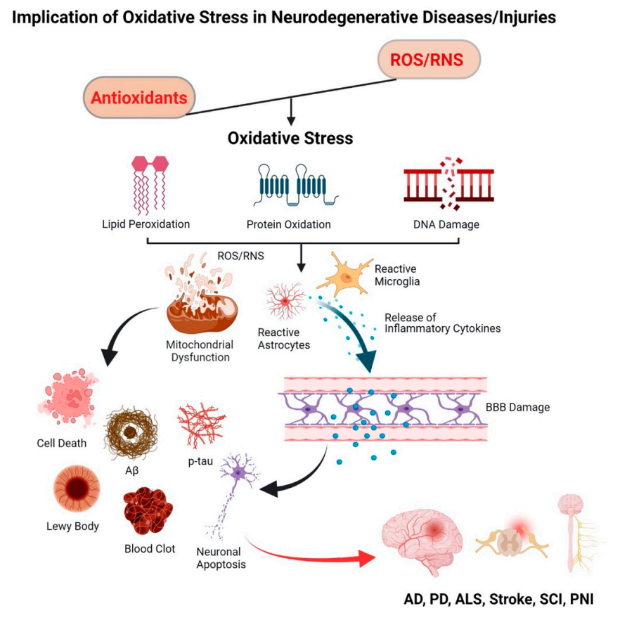
[49]. Prior to the development of plaque pathology, oxidative stress has been recognized as the key player in the etiology of AD, contributing to mitochondrial dysfunction in synapses and neurons, and in Aβ production [50][51]. In fact, the concept of oxidative stress in AD was originally derived from the “free radical theory of aging”, meaning that free radicals play a central role in the aging process [52]. Mitochondrial dysfunction in AD includes impaired mitochondrial complexes [53][54][55][56], malfunctioning of F1Fo adenosine triphosphate (ATP) synthase, which is involved in oxidative phosphorylation [57][58], and damage to the promoter of the mitochondrial ATP synthase gene that controls ATP generation [59][60]. Further, dysfunctional mitochondria produce 4-HNE that upregulates γ-secretase complex and promotes cleavage of the amyloid precursor protein (APP), leading to Aβ accumulation [61][62]. In addition, increased Ca2+ and ROS levels lead to a buildup of p-tau aggregates which are toxic and are considered as one of the defining pathological hallmarks of AD patients [63]. ROS also play a pivotal role in the stress kinases like the phospho-c-Jun N-terminal kinase 1 (p-JNK) pathway which is linked to tau hyperphosphorylation and cell death in response to Aβ accumulation 
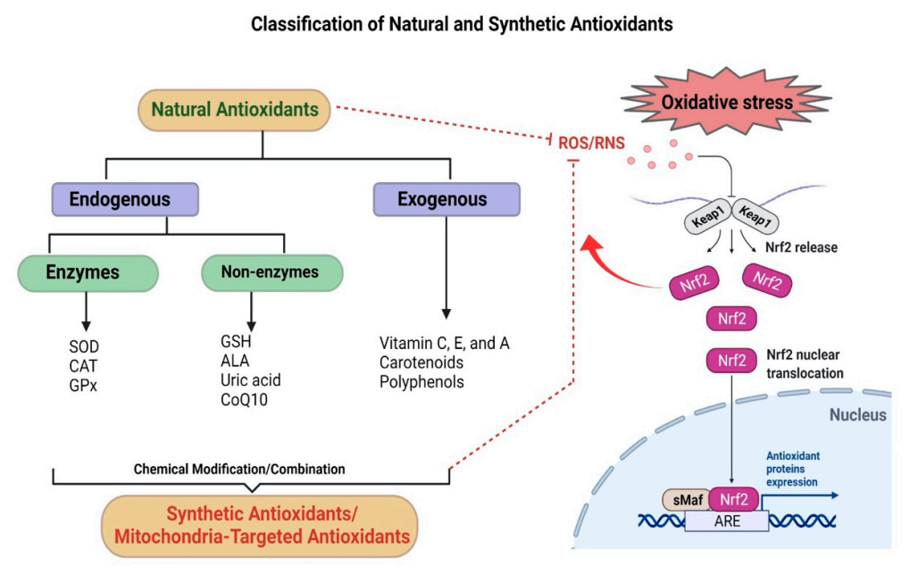
[64]. Further, oxidative stress reduces the activities of antioxidants, i.e., superoxide dismutase (SOD), catalase (CAT), and glutathione S-transferase (GST), thus weakening the endogenous antioxidant defense of the CNS [65]. Further, oxidative stress increases The increased levels of LPO under oxidative stress are strongly associated with neurotoxicity in AD [50] as it leads to an increase in amyloidogenesis through upregulation of β-secretase expression [66]. Although there are several downstream degenerative events, it appears that mitochondrial dysfunction and oxidative stress are the key triggering factors in the pathogenesis of AD.

2.1.2. Parkinson’s Disease (PD)

PD is the second-most common neurodegenerative disease after AD that causes both motor and nonmotor symptoms [67]. The pathology of PD is driven by the accumulation and aggregation of α-synuclein, a presynaptic neuronal protein in the nervous system [68]. The mechanisms associated with the pathogenesis of PD include aberrant protein homeostasis, bioenergetic impairment, and oxidative stress [69]. Oxidative stress is associated with α-synuclein protein aggregation [64]. The cascade of events leading to degeneration of dopaminergic neurons in PD is also linked to oxidative stress [70]. Analysis of the postmortem brain tissue of the victims of PD shows elevated levels of oxidative stress markers such as 4-HNE, protein carbonyl, 8-hydroxy-2′-deoxyguanosine, and 8-hydroxy-guanosine [71]. In addition, oxidative stress is associated with the formation of Lewy bodies, which are the clumps of protein in the PD brain [72]. Experimental evidence in PD models suggests that oxidative stress in the dopaminergic neurons activates p38 mitogen-activated protein kinase (p38 MAPK) pathway that ultimately leads to apoptosis of the brain cells [73].

2.1.3. Amyotrophic Lateral Sclerosis (ALS)

ALS is also known as Lou Gehrig’s disease, in which motor neurons in the brain, brain stem, and spinal cord are damaged, resulting in muscle weakness, atrophy, paralysis, and premature death [74]. Oxidative stress, mitochondrial dysfunction, and mutations in the genes that act on mitochondrial processes are involved in the pathophysiology of ALS [75][76]. Most of the familial ALS patients (15–20%) have mutations in the superoxide dismutase 1 (SOD1) gene, which plays an important role in the defense mechanism against oxidative stress [77]. More than 150 ALS-related SOD1 gene mutations have been discovered in various parts of the enzyme, which result in protein misfolding and aggregation, increased ROS production, and redox system disequilibrium, ultimately resulting in nerve cell loss [77][78]. ALS is also linked to several interrelated risk factors, such as neuroinflammation, excitotoxicity, mitochondrial dysfunction/dysregulation, and endoplasmic reticulum stress [79][80][81]. Considerably high oxidative stress biomarkers such as MDA, 8-hydroxyguanosine, and advanced oxidation protein products are found in ALS patients [82]. In sporadic ALS patients, cystine/glutamate antiporter overexpression was observed that causes increased oxidative stress and extracellular glutamate accumulation [83]. In addition, dysregulation of the retinoic acid (RA) signaling pathway, a product of vitamin A, contributes to the death of motor neurons [84].

2.2. Injury-Induced Oxidative Stress

Neuronal tissue injury, physical or due to ischemic condition, is known to induce oxidative stress that triggers progressive degeneration, known as secondary injury.

2.2.1. Stroke

In stroke, thrombus formation in cerebral blood vessels creates an ischemic condition, triggering free radical formation and tissue damage (Figure 2) [85]. Resumption of blood supply to the ischemic region further exuberates the condition as more free radicals are formed, termed “reperfusion injury or reoxygenation injury” [86]. Collectively, it is referred to as the ischemia/reperfusion (I/R) injury [86]. Oxidative stress leads to mitochondrial dysfunction, neuroinflammation, and glutamate excitotoxicity, resulting in the blood-brain barrier (BBB) damage, apoptosis/necrosis of neurons, and supporting cellular elements (glial cells and vessels) [87][88][89]. These are the prominent features of neurodegeneration in stroke-related cerebral pathology [90][91][92][93]. Further, excessive ROS production or impaired ROS degradation [94][95] stimulates vasoconstriction, increased platelet aggregation, and endothelial cell permeability, thereby affecting cerebral blood circulation [96]. Activation of matrix metalloproteinases (MMPs) disrupts the cerebral extracellular matrix (ECM), which causes immunocyte infiltration and neuroinflammation, culminating in the breakdown of the neurovascular unit (NVU), leading to hemorrhage and edema [97][98].
Figure 2. ROS-mediated degenerative events during a stroke. Excessive production of ROS during I/R injury leads to mechanical damage to the brain due to breakdown of the BBB and hemorrhage and edema, causing a build-up of intracranial pressure (ICP). The biochemical changes lead to inflammation and progression of apoptosis. Therefore, excess ROS formed during I/R is considered a target to inhibit the progression of secondary brain damage.

2.2.2. Spinal Cord Injury (SCI)

SCI is another common form of neuronal injury that causes neurological dysfunctions [99] and is characterized by an initial primary injury followed by the secondary phase of injury (Figure 3) [100]. Primary injury results immediately from the initial trauma causing damage to the blood vessels and axons [101]. In contrast, secondary injury is the indirect result of the primary injury that involves inflammation and oxidative stress [10]. The secondary injury progression occurs not only at the site of impact, but it spreads along the entire spinal cord, including the faraway segments of the spinal cord that are not impacted, making the condition more devastating and debilitating with time [101]. Following injury, the elevated levels of ROS and the consequent oxidative stress are considered critical events associated with the secondary injury progression [102]. Under oxidative stress condition, dysfunctional mitochondria become the source of ROS [103] that cause a further cascade of degenerative processes, particularly curtailing ATP production required for normal cellular functioning, thus promoting apoptosis [103]. The excess ROS alters cell functions by modulating ion channels, followed by excessive accumulation of intracellular calcium ions that eventually causes excitotoxicity [104]. Oxidative stress damages the microvascular endothelium that reduces the spinal cord white matter blood flow, resulting in ischemic injury [105].
Figure 3. Secondary injury cascade following spinal cord injury. Traumatic injury to the spinal cord leads to secondary injury progression that affects the lesion site and the entire spinal cord, including the cranial and caudal segments of the spinal cord. Following injury, excessive production of ROS is considered to trigger the secondary injury cascade of progressive degeneration that affects the entire spinal cord.

2.2.3. Peripheral Nerve Injury (PNI)

The peripheral nervous system (PNS) is a bundle of long nerve fibers that connect different parts of the body with the CNS. Damage to the peripheral nerves due to trauma and compression can cause impairment in the brain’s communication with the target organs [106]. These injuries affect motor and sensory behaviors, perception, consciousness, and sensations of the skin and joints [106]. The most common symptoms of PNI are defects in sensory and motor function that can lead to complete paralysis of the affected limb or the development of intractable neuropathic pain [107]. Many surgical procedures, such as oral and maxillofacial surgery, can also cause injury to the peripheral nerves [108]. The major component of the mechanism and pathogenies of PNI involves oxidative stress and inflammation that exacerbates neural damages and plays a negative role in the regeneration process [109]. Experimental evidence at the preclinical level has demonstrated that inhibiting oxidative stress could help improve functional recovery by accelerating the repair processes [110][111][112][113][114].
Other neurodegenerative diseases implicated due to oxidative stress are: vascular dementia [115], Down syndrome [116], Autism [117], attention-deficit/hyperactivity disorder (ADHD) [118], Huntington’s disease (HD) [119], multiple sclerosis (MS) [120], depression [121], and epilepsy [122]. Similarly, in traumatic brain injury (TBI) [123], progressive degeneration occurs due to the accumulation of excessive free radicals, glutamate release, Ca2+ overload, mitochondrial dysfunction, leading to apoptosis/necrosis [123].

3. Antioxidants

From the above review of the etiology of different neurodegenerative diseases, oxidative stress is considered as the key component, whether these are chronic neurodegenerative conditions such as AD, PD, or ALS or caused by neuronal tissue injuries, such as in stroke, SCI, or PNI. Dysfunctional mitochondria under oxidative stress become the main source of free radical formation and deplete the energy needed for normal cellular function, leading to inflammation and cell death [124]. Another set of literature data indicates that dysfunctional mitochondria cause oxidative stress [125]. Thus, there is a complexity in understanding the root cause, whether oxidative stress leads to mitochondrial dysfunction, or it is mitochondrial dysfunction that leads to oxidative stress [49][126][127]. Despite ambiguity on the root cause of oxidative stress, it is hypothesized that an effective treatment based on antioxidants can alleviate oxidative stress and regain the redox balance that can attenuate mitochondrial dysfunction and curtail the downstream cascade of degeneration [126]. It is also contemplated that oxidative stress-free condition can promote regeneration and healing by the endogenous mechanisms, such as by promoting migration and differentiation of progenitor and stem cells [127]. In addition, an oxidative stress-free environment could promote differentiation of macrophages preferentially to M2 phenotype, which contains growth factors and can promote healing, rather than to M1 phenotype, which contains degenerative inflammatory cytokines [128]. With this in consideration, natural and synthetic antioxidants have been evaluated in preclinical model studies and clinical trials [129].
Antioxidants can reduce oxidative stress by quenching/scavenging free radical intermediates, thereby preventing oxidative chain reactions from propagating [4]. These antioxidants predominantly include various endogenous antioxidant enzymes with their substrates or coenzymes and nonenzymatic antioxidants, along with exogenous (natural and synthetic) antioxidant sources that maintain the redox equilibrium in the biological system [130]. Endogenous antioxidant activity is directly regulated by nuclear factor erythroid 2-related factor 2 (Nrf2). It is a ubiquitous redox-sensitive transcription factor that stimulates the expression of antioxidant response element (ARE)-containing gene promoters involved in ROS detoxification. These promoters are heme oxygenase 1 (HO-1), glutathione s-transferase (GST), and NADPH quinine oxidoreductase 1 (NQO1) (Figure 4) [131]. Thus, the Nrf2 pathway is an important aspect of the cellular defense mechanism against oxidative stress [132].
Figure 4. Natural and synthetic antioxidants: Classification of natural and synthetic antioxidants and the endogenous Nrf2 pathway, which regulates the activation of ARE genes. Kelch-like ECH-associated protein 1 (Keap1) represents a negative regulator of Nrf2. Under physiological conditions, Keap1 forms a ubiquitin E3 ligase complex with Cullin3 in the cytoplasm that targets Nrf2 for polyubiquitination and rapid proteasomal degradation. During oxidative stress, cysteines in Keap1 are modified and inactivated, and Nrf2 can quickly translocate into the nucleus, where it binds to small musculoaponeurotic fibrosarcoma oncogene homolog (sMaf) proteins, upregulates downstream ARE genes, and maintains redox homeostasis.

4. Antioxidant-Based Nanotherapy

To overcome the limitations of natural and synthetic antioxidants, significant efforts have been made to improve their efficacy using drug delivery approaches. These include exploring nanocarriers of different polymeric materials, conjugates, and complexes [133][134] to improve their stability, half-lives, transport to the CNS, and sustained their effect in the target tissue (Figure 5).
Figure 5. Antioxidant-based nanotherapy. Schematic depicting advantages of delivery of antioxidant-loaded nanoparticles to improve t half-life of antioxidants and their ability to cross the BBB, improve bioavailability, and sustain the effect, thus effectively neutralizing oxidative stress in neurodegenerative diseases.
Due to its broad pharmacological effects, including anti-inflammatory and antioxidant properties, curcumin has been widely investigated in clinical studies. To overcome its low water solubility, poor bioavailability, and rapid metabolism, curcumin is formulated as nanocurcumin using different nanocarriers, such as liposomes, polymers, conjugates, cyclodextrins, micelles, dendrimers, and nanoparticles [135]. Transferrin-conjugated poly (lactic co-glycolic acid) (PLGA) nanoparticles have been demonstrated to improve the bioavailability of curcumin to the brain and reduce Aβ deposition and tau hyperphosphorylation in the AD model [136]. Similarly, different formulations of nanoparticles have been shown to inhibit aggregation of Aβ and reduce depressive-like behavior and oxidative stress in AD models [137][138]. Intra-arterial administration of resveratrol (RES)-encapsulated nanoparticle (RES-NP) in a rat transient middle cerebral artery occlusion (t-MCAO) enhanced the resveratrol bioavailability and its brain-penetration, resulting in reduced infarct volume, and attenuated oxidative stress [139], brain edema, and neuronal apoptosis. The treatment also contributed to neurogenesis, leading to improved neurological recovery [140]. In a cerebral palsy rabbit model, intravenous treatment of dendrimer-based N-acetyl-l-cysteine (NAC) [141], a glutathione precursor with antioxidant and anti-inflammatory properties [142], reduced neuroinflammation and neurological injury, and improved motor function. In general, formulating antioxidants in nanocarriers has enhanced their efficacy due to better stability and/or improved transport to the CNS than free antioxidants [143][144][145][146][147][148][149][150][151][152][153][154][155][156][157][158][159][160][161][162][163][164][165][166][167][168][169]. Nanocurcumin has been evaluated as an add-on therapy to Riluzole in a pilot randomized clinical trial for safety and efficacy in ALS [170] and AD patients as dietary supplements [171]. In another study, solid–lipid curcumin showed significantly improved cognition and mood in a healthy older population [172].
Edaravone-loaded ceria nanoparticles have been demonstrated to cross the BBB via receptor-mediated transcytosis and protect the BBB [173]. In addition to the antioxidant property of ceria nanoparticles, edaravone provided its effect against oxidative stress in a stroke model [173]. Jin et al. demonstrated that the treatment with edaravone-encapsulated agonistic micelles caused rapid infarct volume reduction, prolonged survival, improved axonal remodeling, and reduced behavioral deficits than free edaravone-treated animals [174]. Wang et al. reviewed nanotechnology-based strategies for the treatment of ALS, including antioxidant agents [175]. Nanoparticle-loaded edaravone has been tested on the postoperative effects in patients with cerebral hemorrhage. The nanoparticle-loaded edaravone showed reduced edema as compared to free edaravone treated group, significantly improved neurological function, and reduced the production and release of interleukin and tumor necrosis factor, which was considered beneficial to protect healthy brain tissue and other organs, and conducive to the recovery and healing [176].
Despite promising results in preclinical studies, clinical translation of antioxidants as a therapy to treat neurodegenerative diseases remains elusive. The issues could be their low bioavailability, instability, limited transport to the target tissue, and/or poor antioxidant capacity, requiring repeated and high dosing, which cannot be administered to humans because of dose-limiting toxicity. Nanoparticle-mediated delivery of antioxidant enzymes could potentially address some of the above issues. Apart from being endogenous, the main advantage of antioxidant enzymes is their catalytic mechanism of action; hence, they are significantly more effective at lower doses in detoxifying the deleterious effects of free radicals than nonenzymatic antioxidants.

References

  1. Singh, A.; Kukreti, R.; Saso, L.; Kukreti, S. Oxidative Stress: A Key Modulator in Neurodegenerative Diseases. Molecules 2019, 24, 1583.
  2. Sun, Y.; Lu, Y.; Saredy, J.; Wang, X.; Drummer Iv, C.; Shao, Y.; Saaoud, F.; Xu, K.; Liu, M.; Yang, W.Y.; et al. ROS systems are a new integrated network for sensing homeostasis and alarming stresses in organelle metabolic processes. Redox Biol. 2020, 37, 101696.
  3. Collin, F. Chemical Basis of Reactive Oxygen Species Reactivity and Involvement in Neurodegenerative Diseases. Int. J. Mol. Sci. 2019, 20, 2407.
  4. Sharifi-Rad, M.; Anil Kumar, N.V.; Zucca, P.; Varoni, E.M.; Dini, L.; Panzarini, E.; Rajkovic, J.; Tsouh Fokou, P.V.; Azzini, E.; Peluso, I.; et al. Lifestyle, Oxidative Stress, and Antioxidants: Back and Forth in the Pathophysiology of Chronic Diseases. Front. Physiol. 2020, 11, 694.
  5. Man, A.W.C.; Li, H.; Xia, N. Impact of Lifestyles (Diet and Exercise) on Vascular Health: Oxidative Stress and Endothelial Function. Oxid. Med. Cell Longev. 2020, 2020, 1496462.
  6. Kurutas, E.B. The importance of antioxidants which play the role in cellular response against oxidative/nitrosative stress: Current state. Nutr. J. 2016, 15, 71.
  7. Cunha-Oliveira, T.; Montezinho, L.; Mendes, C.; Firuzi, O.; Saso, L.; Oliveira, P.J.; Silva, F.S.G. Oxidative Stress in Amyotrophic Lateral Sclerosis: Pathophysiology and Opportunities for Pharmacological Intervention. Oxid. Med. Cell Longev. 2020, 2020, 5021694.
  8. Birben, E.; Sahiner, U.M.; Sackesen, C.; Erzurum, S.; Kalayci, O. Oxidative stress and antioxidant defense. World Allergy Organ. J. 2012, 5, 9–19.
  9. Journey through the Diagnosis of Dementia—World Alzheimer Report 2021. Available online: https://www.alzint.org/resource/world-alzheimer-report-2021/ (accessed on 14 January 2022).
  10. Anjum, A.; Yazid, M.D.; Fauzi Daud, M.; Idris, J.; Ng, A.M.H.; Selvi Naicker, A.; Ismail, O.H.R.; Athi Kumar, R.K.; Lokanathan, Y. Spinal Cord Injury: Pathophysiology, Multimolecular Interactions, and Underlying Recovery Mechanisms. Int. J. Mol. Sci. 2020, 21, 7533.
  11. von Arnim, C.A.; Herbolsheimer, F.; Nikolaus, T.; Peter, R.; Biesalski, H.K.; Ludolph, A.C.; Riepe, M.; Nagel, G.; Acti, F.E.U.S.G. Dietary antioxidants and dementia in a population-based case-control study among older people in South Germany. J. Alzheimers Dis. 2012, 31, 717–724.
  12. Urano, S.; Asai, Y.; Makabe, S.; Matsuo, M.; Izumiyama, N.; Ohtsubo, K.; Endo, T. Oxidative injury of synapse and alteration of antioxidative defense systems in rats, and its prevention by vitamin E. Eur. J. Biochem. 1997, 245, 64–70.
  13. Magistretti, P.J.; Allaman, I. A cellular perspective on brain energy metabolism and functional imaging. Neuron 2015, 86, 883–901.
  14. Siraki, A.G.; O’Brien, P.J. Prooxidant activity of free radicals derived from phenol-containing neurotransmitters. Toxicology 2002, 177, 81–90.
  15. Salim, S. Oxidative Stress and the Central Nervous System. J. Pharmacol. Exp. Ther. 2017, 360, 201–205.
  16. Cobley, J.N.; Fiorello, M.L.; Bailey, D.M. 13 reasons why the brain is susceptible to oxidative stress. Redox Biol. 2018, 15, 490–503.
  17. Liguori, I.; Russo, G.; Curcio, F.; Bulli, G.; Aran, L.; Della-Morte, D.; Gargiulo, G.; Testa, G.; Cacciatore, F.; Bonaduce, D.; et al. Oxidative stress, aging, and diseases. Clin. Interv. Aging 2018, 13, 757–772.
  18. Phaniendra, A.; Jestadi, D.B.; Periyasamy, L. Free radicals: Properties, sources, targets, and their implication in various diseases. Indian J. Clin. Biochem. 2015, 30, 11–26.
  19. Popa-Wagner, A.; Mitran, S.; Sivanesan, S.; Chang, E.; Buga, A.M. ROS and brain diseases: The good, the bad, and the ugly. Oxid. Med. Cell Longev. 2013, 2013, 963520.
  20. Selivanov, V.A.; Votyakova, T.V.; Pivtoraiko, V.N.; Zeak, J.; Sukhomlin, T.; Trucco, M.; Roca, J.; Cascante, M. Reactive oxygen species production by forward and reverse electron fluxes in the mitochondrial respiratory chain. PLoS Comput. Biol. 2011, 7, e1001115.
  21. Mailloux, R.J.; McBride, S.L.; Harper, M.E. Unearthing the secrets of mitochondrial ROS and glutathione in bioenergetics. Trends Biochem. Sci. 2013, 38, 592–602.
  22. Walker, C.L.; Pomatto, L.C.D.; Tripathi, D.N.; Davies, K.J.A. Redox Regulation of Homeostasis and Proteostasis in Peroxisomes. Physiol. Rev. 2018, 98, 89–115.
  23. Davies, M.J.; Hawkins, C.L. The Role of Myeloperoxidase in Biomolecule Modification, Chronic Inflammation, and Disease. Antioxid. Redox Signal. 2020, 32, 957–981.
  24. Ballance, W.C.; Qin, E.C.; Chung, H.J.; Gillette, M.U.; Kong, H. Reactive oxygen species-responsive drug delivery systems for the treatment of neurodegenerative diseases. Biomaterials 2019, 217, 119292.
  25. Veith, A.; Moorthy, B. Role of Cytochrome P450s in the Generation and Metabolism of Reactive Oxygen Species. Curr. Opin. Toxicol. 2018, 7, 44–51.
  26. Araujo, J.E.; Jorge, S.; Santos, H.M.; Chiechi, A.; Galstyan, A.; Lodeiro, C.; Diniz, M.; Kleinman, M.T.; Ljubimova, J.Y.; Capelo, J.L. Proteomic changes driven by urban pollution suggest particulate matter as a deregulator of energy metabolism, mitochondrial activity, and oxidative pathways in the rat brain. Sci. Total Environ. 2019, 687, 839–848.
  27. Calderon-Garciduenas, L.; Leray, E.; Heydarpour, P.; Torres-Jardon, R.; Reis, J. Air pollution, a rising environmental risk factor for cognition, neuroinflammation and neurodegeneration: The clinical impact on children and beyond. Rev. Neurol. 2016, 172, 69–80.
  28. Rai, Y.; Anita; Kumari, N.; Singh, S.; Kalra, N.; Soni, R.; Bhatt, A.N. Mild mitochondrial uncoupling protects from ionizing radiation induced cell death by attenuating oxidative stress and mitochondrial damage. Biochim. Biophys. Acta Bioenerg. 2021, 1862, 148325.
  29. Mullenders, L.H.F. Solar UV damage to cellular DNA: From mechanisms to biological effects. Photochem. Photobiol. Sci. 2018, 17, 1842–1852.
  30. Akakin, D.; Tok, O.E.; Anil, D.; Akakin, A.; Sirvanci, S.; Sener, G.; Ercan, F. Electromagnetic Waves from Mobile Phones may Affect Rat Brain During Development. Turk. Neurosurg. 2021, 31, 412–421.
  31. Choi, S.; Krishnan, J.; Ruckmani, K. Cigarette smoke and related risk factors in neurological disorders: An update. Biomed. Pharmacother. 2017, 85, 79–86.
  32. Naha, N.; Gandhi, D.N.; Gautam, A.K.; Prakash, J.R. Nicotine and cigarette smoke modulate Nrf2-BDNF-dopaminergic signal and neurobehavioral disorders in adult rat cerebral cortex. Hum. Exp. Toxicol. 2018, 37, 540–556.
  33. Rodriguez-Martinez, E.; Nava-Ruiz, C.; Escamilla-Chimal, E.; Borgonio-Perez, G.; Rivas-Arancibia, S. The Effect of Chronic Ozone Exposure on the Activation of Endoplasmic Reticulum Stress and Apoptosis in Rat Hippocampus. Front. Aging Neurosci. 2016, 8, 245.
  34. Gargouri, B.; Bhatia, H.S.; Bouchard, M.; Fiebich, B.L.; Fetoui, H. Inflammatory and oxidative mechanisms potentiate bifenthrin-induced neurological alterations and anxiety-like behavior in adult rats. Toxicol. Lett. 2018, 294, 73–86.
  35. Lahouel, A.; Kebieche, M.; Lakroun, Z.; Rouabhi, R.; Fetoui, H.; Chtourou, Y.; Djamila, Z.; Soulimani, R. Neurobehavioral deficits and brain oxidative stress induced by chronic low dose exposure of persistent organic pollutants mixture in adult female rat. Environ. Sci. Pollut. Res. Int. 2016, 23, 19030–19040.
  36. Cruces-Sande, A.; Rodriguez-Perez, A.I.; Herbello-Hermelo, P.; Bermejo-Barrera, P.; Mendez-Alvarez, E.; Labandeira-Garcia, J.L.; Soto-Otero, R. Copper Increases Brain Oxidative Stress and Enhances the Ability of 6-Hydroxydopamine to Cause Dopaminergic Degeneration in a Rat Model of Parkinson’s Disease. Mol. Neurobiol. 2019, 56, 2845–2854.
  37. Yauger, Y.J.; Bermudez, S.; Moritz, K.E.; Glaser, E.; Stoica, B.; Byrnes, K.R. Iron accentuated reactive oxygen species release by NADPH oxidase in activated microglia contributes to oxidative stress in vitro. J. Neuroinflamm. 2019, 16, 41.
  38. Langley, M.R.; Ghaisas, S.; Ay, M.; Luo, J.; Palanisamy, B.N.; Jin, H.; Anantharam, V.; Kanthasamy, A.; Kanthasamy, A.G. Manganese exposure exacerbates progressive motor deficits and neurodegeneration in the MitoPark mouse model of Parkinson’s disease: Relevance to gene and environment interactions in metal neurotoxicity. Neurotoxicology 2018, 64, 240–255.
  39. Ashok, A.; Rai, N.K.; Tripathi, S.; Bandyopadhyay, S. Exposure to As-, Cd-, and Pb-mixture induces Abeta, amyloidogenic APP processing and cognitive impairments via oxidative stress-dependent neuroinflammation in young rats. Toxicol. Sci 2015, 143, 64–80.
  40. Di Meo, S.; Reed, T.T.; Venditti, P.; Victor, V.M. Role of ROS and RNS Sources in Physiological and Pathological Conditions. Oxid. Med. Cell Longev. 2016, 2016, 1245049.
  41. Forstermann, U.; Sessa, W.C. Nitric oxide synthases: Regulation and function. Eur. Heart J. 2012, 33, 829–837.
  42. Rahal, A.; Kumar, A.; Singh, V.; Yadav, B.; Tiwari, R.; Chakraborty, S.; Dhama, K. Oxidative stress, prooxidants, and antioxidants: The interplay. Biomed. Res. Int. 2014, 2014, 761264.
  43. Sies, H. Oxidative stress: Oxidants and antioxidants. Exp. Physiol. 1997, 82, 291–295.
  44. Juan, C.A.; Perez de la Lastra, J.M.; Plou, F.J.; Perez-Lebena, E. The Chemistry of Reactive Oxygen Species (ROS) Revisited: Outlining Their Role in Biological Macromolecules (DNA, Lipids and Proteins) and Induced Pathologies. Int. J. Mol. Sci. 2021, 22, 4642.
  45. Damiano, M.; Starkov, A.A.; Petri, S.; Kipiani, K.; Kiaei, M.; Mattiazzi, M.; Flint Beal, M.; Manfredi, G. Neural mitochondrial Ca2+ capacity impairment precedes the onset of motor symptoms in G93A Cu/Zn-superoxide dismutase mutant mice. J. Neurochem. 2006, 96, 1349–1361.
  46. Singh, B.K.; Tripathi, M.; Pandey, P.K.; Kakkar, P. Alteration in mitochondrial thiol enhances calcium ion dependent membrane permeability transition and dysfunction in vitro: A cross-talk between mtThiol, Ca(2+), and ROS. Mol. Cell Biochem. 2011, 357, 373–385.
  47. Chiang, S.C.; Meagher, M.; Kassouf, N.; Hafezparast, M.; McKinnon, P.J.; Haywood, R.; El-Khamisy, S.F. Mitochondrial protein-linked DNA breaks perturb mitochondrial gene transcription and trigger free radical-induced DNA damage. Sci. Adv. 2017, 3, e1602506.
  48. Eratne, D.; Loi, S.M.; Farrand, S.; Kelso, W.; Velakoulis, D.; Looi, J.C. Alzheimer’s disease: Clinical update on epidemiology, pathophysiology and diagnosis. Australas. Psychiatry 2018, 26, 347–357.
  49. DeTure, M.A.; Dickson, D.W. The neuropathological diagnosis of Alzheimer’s disease. Mol. Neurodegener. 2019, 14, 32.
  50. Markesbery, W.R. Oxidative stress hypothesis in Alzheimer’s disease. Free Radic. Biol. Med. 1997, 23, 134–147.
  51. Sharma, C.; Kim, S.R. Linking Oxidative Stress and Proteinopathy in Alzheimer’s Disease. Antioxidants 2021, 10, 1231.
  52. Harman, D. Free radical theory of aging. Mutat. Res. 1992, 275, 257–266.
  53. Adav, S.S.; Park, J.E.; Sze, S.K. Quantitative profiling brain proteomes revealed mitochondrial dysfunction in Alzheimer’s disease. Mol. Brain 2019, 12, 8.
  54. Kim, S.H.; Vlkolinsky, R.; Cairns, N.; Lubec, G. Decreased levels of complex III core protein 1 and complex V beta chain in brains from patients with Alzheimer’s disease and Down syndrome. Cell Mol. Life Sci. 2000, 57, 1810–1816.
  55. Kim, S.H.; Vlkolinsky, R.; Cairns, N.; Fountoulakis, M.; Lubec, G. The reduction of NADH ubiquinone oxidoreductase 24- and 75-kDa subunits in brains of patients with Down syndrome and Alzheimer’s disease. Life Sci. 2001, 68, 2741–2750.
  56. Liang, W.S.; Reiman, E.M.; Valla, J.; Dunckley, T.; Beach, T.G.; Grover, A.; Niedzielko, T.L.; Schneider, L.E.; Mastroeni, D.; Caselli, R.; et al. Alzheimer’s disease is associated with reduced expression of energy metabolism genes in posterior cingulate neurons. Proc. Natl. Acad. Sci. USA 2008, 105, 4441–4446.
  57. Gauba, E.; Chen, H.; Guo, L.; Du, H. Cyclophilin D deficiency attenuates mitochondrial F1Fo ATP synthase dysfunction via OSCP in Alzheimer’s disease. Neurobiol. Dis. 2019, 121, 138–147.
  58. Beck, S.J.; Guo, L.; Phensy, A.; Tian, J.; Wang, L.; Tandon, N.; Gauba, E.; Lu, L.; Pascual, J.M.; Kroener, S.; et al. Deregulation of mitochondrial F1FO-ATP synthase via OSCP in Alzheimer’s disease. Nat. Commun. 2016, 7, 11483.
  59. Lu, T.; Pan, Y.; Kao, S.Y.; Li, C.; Kohane, I.; Chan, J.; Yankner, B.A. Gene regulation and DNA damage in the ageing human brain. Nature 2004, 429, 883–891.
  60. Reed, T.; Perluigi, M.; Sultana, R.; Pierce, W.M.; Klein, J.B.; Turner, D.M.; Coccia, R.; Markesbery, W.R.; Butterfield, D.A. Redox proteomic identification of 4-hydroxy-2-nonenal-modified brain proteins in amnestic mild cognitive impairment: Insight into the role of lipid peroxidation in the progression and pathogenesis of Alzheimer’s disease. Neurobiol. Dis. 2008, 30, 107–120.
  61. Esposito, L.; Raber, J.; Kekonius, L.; Yan, F.; Yu, G.Q.; Bien-Ly, N.; Puolivali, J.; Scearce-Levie, K.; Masliah, E.; Mucke, L. Reduction in mitochondrial superoxide dismutase modulates Alzheimer’s disease-like pathology and accelerates the onset of behavioral changes in human amyloid precursor protein transgenic mice. J. Neurosci. 2006, 26, 5167–5179.
  62. Mattson, M.P.; Fu, W.; Waeg, G.; Uchida, K. 4-Hydroxynonenal, a product of lipid peroxidation, inhibits dephosphorylation of the microtubule-associated protein tau. Neuroreport 1997, 8, 2275–2281.
  63. Misrani, A.; Tabassum, S.; Yang, L. Mitochondrial Dysfunction and Oxidative Stress in Alzheimer’s Disease. Front. Aging Neurosci. 2021, 13, 617588.
  64. Patten, D.A.; Germain, M.; Kelly, M.A.; Slack, R.S. Reactive oxygen species: Stuck in the middle of neurodegeneration. J. Alzheimers Dis. 2010, 20 (Suppl. 2), S357–S367.
  65. Marcus, D.L.; Thomas, C.; Rodriguez, C.; Simberkoff, K.; Tsai, J.S.; Strafaci, J.A.; Freedman, M.L. Increased peroxidation and reduced antioxidant enzyme activity in Alzheimer’s disease. Exp. Neurol. 1998, 150, 40–44.
  66. Chen, L.; Na, R.; Gu, M.; Richardson, A.; Ran, Q. Lipid peroxidation up-regulates BACE1 expression in vivo: A possible early event of amyloidogenesis in Alzheimer’s disease. J. Neurochem. 2008, 107, 197–207.
  67. Poewe, W.; Seppi, K.; Tanner, C.M.; Halliday, G.M.; Brundin, P.; Volkmann, J.; Schrag, A.E.; Lang, A.E. Parkinson disease. Nat. Rev. Dis. Primers 2017, 3, 17013.
  68. Simon, D.K.; Tanner, C.M.; Brundin, P. Parkinson Disease Epidemiology, Pathology, Genetics, and Pathophysiology. Clin. Geriatr. Med. 2020, 36, 1–12.
  69. Gomez-Benito, M.; Granado, N.; Garcia-Sanz, P.; Michel, A.; Dumoulin, M.; Moratalla, R. Modeling Parkinson’s Disease With the Alpha-Synuclein Protein. Front. Pharmacol. 2020, 11, 356.
  70. Dias, V.; Junn, E.; Mouradian, M.M. The role of oxidative stress in Parkinson’s disease. J. Parkinsons Dis. 2013, 3, 461–491.
  71. Puspita, L.; Chung, S.Y.; Shim, J.W. Oxidative stress and cellular pathologies in Parkinson’s disease. Mol. Brain 2017, 10, 53.
  72. Trist, B.G.; Hare, D.J.; Double, K.L. Oxidative stress in the aging substantia nigra and the etiology of Parkinson’s disease. Aging Cell 2019, 18, e13031.
  73. Tong, H.; Zhang, X.; Meng, X.; Lu, L.; Mai, D.; Qu, S. Simvastatin Inhibits Activation of NADPH Oxidase/p38 MAPK Pathway and Enhances Expression of Antioxidant Protein in Parkinson Disease Models. Front. Mol. Neurosci. 2018, 11, 165.
  74. Park, H.R.; Yang, E.J. Oxidative Stress as a Therapeutic Target in Amyotrophic Lateral Sclerosis: Opportunities and Limitations. Diagnostics 2021, 11, 1546.
  75. Petrov, D.; Daura, X.; Zagrovic, B. Effect of Oxidative Damage on the Stability and Dimerization of Superoxide Dismutase 1. Biophys. J. 2016, 110, 1499–1509.
  76. Richardson, K.; Allen, S.P.; Mortiboys, H.; Grierson, A.J.; Wharton, S.B.; Ince, P.G.; Shaw, P.J.; Heath, P.R. The effect of SOD1 mutation on cellular bioenergetic profile and viability in response to oxidative stress and influence of mutation-type. PLoS ONE 2013, 8, e68256.
  77. Pansarasa, O.; Bordoni, M.; Diamanti, L.; Sproviero, D.; Gagliardi, S.; Cereda, C. SOD1 in Amyotrophic Lateral Sclerosis: “Ambivalent” Behavior Connected to the Disease. Int. J. Mol. Sci. 2018, 19, 1345.
  78. Huai, J.; Zhang, Z. Structural Properties and Interaction Partners of Familial ALS-Associated SOD1 Mutants. Front. Neurol. 2019, 10, 527.
  79. Obrador, E.; Salvador, R.; Lopez-Blanch, R.; Jihad-Jebbar, A.; Valles, S.L.; Estrela, J.M. Oxidative Stress, Neuroinflammation and Mitochondria in the Pathophysiology of Amyotrophic Lateral Sclerosis. Antioxidants 2020, 9, 901.
  80. Blasco, H.; Garcon, G.; Patin, F.; Veyrat-Durebex, C.; Boyer, J.; Devos, D.; Vourc’h, P.; Andres, C.R.; Corcia, P. Panel of Oxidative Stress and Inflammatory Biomarkers in ALS: A Pilot Study. Can. J. Neurol. Sci. 2017, 44, 90–95.
  81. Bellezza, I.; Grottelli, S.; Costanzi, E.; Scarpelli, P.; Pigna, E.; Morozzi, G.; Mezzasoma, L.; Peirce, M.J.; Moresi, V.; Adamo, S.; et al. Peroxynitrite Activates the NLRP3 Inflammasome Cascade in SOD1(G93A) Mouse Model of Amyotrophic Lateral Sclerosis. Mol. Neurobiol. 2018, 55, 2350–2361.
  82. Wang, Z.; Bai, Z.; Qin, X.; Cheng, Y. Aberrations in Oxidative Stress Markers in Amyotrophic Lateral Sclerosis: A Systematic Review and Meta-Analysis. Oxid. Med. Cell Longev. 2019, 2019, 1712323.
  83. Kazama, M.; Kato, Y.; Kakita, A.; Noguchi, N.; Urano, Y.; Masui, K.; Niida-Kawaguchi, M.; Yamamoto, T.; Watabe, K.; Kitagawa, K.; et al. Astrocytes release glutamate via cystine/glutamate antiporter upregulated in response to increased oxidative stress related to sporadic amyotrophic lateral sclerosis. Neuropathology 2020, 40, 587–598.
  84. Kolarcik, C.L.; Bowser, R. Retinoid signaling alterations in amyotrophic lateral sclerosis. Am. J. Neurodegener. Dis. 2012, 1, 130–145.
  85. Sun, M.S.; Jin, H.; Sun, X.; Huang, S.; Zhang, F.L.; Guo, Z.N.; Yang, Y. Free Radical Damage in Ischemia-Reperfusion Injury: An Obstacle in Acute Ischemic Stroke after Revascularization Therapy. Oxid. Med. Cell Longev. 2018, 2018, 3804979.
  86. Kalogeris, T.; Baines, C.P.; Krenz, M.; Korthuis, R.J. Cell biology of ischemia/reperfusion injury. Int. Rev. Cell Mol. Biol. 2012, 298, 229–317.
  87. Wu, L.; Xiong, X.; Wu, X.; Ye, Y.; Jian, Z.; Zhi, Z.; Gu, L. Targeting Oxidative Stress and Inflammation to Prevent Ischemia-Reperfusion Injury. Front. Mol. Neurosci. 2020, 13, 28.
  88. Al-Qazzaz, N.K.; Ali, S.H.; Ahmad, S.A.; Islam, S.; Mohamad, K. Cognitive impairment and memory dysfunction after a stroke diagnosis: A post-stroke memory assessment. Neuropsychiatr. Dis. Treat. 2014, 10, 1677–1691.
  89. Ahad, M.A.; Kumaran, K.R.; Ning, T.; Mansor, N.I.; Effendy, M.A.; Damodaran, T.; Lingam, K.; Wahab, H.A.; Nordin, N.; Liao, P.; et al. Insights into the neuropathology of cerebral ischemia and its mechanisms. Rev. Neurosci. 2020, 31, 521–538.
  90. Lorenzano, S.; Rost, N.S.; Khan, M.; Li, H.; Lima, F.O.; Maas, M.B.; Green, R.E.; Thankachan, T.K.; Dipietro, A.J.; Arai, K.; et al. Oxidative Stress Biomarkers of Brain Damage: Hyperacute Plasma F2-Isoprostane Predicts Infarct Growth in Stroke. Stroke 2018, 49, 630–637.
  91. Nakano, Y.; Yamashita, T.; Li, Q.; Sato, K.; Ohta, Y.; Morihara, R.; Hishikawa, N.; Abe, K. Time-dependent change of in vivo optical imaging of oxidative stress in a mouse stroke model. J. Neurosci. Res. 2017, 95, 2030–2039.
  92. Kishimoto, M.; Suenaga, J.; Takase, H.; Araki, K.; Yao, T.; Fujimura, T.; Murayama, K.; Okumura, K.; Ueno, R.; Shimizu, N.; et al. Oxidative stress-responsive apoptosis inducing protein (ORAIP) plays a critical role in cerebral ischemia/reperfusion injury. Sci. Rep. 2019, 9, 13512.
  93. Sacco, R.L.; Kasner, S.E.; Broderick, J.P.; Caplan, L.R.; Connors, J.J.; Culebras, A.; Elkind, M.S.; George, M.G.; Hamdan, A.D.; Higashida, R.T.; et al. An updated definition of stroke for the 21st century: A statement for healthcare professionals from the American Heart Association/American Stroke Association. Stroke 2013, 44, 2064–2089.
  94. Lou, Z.; Wang, A.P.; Duan, X.M.; Hu, G.H.; Song, G.L.; Zuo, M.L.; Yang, Z.B. Upregulation of NOX2 and NOX4 Mediated by TGF-beta Signaling Pathway Exacerbates Cerebral Ischemia/Reperfusion Oxidative Stress Injury. Cell Physiol. Biochem. 2018, 46, 2103–2113.
  95. Xu, N.; Meng, H.; Liu, T.; Feng, Y.; Qi, Y.; Wang, H. TRPC1 Deficiency Exacerbates Cerebral Ischemia/Reperfusion-Induced Neurological Injury by Potentiating Nox4-Derived Reactive Oxygen Species Generation. Cell Physiol. Biochem. 2018, 51, 1723–1738.
  96. Staiculescu, M.C.; Foote, C.; Meininger, G.A.; Martinez-Lemus, L.A. The role of reactive oxygen species in microvascular remodeling. Int. J. Mol. Sci. 2014, 15, 23792–23835.
  97. Rempe, R.G.; Hartz, A.M.S.; Bauer, B. Matrix metalloproteinases in the brain and blood-brain barrier: Versatile breakers and makers. J. Cereb. Blood Flow Metab. 2016, 36, 1481–1507.
  98. Chen, S.; Shao, L.; Ma, L. Cerebral Edema Formation After Stroke: Emphasis on Blood-Brain Barrier and the Lymphatic Drainage System of the Brain. Front. Cell Neurosci. 2021, 15, 716825.
  99. van Den Hauwe, L.; Sundgren, P.C.; Flanders, A.E. Spinal Trauma and Spinal Cord Injury (SCI). In Diseases of the Brain, Head and Neck, Spine 2020–2023: Diagnostic Imaging; Hodler, J., Kubik-Huch, R.A., von Schulthess, G.K., Eds.; IDKD Springer Series: Cham, Switzerland, 2020.
  100. Eckert, M.J.; Martin, M.J. Trauma: Spinal Cord Injury. Surg. Clin. N. Am. 2017, 97, 1031–1045.
  101. Ahuja, C.S.; Nori, S.; Tetreault, L.; Wilson, J.; Kwon, B.; Harrop, J.; Choi, D.; Fehlings, M.G. Traumatic Spinal Cord Injury-Repair and Regeneration. Neurosurgery 2017, 80, S9–S22.
  102. Lin, J.; Xiong, Z.; Gu, J.; Sun, Z.; Jiang, S.; Fan, D.; Li, W. Sirtuins: Potential Therapeutic Targets for Defense against Oxidative Stress in Spinal Cord Injury. Oxid. Med. Cell Longev. 2021, 2021, 7207692.
  103. Bock, F.J.; Tait, S.W.G. Mitochondria as multifaceted regulators of cell death. Nat. Rev. Mol. Cell Biol. 2020, 21, 85–100.
  104. Rabchevsky, A.G.; Michael, F.M.; Patel, S.P. Mitochondria focused neurotherapeutics for spinal cord injury. Exp. Neurol. 2020, 330, 113332.
  105. Popa, C.; Popa, F.; Grigorean, V.T.; Onose, G.; Sandu, A.M.; Popescu, M.; Burnei, G.; Strambu, V.; Sinescu, C. Vascular dysfunctions following spinal cord injury. J. Med. Life 2010, 3, 275–285.
  106. Hussain, G.; Wang, J.; Rasul, A.; Anwar, H.; Qasim, M.; Zafar, S.; Aziz, N.; Razzaq, A.; Hussain, R.; de Aguilar, J.G.; et al. Current Status of Therapeutic Approaches against Peripheral Nerve Injuries: A Detailed Story from Injury to Recovery. Int. J. Biol. Sci. 2020, 16, 116–134.
  107. Walco, G.A.; Dworkin, R.H.; Krane, E.J.; LeBel, A.A.; Treede, R.D. Neuropathic pain in children: Special considerations. Mayo Clin. Proc. 2010, 85, S33–S41.
  108. Agbaje, J.O.; Van de Casteele, E.; Hiel, M.; Verbaanderd, C.; Lambrichts, I.; Politis, C. Neuropathy of Trigeminal Nerve Branches After Oral and Maxillofacial Treatment. J. Maxillofac. Oral Surg. 2016, 15, 321–327.
  109. Menorca, R.M.; Fussell, T.S.; Elfar, J.C. Nerve physiology: Mechanisms of injury and recovery. Hand Clin. 2013, 29, 317–330.
  110. Khezri, M.K.; Turkkan, A.; Koc, C.; Salman, B.; Levent, P.; Cakir, A.; Kafa, I.M.; Cansev, M.; Bekar, A. Anti-Apoptotic and Anti-Oxidant Effects of Systemic Uridine Treatment in an Experimental Model of Sciatic Nerve Injury. Turk. Neurosurg. 2021, 31, 373–378.
  111. Lattimore, M.R., Jr.; Varr, W.F., 3rd. Disposable soft lens ulcerative keratitis in an Army aviator: A case report. Aviat. Space Environ. Med. 1991, 62, 888–889.
  112. Costa, L.S.; Aidar, F.J.; Matos, D.G.; Oliveira, J.U.; Santos, J.L.D.; Almeida-Neto, P.F.; Souza, R.F.; Pereira, D.D.; Garrido, N.D.; Nunes-Silva, A.; et al. Effects of Resistance Training and Bowdichia virgilioides Hydroethanolic Extract on Oxidative Stress Markers in Rats Submitted to Peripheral Nerve Injury. Antioxidants 2020, 9, 941.
  113. Zafar, S.; Anwar, H.; Qasim, M.; Irfan, S.; Maqbool, J.; Sajid, F.; Naqvi, S.A.R.; Hussain, G. Calotropis procera (root) escalates functions rehabilitation and attenuates oxidative stress in a mouse model of peripheral nerve injury. Pak. J. Pharm. Sci. 2020, 33, 2801–2807.
  114. Tang, C.; Han, R.; Wu, J.; Fang, F. Effects of baicalin capsules combined with alpha-lipoic acid on nerve conduction velocity, oxidative stress and inflammatory injury in patients wi.ith diabetic peripheral neuropathy. Am. J. Transl. Res. 2021, 13, 2774–2783.
  115. Luca, M.; Luca, A.; Calandra, C. The Role of Oxidative Damage in the Pathogenesis and Progression of Alzheimer’s Disease and Vascular Dementia. Oxid. Med. Cell Longev. 2015, 2015, 504678.
  116. Perluigi, M.; Butterfield, D.A. Oxidative Stress and Down Syndrome: A Route toward Alzheimer-Like Dementia. Curr. Gerontol. Geriatr. Res. 2012, 2012, 724904.
  117. Pangrazzi, L.; Balasco, L.; Bozzi, Y. Oxidative Stress and Immune System Dysfunction in Autism Spectrum Disorders. Int. J. Mol. Sci. 2020, 21, 3293.
  118. Oztop, D.; Altun, H.; Baskol, G.; Ozsoy, S. Oxidative stress in children with attention deficit hyperactivity disorder. Clin. Biochem 2012, 45, 745–748.
  119. Paul, B.D.; Snyder, S.H. Impaired Redox Signaling in Huntington’s Disease: Therapeutic Implications. Front. Mol. Neurosci. 2019, 12, 68.
  120. Ohl, K.; Tenbrock, K.; Kipp, M. Oxidative stress in multiple sclerosis: Central and peripheral mode of action. Exp. Neurol. 2016, 277, 58–67.
  121. Bhatt, S.; Nagappa, A.N.; Patil, C.R. Role of oxidative stress in depression. Drug Discov. Today 2020, 25, 1270–1276.
  122. Geronzi, U.; Lotti, F.; Grosso, S. Oxidative stress in epilepsy. Expert Rev. Neurother. 2018, 18, 427–434.
  123. Khatri, N.; Thakur, M.; Pareek, V.; Kumar, S.; Sharma, S.; Datusalia, A.K. Oxidative Stress: Major Threat in Traumatic Brain Injury. CNS Neurol. Disord. Drug Targets 2018, 17, 689–695.
  124. Elfawy, H.A.; Das, B. Crosstalk between mitochondrial dysfunction, oxidative stress, and age related neurodegenerative disease: Etiologies and therapeutic strategies. Life Sci. 2019, 218, 165–184.
  125. Guo, C.; Sun, L.; Chen, X.; Zhang, D. Oxidative stress, mitochondrial damage and neurodegenerative diseases. Neural Regen. Res. 2013, 8, 2003–2014.
  126. Kumar, A.; Singh, A. A review on mitochondrial restorative mechanism of antioxidants in Alzheimer’s disease and other neurological conditions. Front. Pharmacol. 2015, 6, 206.
  127. Gerhke, S.A.; Shibli, J.A.; Salles, M.B. Potential of the use of an antioxidant compound to promote peripheral nerve regeneration after injury. Neural Regen. Res. 2015, 10, 1063–1064.
  128. Tan, H.Y.; Wang, N.; Li, S.; Hong, M.; Wang, X.; Feng, Y. The Reactive Oxygen Species in Macrophage Polarization: Reflecting Its Dual Role in Progression and Treatment of Human Diseases. Oxid. Med. Cell Longev. 2016, 2016, 2795090.
  129. Amato, A.; Terzo, S.; Mule, F. Natural Compounds as Beneficial Antioxidant Agents in Neurodegenerative Disorders: A Focus on Alzheimer’s Disease. Antioxidants 2019, 8, 608.
  130. He, L.; He, T.; Farrar, S.; Ji, L.; Liu, T.; Ma, X. Antioxidants Maintain Cellular Redox Homeostasis by Elimination of Reactive Oxygen Species. Cell Physiol. Biochem. 2017, 44, 532–553.
  131. Vasconcelos, A.R.; Dos Santos, N.B.; Scavone, C.; Munhoz, C.D. Nrf2/ARE Pathway Modulation by Dietary Energy Regulation in Neurological Disorders. Front. Pharmacol. 2019, 10, 33.
  132. Villavicencio Tejo, F.; Quintanilla, R.A. Contribution o.of the Nrf2 Pathway on Oxidative Damage and Mitochondrial Failure in Parkinson and Alzheimer’s Disease. Antioxidants 2021, 10, 1069.
  133. Li, M.; Al-Jamal, K.T.; Kostarelos, K.; Reineke, J. Physiologically based pharmacokinetic modeling of nanoparticles. ACS Nano 2010, 4, 6303–6317.
  134. De Matteis, V.; Rinaldi, R. Toxicity Assessment in the Nanoparticle Era. Adv. Exp. Med. Biol. 2018, 1048, 1–19.
  135. Karthikeyan, A.; Senthil, N.; Min, T. Nanocurcumin: A Promising Candidate for Therapeutic Applications. Front. Pharmacol. 2020, 11, 487.
  136. Fan, S.; Zheng, Y.; Liu, X.; Fang, W.; Chen, X.; Liao, W.; Jing, X.; Lei, M.; Tao, E.; Ma, Q.; et al. Curcumin-loaded PLGA-PEG nanoparticles conjugated with B6 peptide for potential use in Alzheimer’s disease. Drug Deliv. 2018, 25, 1091–1102.
  137. Fidelis, E.M.; Savall, A.S.P.; da Luz Abreu, E.; Carvalho, F.; Teixeira, F.E.G.; Haas, S.E.; Bazanella Sampaio, T.; Pinton, S. Curcumin-Loaded Nanocapsules Reverses the Depressant-Like Behavior and Oxidative Stress Induced by beta-Amyloid in Mice. Neuroscience 2019, 423, 122–130.
  138. Huo, X.; Zhang, Y.; Jin, X.; Li, Y.; Zhang, L. A novel synthesis of selenium nanoparticles encapsulated PLGA nanospheres with curcumin molecules for the inhibition of amyloid beta aggregation in Alzheimer’s disease. J. Photochem. Photobiol. B 2019, 190, 98–102.
  139. Ashafaq, M.; Intakhab Alam, M.; Khan, A.; Islam, F.; Khuwaja, G.; Hussain, S.; Ali, R.; Alshahrani, S.; Antar Makeen, H.; Alhazmi, H.A.; et al. Nanoparticles of resveratrol attenuates oxidative stress and inflammation after ischemic stroke in rats. Int. Immunopharmacol. 2021, 94, 107494.
  140. Lu, X.; Dong, J.; Zheng, D.; Li, X.; Ding, D.; Xu, H. Reperfusion combined with intraarterial administration of resveratrol-loaded nanoparticles improved cerebral ischemia-reperfusion injury in rats. Nanomedicine 2020, 28, 102208.
  141. Kannan, S.; Dai, H.; Navath, R.S.; Balakrishnan, B.; Jyoti, A.; Janisse, J.; Romero, R.; Kannan, R.M. Dendrimer-based postnatal therapy for neuroinflammation and cerebral palsy in a rabbit model. Sci. Transl. Med. 2012, 4, 130ra46.
  142. Tardiolo, G.; Bramanti, P.; Mazzon, E. Overview on the Effects of N-Acetylcysteine in Neurodegenerative Diseases. Molecules 2018, 23, 3305.
  143. Yang, M.; Jin, L.; Wu, Z.; Xie, Y.; Zhang, P.; Wang, Q.; Yan, S.; Chen, B.; Liang, H.; Naman, C.B.; et al. PLGA-PEG Nanoparticles Facilitate In Vivo Anti-Alzheimer’s Effects of Fucoxanthin, a Marine Carotenoid Derived from Edible Brown Algae. J. Agric. Food Chem. 2021, 69, 9764–9777.
  144. Dhas, N.; Mehta, T. Cationic biopolymer functionalized nanoparticles encapsulating lutein to attenuate oxidative stress in effective treatment of Alzheimer’s disease: A non-invasive approach. Int. J. Pharm. 2020, 586, 119553.
  145. Singh, N.A.; Mandal, A.K.A.; Khan, Z.A. Inhibition of Al(III)-Induced Abeta42 Fibrillation and Reduction of Neurotoxicity by Epigallocatechin-3-Gallate Nanoparticles. J. Biomed. Nanotechnol. 2018, 14, 1147–1158.
  146. Liu, Z.; Li, X.; Wu, X.; Zhu, C. A dual-inhibitor system for the effective antifibrillation of Abeta40 peptides by biodegradable EGCG-Fe(iii)/PVP nanoparticles. J. Mater. Chem. B 2019, 7, 1292–1299.
  147. Liu, H.; Yu, L.; Dong, X.; Sun, Y. Synergistic effects of negatively charged hydrophobic nanoparticles and (-)-epigallocatechin-3-gallate on inhibiting amyloid beta-protein aggregation. J. Colloid Interface Sci. 2017, 491, 305–312.
  148. Cano, A.; Ettcheto, M.; Chang, J.H.; Barroso, E.; Espina, M.; Kuhne, B.A.; Barenys, M.; Auladell, C.; Folch, J.; Souto, E.B.; et al. Dual-drug loaded nanoparticles of Epigallocatechin-3-gallate (EGCG)/Ascorbic acid enhance therapeutic efficacy of EGCG in a APPswe/PS1dE9 Alzheimer’s disease mice model. J. Control. Release 2019, 301, 62–75.
  149. Amin, F.U.; Shah, S.A.; Badshah, H.; Khan, M.; Kim, M.O. Anthocyanins encapsulated by nanoparticles potentially improved its free radical scavenging capabilities via p38/JNK pathway against Abeta1-42-induced oxidative stress. J. Nanobiotechnology 2017, 15, 12.
  150. Loureiro, J.A.; Andrade, S.; Duarte, A.; Neves, A.R.; Queiroz, J.F.; Nunes, C.; Sevin, E.; Fenart, L.; Gosselet, F.; Coelho, M.A.; et al. Resveratrol and Grape Extract-loaded Solid Lipid Nanoparticles for the Treatment of Alzheimer’s Disease. Molecules 2017, 22, 277.
  151. Amanzadeh Jajin, E.; Esmaeili, A.; Rahgozar, S.; Noorbakhshnia, M. Quercetin-Conjugated Superparamagnetic Iron Oxide Nanoparticles Protect AlCl3-Induced Neurotoxicity in a Rat Model of Alzheimer’s Disease via Antioxidant Genes, APP Gene, and miRNA-101. Front. Neurosci. 2020, 14, 598617.
  152. Halevas, E.; Mavroidi, B.; Nday, C.M.; Tang, J.; Smith, G.C.; Boukos, N.; Litsardakis, G.; Pelecanou, M.; Salifoglou, A. Modified magnetic core-shell mesoporous silica nano-formulations with encapsulated quercetin exhibit anti-amyloid and antioxidant activity. J. Inorg. Biochem. 2020, 213, 111271.
  153. Pinheiro, R.G.R.; Granja, A.; Loureiro, J.A.; Pereira, M.C.; Pinheiro, M.; Neves, A.R.; Reis, S. Quercetin lipid nanoparticles functionalized with transferrin for Alzheimer’s disease. Eur. J. Pharm. Sci. 2020, 148, 105314.
  154. Sun, D.; Li, N.; Zhang, W.; Zhao, Z.; Mou, Z.; Huang, D.; Liu, J.; Wang, W. Design of PLGA-functionalized quercetin nanoparticles for potential use in Alzheimer’s disease. Colloids Surf. B Biointerfaces 2016, 148, 116–129.
  155. Rifaai, R.A.; Mokhemer, S.A.; Saber, E.A.; El-Aleem, S.A.A.; El-Tahawy, N.F.G. Neuroprotective effect of quercetin nanoparticles: A possible prophylactic and therapeutic role in alzheimer’s disease. J. Chem. Neuroanat. 2020, 107, 101795.
  156. Moreno, L.; Puerta, E.; Suarez-Santiago, J.E.; Santos-Magalhaes, N.S.; Ramirez, M.J.; Irache, J.M. Effect of the oral administration of nanoencapsulated quercetin on a mouse model of Alzheimer’s disease. Int. J. Pharm. 2017, 517, 50–57.
  157. Ramires Junior, O.V.; Alves, B.D.S.; Barros, P.A.B.; Rodrigues, J.L.; Ferreira, S.P.; Monteiro, L.K.S.; Araujo, G.M.S.; Fernandes, S.S.; Vaz, G.R.; Dora, C.L.; et al. Nanoemulsion Improves the Neuroprotective Effects of Curcumin in an Experimental Model of Parkinson’s Disease. Neurotox. Res. 2021, 39, 787–799.
  158. Kundu, P.; Das, M.; Tripathy, K.; Sahoo, S.K. Delivery of Dual Drug Loaded Lipid Based Nanoparticles across the Blood-Brain Barrier Impart Enhanced Neuroprotection in a Rotenone Induced Mouse Model of Parkinson’s Disease. ACS Chem. Neurosci. 2016, 7, 1658–1670.
  159. Fernandes, E.J.; Poetini, M.R.; Barrientos, M.S.; Bortolotto, V.C.; Araujo, S.M.; Santos Musachio, E.A.; De Carvalho, A.S.; Leimann, F.V.; Goncalves, O.H.; Ramborger, B.P.; et al. Exposure to lutein-loaded nanoparticles attenuates Parkinson’s model-induced damage in Drosophila melanogaster: Restoration of dopaminergic and cholinergic system and oxidative stress indicators. Chem. Biol. Interact. 2021, 340, 109431.
  160. Palle, S.; Neerati, P. Improved neuroprotective effect of resveratrol nanoparticles as evinced by abrogation of rotenone-induced behavioral deficits and oxidative and mitochondrial dysfunctions in rat model of Parkinson’s disease. Naunyn. Schmiedebergs Arch. Pharmacol. 2018, 391, 445–453.
  161. Gaba, B.; Khan, T.; Haider, M.F.; Alam, T.; Baboota, S.; Parvez, S.; Ali, J. Vitamin E Loaded Naringenin Nanoemulsion via Intranasal Delivery for the Management of Oxidative Stress in a 6-OHDA Parkinson’s Disease Model. Biomed. Res. Int. 2019, 2019, 2382563.
  162. Trapani, A.; Guerra, L.; Corbo, F.; Castellani, S.; Sanna, E.; Capobianco, L.; Monteduro, A.G.; Manno, D.E.; Mandracchia, D.; Di Gioia, S.; et al. Cyto/Biocompatibility of Dopamine Combined with the Antioxidant Grape Seed-Derived Polyphenol Compounds in Solid Lipid Nanoparticles. Molecules 2021, 26, 916.
  163. Chen, L.; Watson, C.; Morsch, M.; Cole, N.J.; Chung, R.S.; Saunders, D.N.; Yerbury, J.J.; Vine, K.L. Improving the Delivery of SOD1 Antisense Oligonucleotides to Motor Neurons Using Calcium Phosphate-Lipid Nanoparticles. Front. Neurosci. 2017, 11, 476.
  164. Medina, D.X.; Chung, E.P.; Teague, C.D.; Bowser, R.; Sirianni, R.W. Intravenously Administered, Retinoid Activating Nanoparticles Increase Lifespan and Reduce Neurodegeneration in the SOD1(G93A) Mouse Model of ALS. Front. Bioeng. Biotechnol. 2020, 8, 224.
  165. Mauricio, M.D.; Guerra-Ojeda, S.; Marchio, P.; Valles, S.L.; Aldasoro, M.; Escribano-Lopez, I.; Herance, J.R.; Rocha, M.; Vila, J.M.; Victor, V.M. Nanoparticles in Medicine: A Focus on Vascular Oxidative Stress. Oxid. Med. Cell Longev. 2018, 2018, 6231482.
  166. Mei, T.; Kim, A.; Vong, L.B.; Marushima, A.; Puentes, S.; Matsumaru, Y.; Matsumura, A.; Nagasaki, Y. Encapsulation of tissue plasminogen activator in pH-sensitive self-assembled antioxidant nanoparticles for ischemic stroke treatment—Synergistic effect of thrombolysis and antioxidant. Biomaterials 2019, 215, 119209.
  167. Marques, M.S.; Cordeiro, M.F.; Marinho, M.A.G.; Vian, C.O.; Vaz, G.R.; Alves, B.S.; Jardim, R.D.; Hort, M.A.; Dora, C.L.; Horn, A.P. Curcumin-loaded nanoemulsion improves haemorrhagic stroke recovery in wistar rats. Brain Res. 2020, 1746, 147007.
  168. Chen, W.; Zhao, Z.; Zhao, S.; Zhang, L.; Song, Q. Resveratrol and Puerarin loaded polymeric nanoparticles to enhance the chemotherapeutic efficacy in spinal cord injury. Biomed. Microdevices 2020, 22, 69.
  169. Luan, X.; Sansanaphongpricha, K.; Myers, I.; Chen, H.; Yuan, H.; Sun, D. Engineering exosomes as refined biological nanoplatforms for drug delivery. Acta Pharmacol. Sin. 2017, 38, 754–763.
  170. Ahmadi, M.; Agah, E.; Nafissi, S.; Jaafari, M.R.; Harirchian, M.H.; Sarraf, P.; Faghihi-Kashani, S.; Hosseini, S.J.; Ghoreishi, A.; Aghamollaii, V.; et al. Safety and Efficacy of Nanocurcumin as Add-On Therapy to Riluzole in Patients With Amyotrophic Lateral Sclerosis: A Pilot Randomized Clinical Trial. Neurotherapeutics 2018, 15, 430–438.
  171. NCT01001637; Efficacy and Safety of Curcumin Formulation in Alzheimer’s Disease. US National Library of Medicine: Bethesda, MD, USA, 2009.
  172. Cox, K.H.; Pipingas, A.; Scholey, A.B. Investigation of the effects of solid lipid curcumin on cognition and mood in a healthy older population. J. Psychopharmacol. 2015, 29, 642–651.
  173. Bao, Q.; Hu, P.; Xu, Y.; Cheng, T.; Wei, C.; Pan, L.; Shi, J. Simultaneous Blood-Brain Barrier Crossing and Protection for Stroke Treatment Based on Edaravone-Loaded Ceria Nanoparticles. ACS Nano 2018, 12, 6794–6805.
  174. Jin, Q.; Cai, Y.; Li, S.; Liu, H.; Zhou, X.; Lu, C.; Gao, X.; Qian, J.; Zhang, J.; Ju, S.; et al. Edaravone-Encapsulated Agonistic Micelles Rescue Ischemic Brain Tissue by Tuning Blood-Brain Barrier Permeability. Theranostics 2017, 7, 884–898.
  175. Wang, G.Y.; Rayner, S.L.; Chung, R.; Shi, B.Y.; Liang, X.J. Advances in nanotechnology-based strategies for the treatments of amyotrophic lateral sclerosis. Mater. Today Bio 2020, 6, 100055.
  176. Dang, L.; Dong, X.; Yang, J. Influence of Nanoparticle-Loaded Edaravone on Postoperative Effects in Patients with Cerebral Hemorrhage. J. Nanosci. Nanotechnol. 2021, 21, 1202–1211.
More
Information
Subjects: Neurosciences
Contributor MDPI registered users' name will be linked to their SciProfiles pages. To register with us, please refer to https://encyclopedia.pub/register :
View Times: 1.0K
Entry Collection: Neurodegeneration
Revisions: 2 times (View History)
Update Date: 24 Feb 2022
1000/1000
Video Production Service