Submitted Successfully!
To reward your contribution, here is a gift for you: A free trial for our video production service.
Thank you for your contribution! You can also upload a video entry or images related to this topic.
Version Summary Created by Modification Content Size Created at Operation
1 + 2956 word(s) 2956 2021-09-24 11:37:52 |
2 Format change -69 word(s) 2887 2021-11-22 03:12:10 |

Video Upload Options

Do you have a full video?

Confirm

Are you sure to Delete?
Cite
If you have any further questions, please contact Encyclopedia Editorial Office.
Paradiso, F. Third Dimension in Cancer. Encyclopedia. Available online: https://encyclopedia.pub/entry/16210 (accessed on 18 April 2024).
Paradiso F. Third Dimension in Cancer. Encyclopedia. Available at: https://encyclopedia.pub/entry/16210. Accessed April 18, 2024.
Paradiso, Francesca. "Third Dimension in Cancer" Encyclopedia, https://encyclopedia.pub/entry/16210 (accessed April 18, 2024).
Paradiso, F. (2021, November 19). Third Dimension in Cancer. In Encyclopedia. https://encyclopedia.pub/entry/16210
Paradiso, Francesca. "Third Dimension in Cancer." Encyclopedia. Web. 19 November, 2021.
Third Dimension in Cancer
Edit

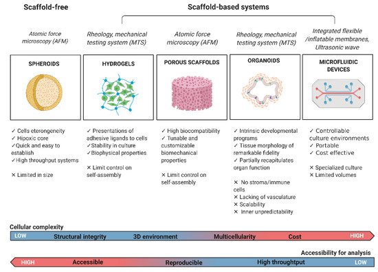
From the development of self-aggregating, scaffold-free multicellular spheroids to the inclusion of scaffold systems, 3D models have progressively increased in complexity to better mimic native tissues. The inclusion of a third dimension in cancer models allows researchers to zoom out from a significant but limited cancer cell research approach to a wider investigation of the tumor microenvironment. This model can include multiple cell types and many elements from the extracellular matrix (ECM), which provides mechanical support for the tissue, mediates cell-microenvironment interactions, and plays a key role in cancer cell invasion. Both biochemical and biophysical signals from the extracellular space strongly influence cell fate, the epigenetic landscape, and gene expression. Specifically, a detailed mechanistic understanding of tumor cell-ECM interactions, especially during cancer invasion, is lacking.

3D model cancer microenvironment biomaterials

1. Introduction

1.1. Mechanosensing: A Biophysical Signal Travels from the Outside to the Inside of the Cells

Today, tumors are depicted as organs, with a higher complexity compared to normal healthy tissue [1]. The complexity of a neoplastic disease relies on individual specialized cells within the tumor tissue that sustain the ability of cancer cells to build a flourishing niche in order to grow and spread, called the tumor microenvironment (TME) [2]. Tumor cells and mesenchymal cells, forming the tumor-associated stroma, collaborate in a multistep process to create the overall tumor tissue (composed of cells and extracellular matrix [ECM]), which is very different from its healthy counterpart in biological, mechanical, chemical, and topographic characteristics [3][4]. During tumor development, cancer cells and stromal cells remodel the surrounding microenvironment and consequently affect and are affected by both biochemical and biophysical signals derived from it [5][6]. For example, the biophysical signal derived from changes in ECM stiffness is a solid tumor-specific trigger that enhances epithelial cells to transition into a malignant phenotype [7]. Alterations in tumor mechanics can derive from imbalances between matrix deposition and degradation [8][9][10]; increases in matrix crosslinking [11][12]; defective lymphatic drainage and leaking from blood vessels (increase in oncotic and fluid pressure) [13][14]; and deregulated growth and high cell densities that generate solid pressure and cell “jamming”, which prevents cell movements [15][16]. These mechanical signals are transduced into biochemical signals from the cytoskeleton to the nucleus, actively affecting tumor cell behavior (Figure 1). Biophysical signals are mostly conveyed by membrane-spanning dimers called integrin receptors, which bind to the surrounding ECM and physically bridge them to the cell cytoskeleton, translating mechanical signals into biochemical ones [17][18][19]. In this way, focal adhesions and integrin receptors serve as biochemical signaling hubs to initiate mechanoresponsive signaling pathways by concentrating and directing signaling proteins involved in cell polarity, response to tensile strength, migration, and invasion [20][21]. Mechanical signals transduced from the external microenvironment to the intracellular cytoskeleton can be further transmitted into the nucleus, where they can affect nuclear architecture and chromosome and chromatin organization, resulting in both genetic and epigenetic landscape changes [22]. Analogous to focal adhesions, linkers of nucleoskeleton and cytoskeleton protein (LINC) complexes on the nuclear membrane physically connect the cytoskeleton to the nucleoskeleton [23][24]. This complex comprises two families: the KASH domain proteins (nesprins) bind to various cytoskeletal constituents, whereas the SUN domain proteins associate with the nuclear lamina (laminins) and nuclear pore complexes (NPCs) [25]. Laminins connect directly and indirectly through emerin and lamin B receptor (LBR) binding to different regulatory proteins that are involved in chromatin modification, transcriptional regulation, and mRNA processing, and to BAF protein (or BANF1), which binds directly to double-stranded DNA [26]. These anchor complexes that connect the ECM to the nucleus through the cytoskeleton can alter the DNA regulatory complex activity with consequent changes in chromatin organization [27], gene transcription [28], RNA splicing, or chromatin modification [26][29].
Figure 1. Mechanical coupling of the extracellular matrix with the nucleus: Mechanical signal propagation through molecular pathways inside the cytosol and nucleus. Made with Biorender.

1.2. Mechanomodeling: Inclusion of Biophysical Signals in 3D Cancer Systems

Dissection of the effect of biophysical signals on cancer cells during tumor development has become the focus of many in vitro studies. The simplicity, low cost, and reproducibility of 2D models made them the mainstay of biological research, but in vivo tissue complexity can only be approached using 3D systems. In fact, in tridimensional systems, the mechanical properties can be tuned so that models mimic a wide range of tissue stiffness [30][31][32]. Moreover, cell adhesion, spreading, and migration is not constrained to a single layer [33][34][35]; sequestration/gradients of soluble biomolecules can be modulated to finely control cell fate and differentiation [36][37]; and ECM can be customized to reproduce the in vivo cell experience through different sets of chemical and mechanical signals [38][39][40]. Many processes are intrinsically tied to cell–cell and cell–matrix interactions, whether through synthesis, degradation, directed migration, or mechanical cues, and cannot be fully reproduced in conventional 2D cell culture [41][42][43][44], e.g., cancer metastasis or cancer–stroma interaction [45][46][47][48].
The inclusion of mechanical constraints in designing an in vitro model requires the use of 3D culture platforms (scaffold-free or scaffold-based approaches) to fully mimic native tumor tissue biology as well as mechanical and biochemical properties [49]. The use of scaffold-based approaches to growing cells in a 3D environment is very common in tissue engineering [50]. The challenges of reproducing microenvironment features in a 3D model fueled the scientific community to develop a wide variety of platforms to address different levels of complexity, e.g., cells can be seeded on pre-formed porous scaffolds/fibrous materials (obtained by two-phase emulsion, freeze-drying, or electro-spinning techniques) or encapsulated in biomaterials made of water-soluble polymers called hydrogels [51][52][53]. Another promising trend is the use of native ECM obtained by tissue decellularization, employed as a scaffold for cell seeding or as an additive component of 3D gels in order to mimic in vitro the ECM architecture and chemical/biological properties [54][55][56]. Furthermore, tissue physiology can be reproduced with the use of adult or pluripotent stem cell-derived organoids, which are self-organized 3D tissue cultures crafted from stem cells to replicate part or much of the complexity of an organ; alternatively, cells can be grown in a multi-channel 3D microfluidic cell culture chip that simulates mechanics, activities, and physiological response of specific organs or systems. All these platforms have different levels of complexity and can reproduce certain mechanical features from the native tissue that will be discussed in the following sections (Figure 2).
Figure 2. Classification of the most common cancer study models with their strengths and limitations. The primary mechanical tests performed on each platform are reported in italics. Biological and technical characteristics of the cancer model are highlighted. On the bottom, 3D in vitro systems cellular complexity and accessibility of imaging and analysis. Made with Biorender.

1.3. Mechanotesting: Technologies to Approach Biophysical Studies in 3D Cancer Modeling

The studies of mechanics in 3D systems for cancer research have enabled researchers to develop better technologies and adapt protocols from material science to cancer biology. So far, at a cellular level, mechanics can be measured with different methods probing stiffness at the nano-scale and micro-scale, including micropipette aspiration [57][58][59] and optical stretcher [60] for measuring mechanics of whole cells in suspensions, and atomic force microscopy (AFM) for the investigation of single adherent cells [61][62][63][64]. For example, many studies used AFM on different types of epithelial cancer cells, showing that cancer cells are generally softer and display lower intrinsic variability in cell stiffness than non-malignant cells [65][66]. Furthermore, AFM was successfully applied to measure the mechanics of tumor spheroids to promote understanding of tumor growth in confined environments, showing that tumor spheroids grown in stiff hydrogels were significantly stiffer than those grown in compliant hydrogels [67]. In addition, AFM can reveal the mechanical dynamics of the basement membrane during the invasion process of tumor cells. Morphological imaging by AFM showed that the basement membrane cultured with cancer and stromal cells had higher roughness and more holes during the tumor breaching process but became softer upon cancer cell and fibroblast growth, clearly suggesting basement membrane mechanics are dynamic during cancer invasion and metastasis [68].
Elastic and viscoelastic properties of tissues, cells, and ECM are typically measured using rheology, the study of flow and deformation of matter, to characterize both the elastic (Gʹ, storage modulus) and viscous (Gʹʹ, loss modulus) behavior. Rheology measurements provide an interesting tool to study the interaction between forces and the flow/deformation of materials that exhibit a combination of elastic, viscous, and plastic behavior, like normal and tumor tissues [69][70]. Tissues are composed of colloidal particles, filamentous polymers, and other supra-molecular arrangements, leading to complicated deformations in response to mechanical stress. Rheological measurement probes the mechanical responses of viscoelastic media but also establishes predictions for mechanical behavior based on the micro- or nanostructure of the material [71]. Finally, bulk compression or tension analysis is used to measure the elastic modulus (E; Young’s modulus), which relates to the architecture of the bulk tissue or the 3D model under investigation [72][73][74].
The overall mechanical properties of a tissue are meaningful in assessing its physical characteristics during processes such as cancer development [75]. Although the TME is of critical importance during the initiation and spread of cancer, relatively little is known about its biophysical evolution and how it impacts nuclear processes inside the cells, such as epigenetic regulation, which have direct consequences on gene expression.

2. Current Insight on 3D Model in Cancer

Tumor development is a dynamic process orchestrated by cellular crosstalk and interaction with the surrounding matrix in a 3D context. The complexity of these mechanisms relies both on the number of players exchanging signals and the different nature of those signals (biochemical, biological, and biophysical), which influence cancer cell fate. Understanding the key underlying changes happening during tumor initiation and progression is necessary to develop efficient diagnostic methods and treatments. To achieve this goal, it need to deconstruct complexity into simpler and more predictable systems. Widely used as in vitro models, 2D culture systems fail at reproducing physiological conditions, leading to experimental inconsistency, lack of reproducibility, and a very poor level of complexity. To avoid inconclusive and misleading results, a third dimension was included in in vitro models, sometimes coupled with the use of additional substrate material, which may or may not be biologically active, as structural support for cell growth.
The first steps have been focused on the generation of multicellular spheroids that allow the formation of a core with hypoxic and quiescent cells. Growing as independent cellular aggregates, they mimic anticancer drug resistance compared to conventional cultures [76] but fail to reproduce cancer–environment interactions. In this framework, ECM components have been introduced to expose cells to appropriate physical, chemical, and mechanical cues. These cultures have reported significant phenotypic and behavioral differences between normal and metastatic epithelial cells.
Different materials are exploited to model different tumor stages. Collagen, Matrigel, and hyaluronic acid materials have been the most common natural materials used for modeling and studying both primary and invasive tumors; as platforms to mimic either the processes like ECM degradation, migration, and the epithelial–mesenchymal transition; and the advanced process of intravasation, extravasation, and metastasis through the mesenchymal–epithelial transition (MET). Synthetic polymers, such as PEG or nanofiber scaffolds (RAD16-I) functionalized with adhesive/recognition sites for integrin binding or protease degradation, are also useful to study the effect of tumors on ECM [77]. Perhaps the most exciting developments have been the recent OoC methods, which allow for the construction of connected chambers that mimic different organ compartments, for example, liver ducts and blood vasculature [78]. Tumor-on-chip platforms have been designed to recreate controllable culture environments, mainly to investigate the blood circulation, drug delivery, intravasation, and extravasation processes occurring during tumor progression. Unfortunately, the extensive user training required for multistep fabrication, specific set-up equipment, small-volume culture and staining protocols, and difficulties in recovering seeded cells for further characterization represent a few of the disadvantages of using such platforms.
Overall, 3D cultures could represent a highly informative and effective model to optimize drug candidates, mimicking native tissue distribution, and reduce animal testing, improving cost-effectiveness and avoiding ethical concerns. To this aim, 3D models need to gain high-throughput applicability, simple and standardized culture protocols and analysis techniques, and high-resolution imaging (Figure 2).
Indeed, many microscopy techniques can only be applied to imaging transparent matrix gels. More advanced analysis approaches need to be developed and applied for non-transparent scaffolds. Furthermore, 3D microenvironments lack vasculature and hence both nutrient supply and the normal transport of small molecules; unlike continuous in vivo models, they mimic static or short-term conditions and do not model interactions with other cell types or the influence of the latter upon the culture. Related to this, and fundamental to mimic a living tissue and a more natural ECM secretion, is the inclusion of cell types such as fibroblasts, endothelial cells, and mesenchymal stem cells in coculture with cancer cells to enable the production of endogenous ECM by the stromal cells. Furthermore, the inclusion of primary patient-derived cells will enable the development of more accurate 3D models, retaining the patient and tumor characteristics and more appropriately reflecting tumor heterogeneity in the population [79]. Interestingly, biobanks of patient-derived 3D cancer models could refine our understanding of interpatient as well as intrapatient heterogeneity, paving the way for personalized cancer therapies.
Finally, matrix chemical composition and physical properties should be optimized with reference to natural tissue properties. Three-dimensional system design should progress to highly resemble the tissue being studied in order to fill the gap between in vitro and in vivo models. A good 3D platform will evaluate the efficacy of anticancer agents, discover potential target genes for therapy, and reveal signaling pathways relevant for tumor progression.
We explored different 3D culture techniques in this review, showing their potential in the study of mechanosensing, although the selection of one model over another is highly contextual and depends on the studied biological questions (Figure 2).

3. Future Directions

Cancer models span from uni-/multicellular spheroids, cancer-on-a-chip, organotypic slices of cancer tissues, and hydrogel- and scaffold-based systems to self-assembly (no exogenous scaffold needed) techniques (Figure 2). Although many questions and hurdles remain about 3D models’ accessibility, reproducibility, and integrity, increasing the complexity of the TME represents one of the next important challenges in the field. The multicellularity required to model this environment necessitates the incorporation of stromal cells and immune cells together with a vascular/lymphatic network simulating dynamic blood flow and providing mechanical signals regulating tumor development and function.
The future of medicine appears to be precision and personalized medicine. Precision medicine approaches patient care on the basis of a genetic understanding of their disease (e.g., blood transfusion according to blood typing and autologous grafting), targeting specific disease variants. Personalized medicine focuses on providing patient-tailored therapies. In this framework, patient-based 3D models display unique features as a result of the cell donor’s genetic and epigenetic backgrounds, lifestyle, and medical history, so the models can be used to evaluate drug efficacy and responses as a precision medicine approach.
The inclusion of 3D model systems in bioreactors will provide close control and monitoring of the environment (e.g., temperature, pH, nutrient supply, and waste removal), together with higher reproducibility and automation. Media flow systems will allow for circulation of nutrients, removal of wastes, and homogeneity of the environment within the reactor, ideal for high-volume cell production and ex vivo tissue engineering applications. Alongside bioreactors, progresses in 3D bio-printing will improve the diversity, fidelity, and capacity of 3D culture models in cancer research. Three-dimensional bio-printing techniques can generate geometric constructs containing viable cells but can also simplify high-throughput applications with precise reproducibility [80].
Sometimes, the scaffold itself can be an innovative therapeutic strategy. The concept of creating an artificial niche can be integrated with the potential to capture tumor cells actively disseminating in the peritoneal cavity to create a therapeutic strategy modulating the interactions of metastatic cells with the ECM. This idea was tested with the aim of transforming a disseminated disease into a focal disease. Researchers developed a “biomimetic” ECM composed of a non-resorbable 3D scaffold with collagen coating on different murine preclinical models of advanced ovarian cancer, showing the possibility to control peritoneal carcinomatosis upon primary ovarian debulking surgery and to expand the percentage of patients who are candidates for second rescue surgeries at the time of relapse [81].
Furthermore, investigations into these systems will provide new insight into the tumor matrix, creating a common ground to tackle solid cancers, reconfiguring the cancer matrisome while identifying new matrix-related targets for drug delivery to the tumor site. Finally, 3D culture systems paired with patient-derived xenografts or patient-derived organoids could represent a clinically relevant platform toward truly personalized research, therapies, and drug development for cancer patients [82].
Overall, these platforms represent a novel, reliable preclinical patient-specific platform to bridge the gap between in vitro and in vivo drug testing assays, providing preclinical evaluation of drug cytotoxicity, efficacy, and efficiency for effective cancer treatment [83][84][85][86][87][88][89][90]. In nanomedicine, these models can be used to study nanoparticle drug delivery, mechanical modulation and imaging, ECM–nanoparticle interactions, nanoparticle diffusion in the ECM, and which cell types internalize the nanoparticles [91][92][93].
Not only cancer research but also fields such as regenerative medicine will benefit from the use of different 3D biomaterials to effectively support cell culture, improve cell transplantation, and as platforms for drug research on drug screening [94].
Three-dimensional cell culture approaches hold great promise for various purposes, ranging from disease modeling to drug discovery and cancer-targeted therapy. With all the advantages of 3D monoculture and coculture systems, the insights they provide will increase our understanding of the tumor micro-milieu while developing and testing new cancer therapies in vitro, attacking two possible targets: the tumor cell and its environment.

References

  1. Maman, S.; Witz, I.P. A history of exploring cancer in context. Nat. Rev. Cancer 2018, 18, 359–376.
  2. Balkwill, F.R.; Capasso, M.; Hagemann, T. The tumor microenvironment at a glance. J. Cell Sci. 2012, 125, 5591–5596.
  3. Bussard, K.M.; Mutkus, L.; Stumpf, K.; Gomez-Manzano, C.; Marini, F.C. Tumor-Associated stromal cells as key contributors to the tumor microenvironment. Breast Cancer Res. 2016, 18, 84.
  4. Hanahan, D.; Weinberg, R.A. The hallmarks of cancer. Cell 2000, 100, 57–70.
  5. Quail, D.F.; Joyce, J.A. Microenvironmental regulation of tumor progression and metastasis. Nat. Med. 2013, 19, 1423–1437.
  6. Tomás-Bort, E.; Kieler, M.; Sharma, S.; Candido, J.B.; Loessner, D. 3D approaches to model the tumor microenvironment of pancreatic cancer. Theranostics 2020, 10, 5074–5089.
  7. Chaudhuri, O.; Koshy, S.; Da Cunha, C.B.; Shin, J.-W.; Verbeke, C.S.; Allison, K.H.; Mooney, D. Extracellular matrix stiffness and composition jointly regulate the induction of malignant phenotypes in mammary epithelium. Nat. Mater. 2014, 13, 970–978.
  8. Lu, P.; Takai, K.; Weaver, V.M.; Werb, Z. Extracellular matrix degradation and remodeling in development and disease. Cold Spring Harb. Perspect. Biol. 2011, 3, a005058.
  9. Daley, W.P.; Peters, S.; Larsen, M. Extracellular matrix dynamics in development and regenerative medicine. J. Cell Sci. 2008, 121, 255–264.
  10. Romani, P.; Valcarcel-Jimenez, L.; Frezza, C.; Dupont, S. Crosstalk between mechanotransduction and metabolism. Nat. Rev. Mol. Cell Biol. 2020, 22, 22–38.
  11. Levental, K.; Yu, H.; Kass, L.; Lakins, J.N.; Egeblad, M.; Erler, J.; Fong, S.F.; Csiszar, K.; Giaccia, A.; Weninger, W.; et al. Matrix crosslinking forces tumor progression by enhancing integrin signaling. Cell 2009, 139, 891–906.
  12. Winkler, J.; Abisoye-Ogunniyan, A.; Metcalf, K.J.; Werb, Z. Concepts of extracellular matrix remodelling in tumour progression and metastasis. Nat. Commun. 2020, 11, 5120.
  13. Garnier, L.; Gkountidi, A.-O.; Hugues, S. Tumor-Associated lymphatic vessel features and immunomodulatory functions. Front. Immunol. 2019, 10, 720.
  14. Padera, T.P.; Meijer, E.; Munn, L.L. The lymphatic system in disease processes and cancer progression. Annu. Rev. Biomed. Eng. 2016, 18, 125–158.
  15. Mohammadi, H.; Sahai, E. Mechanisms and impact of altered tumour mechanics. Nature 2018, 20, 766–774.
  16. Coban, B.; Bergonzini, C.; Zweemer, A.J.M.; Danen, E.H.J. Metastasis: Crosstalk between tissue mechanics and tumour cell plasticity. Br. J. Cancer 2020, 124, 49–57.
  17. Wang, N. Instant integrin mechanosensing. Nat. Mater. 2017, 16, 1173–1174.
  18. Sun, Z.; Guo, S.S.; Fässler, R. Integrin-mediated mechanotransduction. J. Cell Biol. 2016, 215, 445–456.
  19. Jang, I.; Beningo, K. Integrins, CAFs and mechanical forces in the progression of cancer. Cancers 2019, 11, 721.
  20. Revach, O.-Y.; Grosheva, I.; Geiger, B. Biomechanical regulation of focal adhesion and invadopodia formation. J. Cell Sci. 2020, 133.
  21. Hirata, H.; Sokabe, M.; Lim, C.T. Molecular mechanisms underlying the force-dependent regulation of actin-to-ECM linkage at the focal adhesions. Prog. Mol. Biol. Transl. Sci. 2014, 126, 135–154.
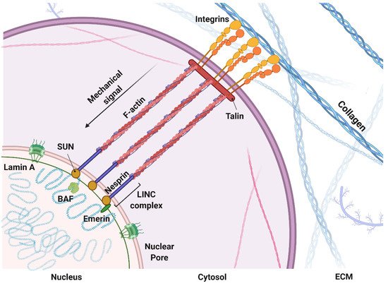
  22. Kirby, T.; Lammerding, J. Emerging views of the nucleus as a cellular mechanosensor. Nature 2018, 20, 373–381.
  23. Fedorchak, G.R.; Kaminski, A.; Lammerding, J. Cellular mechanosensing: Getting to the nucleus of it all. Prog. Biophys. Mol. Biol. 2014, 115, 76–92.
  24. Enyedi, B.; Niethammer, P. A case for the nuclear membrane as a mechanotransducer. Cell. Mol. Bioeng. 2016, 9, 247–251.
  25. Alam, S.; Lovett, D.B.; Dickinson, R.B.; Roux, K.J.; Lele, T.P. Nuclear forces and cell mechanosensing. Prog. Mol. Biol. Transl. Sci. 2014, 126, 205–215.
  26. Wang, N.; Tytell, J.D.; Ingber, D.E. Mechanotransduction at a distance: Mechanically coupling the extracellular matrix with the nucleus. Nat. Rev. Mol. Cell Biol. 2009, 10, 75–82.
  27. Hampoelz, B.; Azou-Gros, Y.; Fabre, R.; Markova, O.; Puech, P.-H.; Lecuit, T. Microtubule-Induced nuclear envelope fluctuations control chromatin dynamics in Drosophila embryos. Development 2011, 138, 3377–3386.
  28. Li, Y.; Chu, J.S.; Kurpinski, K.; Li, X.; Bautista, D.M.; Yang, L.; Sung, K.-L.P.; Li, S. Biophysical regulation of histone acetylation in mesenchymal stem cells. Biophys. J. 2011, 100, 1902–1909.
  29. Cho, S.; Irianto, J.; Discher, D.E. Mechanosensing by the nucleus: From pathways to scaling relationships. J. Cell Biol. 2017, 216, 305–315.
  30. Riehl, B.D.; Kim, E.; Bouzid, T.; Lim, J.Y. The role of microenvironmental cues and mechanical loading milieus in breast cancer cell progression and metastasis. Front. Bioeng. Biotechnol. 2021, 8, 1571.
  31. Alcoser, T.A.; Bordeleau, F.; Carey, S.P.; Lampi, M.C.; Kowal, D.R.; Somasegar, S.; Varma, S.; Shin, S.J.; Reinhart-King, C.A. Probing the biophysical properties of primary breast tumor-derived fibroblasts. Cell. Mol. Bioeng. 2014, 8, 76–85.
  32. Azadi, S.; Shadpour, M.T.; Warkiani, M.E. Characterizing the effect of substrate stiffness on the extravasation potential of breast cancer cells using a 3D microfluidic model. Biotechnol. Bioeng. 2020, 118, 823–835.
  33. Vitale, C.; Fedi, A.; Marrella, A.; Varani, G.; Fato, M.; Scaglione, S. 3D perfusable hydrogel recapitulating the cancer dynamic environment to In Vitro investigate metastatic colonization. Polymers 2020, 12, 2467.
  34. Pasini, A.; Lovecchio, J.; Cortesi, M.; Liverani, C.; Spadazzi, C.; Mercatali, L.; Ibrahim, T.; Giordano, E. Perfusion flow enhances viability and migratory phenotype in 3D-cultured breast cancer cells. Ann. Biomed. Eng. 2021, 1–11.
  35. Bascetin, R.; Laurent-Issartel, C.; Blanc-Fournier, C.; Vendrely, C.; Kellouche, S.; Carreiras, F.; Gallet, O.; Leroy-Dudal, J. A biomimetic model of 3D fluid extracellular macromolecular crowding microenvironment fine-tunes ovarian cancer cells dissemination phenotype. Biomaterials 2020, 269, 120610.
  36. Deszcz, I.; Lis-Nawara, A.; Grelewski, P.; Dragan, S.; Bar, J. Utility of direct 3D co-culture model for chondrogenic differentiation of mesenchymal stem cells on hyaluronan scaffold (Hyaff-11). Regen. Biomater. 2020, 7, 543–552.
  37. Gogoi, M.; Tewari, R.P. 3D printed chitosan composite scaffold for chondrocytes differentiation. Curr. Med. Imaging 2021, 17, 832–842.
  38. Norouzi, M.; Nazari, B.; Miller, D.W. Injectable hydrogel-based drug delivery systems for local cancer therapy. Drug Discov. Today 2016, 21, 1835–1849.
  39. Nii, T.; Makino, K.; Tabata, Y. A cancer invasion model combined with cancer-associated fibroblasts aggregates incorporating gelatin hydrogel microspheres containing a p53 inhibitor. Tissue Eng. Part C Methods 2019, 25, 711–720.
  40. Cao, H.; Lee, M.K.H.; Yang, H.; Sze, S.K.; Tan, N.S.; Tay, C.Y. Mechanoregulation of cancer-associated fibroblast phenotype in three-dimensional interpenetrating hydrogel networks. Langmuir 2018, 35, 7487–7495.
  41. Jensen, C.; Teng, Y. Is it time to start transitioning from 2D to 3D cell culture? Front. Mol. Biosci. 2020, 7, 33.
  42. Brock, E.J.; Ji, K.; Shah, S.; Mattingly, R.R.; Sloane, B.F. In Vitro models for studying invasive transitions of ductal carcinoma In Situ. J. Mammary Gland. Biol. Neoplasia 2018, 24, 1–15.
  43. Lintz, M.; Muñoz, A.; Reinhart-King, C.A. The mechanics of single cell and collective migration of tumor cells. J. Biomech. Eng. 2017, 139, 021005–0210059.
  44. Friedl, P.; Bröcker, E.B. The biology of cell locomotion within three-dimensional extracellular matrix. Cell Mol. Life Sci. 2000, 57, 41–64.
  45. Liu, C.; Mejia, D.L.; Chiang, B.; Luker, K.E.; Luker, G.D. Hybrid collagen alginate hydrogel as a platform for 3D tumor spheroid invasion. Acta Biomater. 2018, 75, 213–225.
  46. Kar, S.; Molla, M.S.; Katti, D.R.; Katti, K.S. Tissue-engineered nanoclay-based 3D in vitro breast cancer model for studying breast cancer metastasis to bone. J. Tissue Eng. Regen. Med. 2018, 13, 119–130.
  47. Holen, I.; Nutter, F.; Wilkinson, J.M.; Evans, C.A.; Avgoustou, P.; Ottewell, P.D. Human breast cancer bone metastasis In Vitro and In Vivo: A novel 3D model system for studies of tumour cell-bone cell interactions. Clin. Exp. Metastasis 2015, 32, 689–702.
  48. Devarasetty, M.; Dominijanni, A.; Herberg, S.; Shelkey, E.; Skardal, A.; Soker, S. Simulating the human colorectal cancer microenvironment in 3D tumor-stroma co-cultures In Vitro and In Vivo. Sci. Rep. 2020, 10, 9832.
  49. Chhetri, A.; Chittiboyina, S.; Atrian, F.; Bai, Y.; Delisi, D.A.; Rahimi, R.; Garner, J.; Efremov, Y.; Park, K.; Talhouk, R.; et al. Cell culture and coculture for oncological research in appropriate microenvironments. Curr. Protoc. Chem. Biol. 2019, 11, e65.
  50. Shimojo, A.A.M.; Rodrigues, I.C.P.; Perez, A.G.M.; Souto, E.M.B.; Gabriel, L.P.; Webster, T. Scaffolds for Tissue Engineering: A State-of-the-Art Review Concerning Types, Properties, Materials, Processing, and Characterization. In Racing for the Surface: Antimicrobial and Interface Tissue Engineering; Li, B., Blanco, I., Eds.; Springer International Publishing: Berlin/Heidelberg, Germany, 2020; pp. 647–676.
  51. Qiao, H.; Tang, T. Engineering 3D approaches to model the dynamic microenvironments of cancer bone metastasis. Bone Res. 2018, 6, 3.
  52. Díaz, E.C.G.; Sinha, S.; Avedian, R.S.; Yang, F. Tissue-engineered 3D models for elucidating primary and metastatic bone cancer progression. Acta Biomater. 2019, 99, 18–32.
  53. Liaw, C.Y.; Ji, S.; Guvendiren, M. Engineering 3D hydrogels for personalized In Vitro human tissue models. Adv. Healthc. Mater. 2018, 7, 1701165.
  54. García-Gareta, E.; Abduldaiem, Y.; Sawadkar, P.; Kyriakidis, C.; Lali, F.; Greco, K.V. Decellularised scaffolds: Just a framework? Current knowledge and future directions. J. Tissue Eng. 2020, 11, 2041731420942903.
  55. Hoshiba, T. Decellularized extracellular matrix for cancer research. Materials 2019, 12, 1311.
  56. Jin, Q.; Liu, G.; Li, S.; Yuan, H.; Yun, Z.; Zhang, W.; Zhang, S.; Dai, Y.; Ma, Y. Decellularized breast matrix as bioactive microenvironment for In Vitro three-dimensional cancer culture. J. Cell. Physiol. 2018, 234, 3425–3435.
  57. Mohammadkarim, A.; Mokhtari-Dizaji, M.; Kazemian, A.; Saberi, H.; Khani, M.M.; Bakhsh, M. The mechanical characteristics of human endothelial cells in response to single ionizing radiation doses by using micropipette aspiration technique. Mol. Cell. Biomech. 2019, 16, 275–287.
  58. González-Bermúdez, B.; Guinea, G.V.; Plaza, G.R. Advances in micropipette aspiration: Applications in cell biomechanics, models, and extended studies. Biophys. J. 2019, 116, 587–594.
  59. Hogan, B.; Babataheri, A.; Hwang, Y.; Barakat, A.I.; Husson, J. Characterizing cell adhesion by using micropipette aspiration. Biophys. J. 2015, 109, 209–219.
  60. Yousafzai, M.S.; Coceano, G.; Bonin, S.; Niemela, J.; Scoles, G.; Cojoc, D. Investigating the effect of cell substrate on cancer cell stiffness by optical tweezers. J. Biomech. 2017, 60, 266–269.
  61. Huang, H.; Dai, C.; Shen, H.; Gu, M.; Wang, Y.; Liu, J.; Chen, L.; Sun, L. Recent advances on the model, measurement technique, and application of single cell mechanics. Int. J. Mol. Sci. 2020, 21, 6248.
  62. Kim, H.; Yamagishi, A.; Imaizumi, M.; Onomura, Y.; Nagasaki, A.; Miyagi, Y.; Okada, T.; Nakamura, C. Quantitative measurements of intercellular adhesion between a macrophage and cancer cells using a cup-attached AFM chip. Colloids Surf. B Biointerfaces 2017, 155, 366–372.
  63. Hayashi, K.; Iwata, M. Stiffness of cancer cells measured with an AFM indentation method. J. Mech. Behav. Biomed. Mater. 2015, 49, 105–111.
  64. Luo, Q.; Kuang, D.; Zhang, B.; Song, G. Cell stiffness determined by atomic force microscopy and its correlation with cell motility. Biochim. Biophys. Acta-Gen. Subj. 2016, 1860, 1953–1960.
  65. Xu, W.; Mezencev, R.; Kim, B.; Wang, L.; McDonald, J.F.; Sulchek, T. Cell stiffness is a biomarker of the metastatic potential of ovarian cancer cells. PLoS ONE 2012, 7, e46609.
  66. Li, M.; Xi, N.; Wang, Y.-C.; Liu, L.-Q. Atomic force microscopy for revealing micro/nanoscale mechanics in tumor metastasis: From single cells to microenvironmental cues. Acta Pharmacol. Sin. 2020, 42, 323–339.
  67. Taubenberger, A.V.; Girardo, S.; Träber, N.; Fischer-Friedrich, E.; Kräter, M.; Wagner, K.; Kurth, T.; Richter, I.; Haller, B.; Binner, M.; et al. 3D Microenvironment stiffness regulates tumor spheroid growth and mechanics via p21 and ROCK. Adv. Biosyst. 2019, 3, 1900128.
  68. Glentis, A.; Oertle, P.; Mariani, P.; Chikina, A.; Marjou, F.E.; Attieh, Y.; Zaccarini, F.; Lae, M.; Loew, D.; Dingli, F.; et al. Cancer-associated fibroblasts induce metalloprotease-independent cancer cell invasion of the basement membrane. Nat. Commun. 2017, 8, 924.
  69. Picout, D.R.; Ross-Murphy, S.B. Rheology of biopolymer solutions and gels. Sci. World J. 2003, 3, 105–121.
  70. Janmey, P.A.; Schliwa, M. Rheology. Curr. Biol. 2008, 18, R639–R641.
  71. Mathieu, S.; Manneville, J.-B. Intracellular mechanics: Connecting rheology and mechanotransduction. Curr. Opin. Cell Biol. 2018, 56, 34–44.
  72. Andrikakou, P.; Vickraman, K.; Arora, H. On the behaviour of lung tissue under tension and compression. Sci. Rep. 2016, 6, 36642.
  73. Park, S.; Ateshian, G.A. Dynamic response of immature bovine articular cartilage in tension and compression, and nonlinear viscoelastic modeling of the tensile response. J. Biomech. Eng. 2006, 128, 623–630.
  74. Frank, E.H.; Jin, M.; Loening, A.; Levenston, M.; Grodzinsky, A.J. A versatile shear and compression apparatus for mechanical stimulation of tissue culture explants. J. Biomech. 2000, 33, 1523–1527.
  75. Deptuła, P.; Łysik, D.; Pogoda, K.; Cieśluk, M.; Namiot, A.; Mystkowska, J.; Król, G.; Głuszek, S.; Janmey, P.A.; Bucki, R. Tissue rheology as a possible complementary procedure to advance histological diagnosis of colon cancer. ACS Biomater. Sci. Eng. 2020, 6, 5620–5631.
  76. Sutherland, R.M.; McCredie, J.A.; Inch, W.R. Growth of multicell spheroids in tissue culture as a model of nodular carcinomas2. J. Natl. Cancer Inst. 1971, 46, 113–120.
  77. Alemany-Ribes, M.; Semino, C.E. Bioengineering 3D environments for cancer models. Adv. Drug Deliv. Rev. 2014, 79–80, 40–49.
  78. Huh, D.; Hamilton, G.A.; Ingber, D.E. From 3D cell culture to organs-on-chips. Trends Cell Biol. 2011, 21, 745–754.
  79. Roy, V.; Magne, B.; Vaillancourt-Audet, M.; Blais, M.; Chabaud, S.; Grammond, E.; Piquet, L.; Fradette, J.; Laverdière, I.; Moulin, V.J.; et al. Human organ-specific 3D cancer models produced by the stromal self-assembly method of tissue engineering for the study of solid tumors. BioMed Res. Int. 2020, 2020, 6051210.
  80. Pati, F.; Gantelius, J.; Svahn, H.A. 3D bioprinting of tissue/organ models. Angew. Chem. Int. Ed. 2016, 55, 4650–4665.
  81. Alonso-Alconada, L.; de la Fuente, A.; Santacana, M.; Ferreiros, A.; Lopez-Lopez, R.; Matias-Guiu, X.; Abal, M. Biomimetic device and foreign body reaction cooperate for efficient tumour cell capture in murine advanced ovarian cancer. Dis. Model. Mech. 2020, 13, dmm043653.
  82. Fong, E.L.S.; Toh, T.B.; Yu, H.; Chow, E. 3D culture as a clinically relevant model for personalized medicine. SLAS Technol. Transl. Life Sci. Innov. 2017, 22, 245–253.
  83. Sensi, F.; D’Angelo, E.; Piccoli, M.; Pavan, P.; Mastrotto, F.; Caliceti, P.; Biccari, A.; Corallo, D.; Urbani, L.; Fassan, M.; et al. Recellularized colorectal cancer patient-derived scaffolds as In Vitro pre-clinical 3D model for drug screening. Cancers 2020, 12, 681.
  84. D’Angelo, E.; Natarajan, D.; Sensi, F.; Ajayi, O.; Fassan, M.; Mammano, E.; Pilati, P.; Pavan, P.; Bresolin, S.; Preziosi, M.; et al. Patient-Derived scaffolds of colorectal cancer metastases as an organotypic 3D model of the liver metastatic microenvironment. Cancers 2020, 12, 364.
  85. Piccoli, M.; D’Angelo, E.; Crotti, S.; Sensi, F.; Urbani, L.; Maghin, E.; Burns, A.; De Coppi, P.; Fassan, M.; Rugge, M.; et al. Decellularized colorectal cancer matrix as bioactive microenvironment for in vitro 3D cancer research. J. Cell. Physiol. 2017, 233, 5937–5948.
  86. Agarwal, T.; Rustagi, A.; Das, J.; Maiti, T.K. PAMAM dendrimer grafted cellulose paper scaffolds as a novel in vitro 3D liver model for drug screening applications. Colloids Surf. B Biointerfaces 2018, 172, 346–354.
  87. Candini, O.; Grisendi, G.; Foppiani, E.M.; Brogli, M.; Aramini, B.; Masciale, V.; Spano, C.; Petrachi, T.; Veronesi, E.; Conte, P.; et al. A novel 3D In Vitro platform for pre-clinical investigations in drug testing, gene therapy, and immuno-oncology. Sci. Rep. 2019, 9, 7154.
  88. Brancato, V.; Oliveira, J.M.; Correlo, V.M.; Reis, R.L.; Kundu, S.C. Could 3D models of cancer enhance drug screening? Biomaterials 2020, 232, 119744.
  89. Booij, T.H.; Price, L.S.; Danen, E.H.J. 3D cell-based assays for drug screens: Challenges in imaging, image analysis, and high-content analysis. SLAS Discov. Adv. Life Sci. R&D 2019, 24, 615–627.
  90. Augustine, R.; Kalva, S.N.; Ahmad, R.; Zahid, A.A.; Hasan, S.; Nayeem, A.; McClements, L.; Hasan, A. 3D Bioprinted cancer models: Revolutionizing personalized cancer therapy. Transl. Oncol. 2021, 14, 101015.
  91. Van Zundert, I.; Fortuni, B.; Rocha, S. From 2D to 3D cancer cell models—The enigmas of drug delivery research. Nanomaterials 2020, 10, 2236.
  92. Nii, T.; Makino, K.; Tabata, Y. Three-Dimensional culture system of cancer cells combined with biomaterials for drug screening. Cancers 2020, 12, 2754.
  93. Yang, M.; Li, J.; Gu, P.; Fan, X. The application of nanoparticles in cancer immunotherapy: Targeting tumor microenvironment. Bioact. Mater. 2020, 6, 1973–1987.
  94. Nii, T.; Katayama, Y. Biomaterial-Assisted regenerative medicine. Int. J. Mol. Sci. 2021, 22, 8657.
More
Information
Contributor MDPI registered users' name will be linked to their SciProfiles pages. To register with us, please refer to https://encyclopedia.pub/register :
View Times: 436
Entry Collection: Environmental Sciences
Revisions: 2 times (View History)
Update Date: 22 Nov 2021
1000/1000