Submitted Successfully!
To reward your contribution, here is a gift for you: A free trial for our video production service.
Thank you for your contribution! You can also upload a video entry or images related to this topic.
Version Summary Created by Modification Content Size Created at Operation
1 + 3548 word(s) 3548 2021-11-08 04:46:50 |
2 Improved the description of ischemia in the definition section. + 2 word(s) 3550 2021-11-09 20:52:53 | |
3 format correct Meta information modification 3550 2021-11-10 04:42:09 |

Video Upload Options

Do you have a full video?

Confirm

Are you sure to Delete?
Cite
If you have any further questions, please contact Encyclopedia Editorial Office.
Hamon, M.; Hauser, P. Bioprinting Scaffolds for Vascular Tissues. Encyclopedia. Available online: https://encyclopedia.pub/entry/15800 (accessed on 19 April 2024).
Hamon M, Hauser P. Bioprinting Scaffolds for Vascular Tissues. Encyclopedia. Available at: https://encyclopedia.pub/entry/15800. Accessed April 19, 2024.
Hamon, Morgan, Peter Hauser. "Bioprinting Scaffolds for Vascular Tissues" Encyclopedia, https://encyclopedia.pub/entry/15800 (accessed April 19, 2024).
Hamon, M., & Hauser, P. (2021, November 08). Bioprinting Scaffolds for Vascular Tissues. In Encyclopedia. https://encyclopedia.pub/entry/15800
Hamon, Morgan and Peter Hauser. "Bioprinting Scaffolds for Vascular Tissues." Encyclopedia. Web. 08 November, 2021.
Bioprinting Scaffolds for Vascular Tissues
Edit

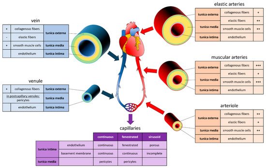
Human organ function and physiology depend on a functional vascular system to facilitate oxygen and nutrient supply, as well as the removal of metabolic products. Ischemia is temporary reduction of blood supply that can cause physiological imbalance due to a lack of oxygen (hypoxia), nutrients, and a failure to eliminate metabolic waste products. Prolonged ischemia is associate with tissue damage and potentially necrosis. In this context, avoiding ischemia time remains critical to preventing hypoxic injury and potential damages to transplant tissues and organs. Despite substantial progress in creating three-dimensional (3D) blood vessels, fabricating a functional vascular multiscale system has remained a challenge . Many techniques have been developed to fabricate vascular networks that can mimic the complexity, the unique structures, and the functionality of human blood vessels.  Among these advancements, 3D bioprinting has become an essential tool for the fabrication of vascularized bioconstructs due to improved control over vascular growth, reproducibility, and scalability of the fabrication process.

vascularization 3D printing bioprinting

1. Vascular System

To fabricate blood vessels and vascularized tissue in vitro, it is critical to understand the biological, physiological, and functional aspects of the in vivo system. The peripheral vascular system (PVS) includes all the blood vessels exiting the heart. Blood vessels are classified in arteries, capillaries, or veins. Arteries transport the blood from the heart to the periphery. They branch into smaller and smaller arteries until they branch into arterioles, metarterioles, and capillaries, the smallest and most numerous blood vessels. The primary function of capillaries is to accommodate the exchange of nutrients, gases, and bi-products between the blood and the cells. They form the connection between arteries and veins, which transport the blood back towards the heart. Capillaries connect to venules merging into increasingly larger veins. It is worth noting that large segments of the PVS are identified as separated anatomical structures while microvasculature (arterioles, metarterioles, capillaries, and venules) are parts of the tissue they vascularized [1][2].
The vessel's structure varies according to the diameter of the vessels, which, in turn, is adapted to the hydrostatic pressure present in its interior (Figure 1). Aside from capillaries, blood vessels are composed of three concentric layers, starting from the lumen they are: the tunica intima, the tunica media, and the adventitia. The tunica intima is composed of a monolayer of endothelial cells (EC) that repose on a basement membrane composed of type IV collagen and laminin. The outer layer of the tunica intima is composed of an elastin layer, also known as the internal elastic lamina. The functions of the tunica intima are to contain cells and fluid within the vessel’s lumen and allow the blood to flow without problems. The tunica media is the middle layer of arteries and veins. It is primarily composed of smooth muscle cells (SMCs), collagen type I and type III, and variable amounts of elastic fibers, depending on the vessel thickness. It supports blood vessels and changes their diameter to regulate blood flow and blood pressure [3]. The external elastic lamina separates the tunica media from the adventitia. The outer layer, the adventitia, is composed of a loose connective tissue of type I collagen and fibroblasts. Its primary function is to restrain the vessel from excessive extension and recoil. These three layers are present in the macro- and micro-vasculature of arteries and veins, except for the capillaries where SMCs are absent and replaced by pericytes. The thickness of the layers is inversely correlated with the degree of branching.
Figure 1. Composition of Vessels in the Cardiovascular System. Graphic and text inserts represent the composition of arteries (red), veins (blue) and capillaries (purple). For each section, the tunica externa (green), the tunica media (orange), and the tunica intima (grey) are indicated, as well as the proportion in collagenous fibers, elastic fibers, smooth muscle cells, and endothelium, ranging from lowest (-) to highest (+++) proportion.
Systemic arteries can be divided into two types, i.e., elastic and muscular, according to their tunica media’s relative composition in elastin and SMCs [4]. Arteries with close proximity to the heart, like the aorta and the pulmonary arteries, are very elastic. Their tunica media contains more elastin than SMCs. This characteristic allows them to maintain a constant pressure gradient, despite the periodic changes in cardiac flow volume. Muscular arteries are medium-sized arteries (e.g., femoral arteries, brachial arteries, radial arteries) and contain more SMCs than elastin in the tunica media. This allows them to contract and expand to adjust their diameter according to the peripheral blood demand, thereby controlling the blood flow in the capillaries. Capillaries are thin-wall vessels with lumen diameters of about 5 to 10 µm. Their thin wall, composed of a single endothelium layer, allows the exchange of nutrients, metabolites, and gases primarily via diffusion. In addition, differences observed in the capillary structure allow different solute permeability in different organs. Accordingly, capillaries are classified as continuous, fenestrated, and sinusoid capillaries [4]. Continuous capillaries have a continuous basement membrane, and their cells are connected by tight cell junctions. They are found in muscles, nervous systems, lungs, and skin [5]. Fenestrated capillaries receive their name from window-like transcellular openings, that generate a mesh-like structure. They are found in exocrine glands, renal glomeruli, and intestinal mucosa [5]. Finally, sinusoid capillaries have large interstitial gaps and an incomplete basement membrane. Sinusoid capillaries are found in the liver, spleen, and bone marrow [5]. Several capillaries join to form a venule. Venules have a diameter of 8 to 100 µm, with few SMCs, little elastin, and a thin adventitia. Venules join to form veins. Like arteries, veins are composed of three layers. However, veins are thin-walled, with a reduced tunica media and less elastic. This particularity allows the venous system to accommodate a large volume of blood (~3/4 of the circulating blood) at relatively low pressure. Unlike arteries, veins contain valves that prevent backflow and allow the blood to reach the heart [1].

2. Printing Methods

Tissue engineering aims to generate functional tissue by combining mechanical engineering, cell biology, robotic and material science. While generally, bioprinting aims to arrange cells in a 3D space according to the cellular and structural anatomy of a modeled tissue, different approaches have been developed to assemble cells to biosimilar tissues. For an overview of printing methods and their advantages and disadvantages, see Table 1. Unlike children’s’ building blocks, cells attach only in a limited fashion and commonly require adhesive components. The combination of cells and an adhesive or carrier material, usually a biopolymer, is called ‘bioink’.
Table 1. Advantages and Disadvantages of the Different 3D Bioprinting Methods.
Materials Application Printer Style Advantages Disadvantages
Extrusion-based Bioplotting wide range of bioinks slow printing process
  Fusion deposition modelling wide range of bioinks limited resolution
only hydrogels
slow
Laser-assisted Stereolithography high resolution possible UV damage to cells
small range of bioinks
Droplet-based Inkjet gentle to printed cells
fast printing
affordable
limitations of cell density
low resolution
Pre-seeding and post-seeding are the two main strategies used to arrange cells in a bioprinted structure. In pre-seeding approaches, cells and the matrix are printed simultaneously, while in post-seeding, a bioprinted matrix is generated and subsequently populated with cells and cultured. Pre-seeding allows better control of the cellular component, as cell can be distributed more homogenous, and it seems to enhance cell retention [6]. On the other hand, post-seeding enables the use of printing conditions that are not conducive to cell viability (heat, UV) and gradual or staged seeding, allowing layering of different cell populations. The technical solutions also differ in how the cells are held in the physical space and cultured to maturation. This review introduces the principles of (i) sacrificial bioprinting and its different methods, (ii) the core/shell method, (iii) and 4D bioprinting.

2.1. Sacrificial

Sacrificial bioprinting is a technique in which a three-dimensional negative or hollow space is generated within a hydrogel or scaffold. The method is intuitively applicable to create a vascular network within a layer or block of engineered tissue [7]. In a first step, the sacrificial degradable hydrogel is deposited to generate a path for a vascular network that can supply the engineered tissue. After that, a hydrogel, or bioink, containing the cells of the future engineered tissue is used to cover the sacrificial deposit. After polymerization, the sacrificial hydrogel is removed by different methods, such as chemical degradation [8], photolithography [9], temperature [10] or mechanical extraction [11]. Finally, endothelial cells are seeded into the hollow channels from which the sacrificial material has been removed, generating a vascular network within the cells embedded in the hydrogel block (Figure 2).
Figure 2. Principle of 3D Bioprinting Scaffold for Vascular Tissue Formation. (A) Sacrificial bioprinting. First step in sacrificial bioprinting is to deposit the sacrificial bioink or material onto a first layer of matrix. The sacrificial material (blue) is then covered with a second layer of matrix. Both matrices may be loaded with cells. After polymerization of the hydrogel, the sacrificial material is removed by chemical degradation, photolithography, temperature, or mechanical extraction, generating a hollow channel within the hydrogel block. In the last step, endothelial cells or endothelial progenitor cells are seeded into the hollow channel. (B) Droplet-based bioprinting. (C) Light- (Laser-)based bioprinting.
The basic method of sacrificial bioprinting can be combined with various strategies to deposit the sacrificial ink or material. Depending on the method used to deposit the sacrificial material, bioprinting is categorized into extrusion based, droplet-based, laser-assisted, and core/shell-based. The advantages and disadvantages of the technology make them more or less suitable for certain applications. Strategies aiming to bioprint a vascular network need to be adapted to the particular vascular system that is to be generated. Techniques that are sufficient to generate simple vessels consisting of a single cell type or layer might require a different printing approach than major blood vessels consisting of several layers and cell types (see Figure 1).

Extrusion-Assisted Techniques

Extrusion-based bioprinting is the most common variant in 3D printing. In extrusion-based bioprinting, a liquid and often viscous solution is extruded, that is pushed from a syringe through a nozzle, and deposited according to the printing layout. Extrusion printing is an economical and versatile method that enables printing of bioinks with high-cell numbers quickly. The force to extrude the bioink through the nozzle is commonly generated pneumatically [12], via a piston [13], or a helical screwblade [14]. Extrusion printing can be divided into direct extrusion, where the bioink is delivered directly from the syringe’s nozzle to the print bed, and indirect extrusion, in which bioink is deposited in combination with supportive material. The challenge for both methods is to create a bioink, with a viscosity low enough to allow printing while rigid enough to hold the shape after printing and being biocompatible for cells to be viable [15]. Extrusion bioprinting is not ideally suited to print blood vessels, as it is challenging to lumen containing tubular structures with multiple layers.
Direct deposition of the bioink, or the sacrificial bioink on the printing bed is a straightforward approach but creating larger or complex structures can be difficult. To overcome the challenges of collapsing or deformed structures, the sacrificial bioink can be printed onto a printing bed or into a carrier matrix that suspends the printed structure. Noor and coworkers used a suspended direct printing approach to create perfusable cardiac patches and hearts [16]. For their personalized medicine approach, they isolated cells from patients’ omentum and reprogrammed and differentiated them into cardiomyocytes and endothelial cells. From the extracellular matrix (ECM) of the omentum they generated a hydrogel, which, combined with cardiomyocytes or endothelial cells, was used to formulate a bioink. Suspended direct extrusion printing was then used to generate vascularized cardiac patches or whole heart-like structures that could be transplanted back to the patient [16]. Extrusion bioprinting is only partially useful for printing vessels, as printing bifurcated vessels might be challenging. As mentioned above, the hydrogel carrier’s physical characteristics of the bioink are of great importance in extrusion-based bioprinting. In addition, the hydrogel should promote the differentiation or maturation of the printed cells.
Leucht et al. studied different modified-gelatin formulations to bioprint a bone and optimize the vascularization of the printed structure [17]. Gelatin can be generated with high viscosity and exhibit strong physical rigidity, but it cannot be crosslinked. Modification of gelatin can increase its suitability for bioprinting. Methacrylation, for example, can improve the printability by reducing viscosity, while acetylation can increase crosslinking. Leucht measured the physical properties of different gelatin, methacrylate-modified gelatin, and acetylated gelatin methacrylate formulations. They encapsulated human dermal microvascular endothelial and adipose-derived stem cells and then analyzed the vascularization of the printed bone. They found that a formulation combining methacrylated gelatin, gelatin, and acetylated gelatin methacrylate was best suitable for generating a vascular network and allowed differentiation of adipose-derived stem cells. Crosstalk between endothelial cells and adipose-derived stem cells promoted the osteogenic differentiation of adipose-derived stem cells [17].
Freeman et al. developed a 3D rotary direct extrusion bioprinter with the purpose of printing vessels of small diameter [18]. Instead of a printing bed, they used a polystyrene rod with a diameter of 4.9 mm that rotated along its rolling axis. On the surface of the rod, they applied a helical layer of bioink, composed of low passage primary neonatal human dermal fibroblasts mixed with fibrinogen and gelatin type A. After printing, the rod was submerged in thrombin to crosslink the fibrinogen and subsequently cultured them for up to 45 days, during which the fibroblasts differentiated and changed their phenotype into a spindle shape. Removal of the rods was followed by a significant reduction of gel length and luminal diameter. On day 7, the thickness of the vessel was ~300 µm when printed with 1 × 10⁶ cells/mL and <125 µm when printed with 3 × 10⁶ cells/mL [18].
Indirect extrusion printing is a sacrificial bioprinting method in which a sacrificial structure is fabricated to support the generation of larger tissues or scaffolds and later removed. It enables the production of complex tissues and further can be adapted for pre- and post-seeding strategies. Recently, indirect extrusion bioprinting has been demonstrated for nerve tissue applications [19], the generation of customized bone-like structures [20], and adipose tissue engineering [21]. Very few articles describe the application of indirect extrusion bioprinting to generate vascular structures or vascularized tissues. However, Bertassoni and colleagues applied an indirect extrusion printing approach to produce a 3D microchannel network within a photo crosslinked hydrogel [22]. They were able to generate perfusable channels within star poly (ethylene glycol-co-lactide) acrylate (SPELA), gelatin methacrylate (GelMA), poly (ethylene glycol) diacrylate (PEGDA), and poly (ethylene glycol) dimethacrylate (PEGDMA) hydrogels. Seeding these channels with cells, they demonstrated osteogenic differentiation and the formation of endothelial monolayers.

2.2. Droplet-Based Bioprinting

Droplet-based Bioprinting (DBB) is a technique to deposit small droplets of biological material, including cells, growth factors, extracellular matrix components, or nucleic acids. It follows the principle of a typical office laser printer, where a small ink bubble is placed at a specific coordinate on the paper in two dimensions. The deposition of bioink bubbles on the top of another, generates the Z-axis of the 3D objects. Precise control over the printing pattern combined with its relative simplicity and the wide range of applications make DBB a very appealing printing technology. For example, DBB allows the fabrication of bifurcated tubular structures and thus enables bioprinting of vascular networks [23]. Currently, droplet sizes of 25 to 300 µm can be controlled [24]. While the DBB is not the first choice to fabricate large objects, the method can be adapted to print different cell types into one layer of a construct. Depending on the precise technology used to generate an ink bubble and deposit it, DBB can be grouped into: (i) inkjet bioprinting, (ii) acoustic-droplet ejection bioprinting, and (iii) microvalve bioprinting, with inkjet bioprinting being the most widely applied technology and with subgroups of his own [24].

Indirect Bioprinting Using DBB

Indirect droplet base bioprinting a blood vessel is generated by depositing endothelial cells in to create a hollow tube. Similar to indirect extrusion printing, indirect droplet-based bioprinting uses a sacrificial structure that is removed after printing is completed. In vascular bioprinting, the sacrificial structure creates the tubular lumen that can be seeded with endothelial cells.
Using an indirect microvalve DBB strategy, Lee et al. generated a perfusable model of a vascular channel. First, they printed a layer of collagen, and after its polymerization, they printed the vascular channel using a mixture of human umbilical vein endothelial cells (HUVECs) and gelatin in the ratio of 1:1. After gelation, the vascular channel was covered with another collagen layer. In a subsequent step, Lee liquified the gelatin at 37 °C and removed the material, thereby leaving a HUVEC seeded vascular channel in the collagen block. After 5 days of low-rate perfusion, the HUVEC had formed a polarized endothelial monolayer that expressed the endothelial junction protein VE-cadherin [25].

2.3. Light-Based Techniques

Originally designed to print electronic components, the method was adapted to print cells from the culture medium directly. Light base techniques can be divided into two separate categories two categories as digital light processing (DLP)-based and laser-based bioprinting [26].

Laser-Induced Forward Transfer

Laser-Induced Forward Transfer (LIFT) is a laser-assisted bioprinting (LAB) in which the bioink is placed at a specific location or substrate using a laser to generate a droplet. The technique has shown high precision and accuracy, enabling the fabrication of complex structures, using different cells and scaffold materials in various combinations. During the LIFT printing process, a stream of bionink is deposited on a metal film [27]. A pulse of energy in the range of 0.5–20 μJ generated by a nanosecond laser is then used to evaporate the bioink stream, thereby generating a droplet that is deposited on the printing bed. UV lasers with wavelengths of 193 nm, 248 nm, or near UV wavelength at 1064 nm have been implemented to create the necessary energy pulse.
Wu et al. followed a LIFT strategy to generate branched vascular structures. With a laser pulsed at 0.5–1.5 μJ/pulse they generated bioink droplets with a diameter of 50 μm and deposited them in 50–150 μm distance from each other to form a branching vascular tree [28]. The bioink was composed of HUVEC and MatrigelTM. As early as one day after printing, the printed HUVEC cells had acquired a stretched phenotype, showing connected HUVEC forming a branching structure. In addition to the printed lumen, the endothelial cells formed a self-assembled secondary continuous lumen. They further found that the addition of vascular endothelial growth factor (VEGF) was essential to protect the integrity of the HUVEC and the lumen.
LIFT technique allows printing of inorganic and organic compounds with high precision in the micrometer range. Guillotin et al. combined the human umbilical vein endothelial cell line Eahy 926 and the rabbit carcinoma cell line B16 and suspended the cells in Dulbecco’s Modified Eagle Medium that was supplemented with varying amounts of glycerol, and sodium alginate or with MatrigelTM, or thrombin [27]. They used LIFT to print the bioink to a network with microvasculature dimensions onto a fibrinogen sheet. Using different concentrations of alginate, the authors were able to control the droplet size and thus the diameter of the printed vasculature. Alginate further functioned as a substitute to the essential extracellular matrix. An important finding was that LAB is a statistical process and that bioinks with a low cell density are unable to generate a network of continuous cells.

2.4. Four-Dimensional (4D) Bioprinting

Common bioprinting is used to generate structures and arrange cells and biomolecules in the three-dimensional space. 4D bioprinting adds the factor time as the 4th dimension to the three spatial bioprinting dimensions. The term applies to bioprinted objects that, after a period of time, can change their functionality or shape as a result of cell-cell fusions or after outside stimulus [29]. For example, structures can be printed as flat objects, seeded with cells, and then rolled up or folded to a 3D shape. The stimulus to trigger the shape change can be a certain pH level, light exposure, the addition of a chemical catalyst, or a combination of similar stimuli [30]. Changing the shape of a bioprinted structure can help to overcome the limitations of regular 3D printing.
Using a 4D printing approach, Kirrilova et al. were able to generate tubes with only 20 µm diameter, which is smaller than any current conventional 3D printing technique can generate [31]. To generate a tube, they printed a layer of mouse bone marrow stromal cells suspended in a hydrogel of methacrylated alginate and hyaluronic acid flat on a glass slide. In a second step, they crosslinked the polymer with green light. Sensitive to Ca2+ ions, the polymer sheets changed their shape and rolled up to generate a tubular structure after being placed in solution. The possibility of generating tubular structures with small diameters renders this technique ideal to generate small vascular structures.
The group of Nakayama developed a unique strategy that allows scaffold-free 3D bioprinting [32]. Instead of a standard printing bed, where the bioink is deposited, this method uses a printing bed that consists of a paralleled array of needles, or ‘Kenzan’, with a diameter of 150 µm. A robotic arm then arranges cell-spheroids in a 3D position in the needle array. After the printing step, the spheroids will fuse, and the printed construct can be removed from the needle array [32]. The Kenzan method has been used to create scaffold-free aortas using spheroids. The printed aorta was then implanted in rat, and tissue remodeling and endothelialization were observed after five days [33]. The advantage of this method is the possibility of generating scaffold- or matrix-free tissue, which could help minimize immune rejection. On the other hand, the method is limited to printing larger constructs due to the size of the needles (150 µm) and the necessary spheroid diameter (500 µm).

References

  1. Levick, J.R. An Introduction to Cardiovascular Physiology; Butterworth-Heinemann: London, UK, 1991; pp. 1–12.
  2. Pugsleya, M.K.; Tabrizchib, R. The vascular system—An overview of structure and function. J. Pharmacol. Toxicol. Methods 2000, 44, 333–340.
  3. Berillis, P. The Role of Collagen in the Aorta’s Structure. Open Circ. Vasc. J. 2013, 6, 1–8.
  4. Tucker, W.D.; Arora, Y.; Mahajan, K. Anatomy, Blood Vessels. In StatPearls; StatPearls Publishing: Treasure Island, FL, USA, 2021.
  5. Okada, H.; Takemura, G.; Suzuki, K.; Oda, K.; Takada, C.; Hotta, Y.; Nagisa, M.; Akiko, T.; Isamu, M.; Yoshiaki, A.; et al. Three-dimensional ultrastructure of capillary endothelial glycocalyx under normal and experimental endotoxemic conditions. Crit. Care 2017, 21, 261.
  6. Zamani, Y.; Mohammadi, J.; Amoabediny, G.; Helder, M.N.; Zandhie-Doulabi, B.; Klein-Nulend, J. Bioprinting of Alginate-Encapsulated Pre-osteoblasts in PLGA/β-TCP Scaffolds Enhances Cell Retention but Impairs Osteogenic Differentiation Compared to Cell Seeding after 3D-Printing. Regen. Eng. Transl. Med. 2020.
  7. Zhang, Y.S.; Oklu, R.; Dokmeci, M.R.; Khademhosseini, A. Three-Dimensional Bioprinting Strategies for Tissue Engineering. Cold Spring Harb. Perspect. Med. 2018, 8, a025718.
  8. Thomas, A.; Orellano, I.; Lam, T.; Noichl, B.; Geiger, M.A.; Amler, A.K.; Kreuder, A.E.; Palmer, C.; Duda, G.; Lauster, R.; et al. Vascular bioprinting with enzymatically degradable bioinks via multi-material projection-based stereolithography. Acta Biomater. 2020, 117, 121–132.
  9. Burdick, J.A.; Chung, C.; Jia, X.; Randolph, M.A.; Langer, R. Controlled degradation and mechanical behavior of photopolymerized hyaluronic acid networks. Biomacromolecules 2005, 6, 386–391.
  10. Liao, C.Y.; Wu, W.J.; Hsieh, C.T.; Yang, H.C.; Tseng, C.S.; Hsu, S.H. Water/ice as sprayable sacrificial materials in low-temperature 3D printing for biomedical applications. Mater. Des. 2018, 60, 624–635.
  11. Zhou, Y. The Application of Ultrasound in 3D Bio-Printing. Molecules 2016, 21, 590.
  12. Rasheed, A.; Azizi, L.; Turkki, P.; Janka, M.; Hytönen, V.P.; Tuukkanen, S. Extrusion-Based Bioprinting of Multilayered Nanocellulose Constructs for Cell Cultivation Using In SituFreezing and Preprint CaCl2 Cross-Linking. ACS Omega 2020, 6, 569–578.
  13. Fisch, P.; Holub, M.; Zenobi-Wong, M. Improved accuracy and precision of bioprinting through progressive cavity pump-controlled extrusion. Biofabrication 2021, 13, 015012.
  14. Justino Netto, J.M.; Idogava, H.T.; Frezzatto Santos, L.E.; de Castro Silveira, Z.; Romio, P.; Lino Alves, J. Screw-assisted 3D printing with granulated materials: A systematic review. Int. J. Adv. Manuf. Technol. 2021, 115, 2711–2727.
  15. Cooke, M.E.; Rosenzweig, D.H. The Rheology of Direct and Suspended Extrusion Bioprinting. APL Bioeng. 2021, 5, 011502.
  16. Noor, N.; Shapira, A.; Edri, R.; Gal, I.; Wertheim, L.; Dvir, T. 3D Printing of Personalized Thick and Perfusable Cardiac Patches and Hearts. Adv. Sci. 2019, 6, 1900344.
  17. Leucht, A.; Volz, A.-C.; Rogal, J.; Borchers, K.; Kluger, P.J. Advanced Gelatin-Based Vascularization Bioinks for Extrusion-Based Bioprinting of Vascularized Bone Equivalents. Sci. Rep. 2020, 10, 5330.
  18. Freeman, S.; Ramos, R.; Alexis Chando, P.; Zhou, L.; Reeser, K.; Jin, S.; Soman, P.; Ye, K. A Bioink Blend for Rotary 3D Bioprinting Tissue Engineered Small-Diameter Vascular Constructs. Acta Biomater. 2019, 95, 152–164.
  19. Naghieh, S.; Sarker, M.D.; Abelseth, E.; Chen, X. Indirect 3D Bioprinting and Characterization of Alginate Scaffolds for Potential Nerve Tissue Engineering Applications. J. Mech. Behav. Biomed. Mater. 2019, 93, 183–193.
  20. Lee, J.-Y.; Choi, B.; Wu, B.; Lee, M. Customized Biomimetic Scaffolds Created by Indirect Three-Dimensional Printing for Tissue Engineering. Biofabrication 2013, 5, 045003.
  21. Contessi Negrini, N.; Celikkin, N.; Tarsini, P.; Farè, S.; Święszkowski, W. Three-Dimensional Printing of Chemically Crosslinked Gelatin Hydrogels for Adipose Tissue Engineering. Biofabrication 2020, 12, 025001.
  22. Bertassoni, L.E.; Cecconi, M.; Manoharan, V.; Nikkhah, M.; Hjortnaes, J.; Cristino, A.L.; Barabaschi, G.; Demarchi, D.; Dokmeci, M.R.; Yang, Y.; et al. Hydrogel Bioprinted Microchannel Networks for Vascularization of Tissue Engineering Constructs. Lab Chip 2014, 14, 2202–2211.
  23. Mae, H.J.; Delgadillo, L.; Wan, J. On-demand modulation of 3D-printed elastomers using programmable droplet inclusions. Proc. Natl. Acad. Sci. USA 2020, 117, 14790–14797.
  24. Gudapati, H.; Dey, M.; Ozbolat, I. A Comprehensive Review on Droplet-Based Bioprinting: Past, Present and Future. Biomaterials 2016, 102, 20–42.
  25. Lee, V.K.; Kim, D.Y.; Ngo, H.; Lee, Y.; Seo, L.; Yoo, S.-S.; Vincent, P.A.; Dai, G. Creating Perfused Functional Vascular Channels Using 3D Bio-Printing Technology. Biomaterials 2014, 35, 8092–8102.
  26. Zhu, W.; Ma, X.; Gou, M.; Mei, D.; Zhang, K.; Chen, S. 3D Printing of Functional Biomaterials for Tissue Engineering. Curr. Opin. Biotechnol. 2016, 40, 103–112.
  27. Guillemot, F.; Souquet, A.; Catros, S.; Guillotin, B. Laser-Assisted Cell Printing: Principle, Physical Parameters versus Cell Fate and Perspectives in Tissue Engineering. Nanomedicine 2010, 5, 507–515.
  28. Wu, P.K.; Ringeisen, B.R. Development of Human Umbilical Vein Endothelial Cell (HUVEC) and Human Umbilical Vein Smooth Muscle Cell (HUVSMC) Branch/Stem Structures on Hydrogel Layers via Biological Laser Printing (BioLP). Biofabrication 2010, 2, 014111.
  29. Morouço, P.; Azimi, B.; Milazzo, M.; Mokhtari, F.; Fernandes, C.; Reis, D.; Danti, S. Four-Dimensional (Bio-)Printing: A Review on Stimuli-Responsive Mechanisms and Their Biomedical Suitability. Appl. Sci. 2020, 10, 9143.
  30. Li, Y.-C.; Zhang, Y.S.; Akpek, A.; Shin, S.R.; Khademhosseini, A. 4D Bioprinting: The next-Generation Technology for Biofabrication Enabled by Stimuli-Responsive Materials. Biofabrication 2016, 9, 012001.
  31. Kirillova, A.; Maxson, R.; Stoychev, G.; Gomillion, C.T.; Ionov, L. 4D Biofabrication Using Shape-Morphing Hydrogels. Adv. Mater. 2017, 29, 1703443.
  32. Moldovan, N.I.; Hibino, N.; Nakayama, K. Principles of the Kenzan Method for Robotic Cell Spheroid-Based Three-Dimensional Bioprinting. Tissue Eng. Part B Rev. 2017, 23, 237–244.
  33. Itoh, M.; Nakayama, K.; Noguchi, R.; Kamohara, K.; Furukawa, K.; Uchihashi, K.; Toda, S.; Oyama, J.-I.; Node, K.; Morita, S. Scaffold-Free Tubular Tissues Created by a Bio-3D Printer Undergo Remodeling and Endothelialization When Implanted in Rat Aortae. PLoS ONE 2015, 10, e0136681.
More
Information
Contributors MDPI registered users' name will be linked to their SciProfiles pages. To register with us, please refer to https://encyclopedia.pub/register : ,
View Times: 401
Revisions: 3 times (View History)
Update Date: 10 Nov 2021
1000/1000