Submitted Successfully!
To reward your contribution, here is a gift for you: A free trial for our video production service.
Thank you for your contribution! You can also upload a video entry or images related to this topic.
Version Summary Created by Modification Content Size Created at Operation
1 + 2641 word(s) 2641 2021-10-25 08:27:56 |
2 Done + 693 word(s) 3334 2021-10-26 04:08:28 |

Video Upload Options

Do you have a full video?

Confirm

Are you sure to Delete?
Cite
If you have any further questions, please contact Encyclopedia Editorial Office.
Granato, M. Nanotechnology in γ-Herpesviruses Treatments. Encyclopedia. Available online: https://encyclopedia.pub/entry/15387 (accessed on 16 April 2024).
Granato M. Nanotechnology in γ-Herpesviruses Treatments. Encyclopedia. Available at: https://encyclopedia.pub/entry/15387. Accessed April 16, 2024.
Granato, Marisa. "Nanotechnology in γ-Herpesviruses Treatments" Encyclopedia, https://encyclopedia.pub/entry/15387 (accessed April 16, 2024).
Granato, M. (2021, October 25). Nanotechnology in γ-Herpesviruses Treatments. In Encyclopedia. https://encyclopedia.pub/entry/15387
Granato, Marisa. "Nanotechnology in γ-Herpesviruses Treatments." Encyclopedia. Web. 25 October, 2021.
Nanotechnology in γ-Herpesviruses Treatments
Edit

Epstein–Barr Virus (EBV) and Kaposi’s sarcoma associated-herpesvirus (KSHV) are γ-herpesviruses that belong to the Herpesviridae family. In the last decade, many studies conducted by scientists and clinicians have indicated that nanotechnology and nanomedicine could improve the outcome of several treatments in γ-herpesvirus-associated diseases. 

γ-herpesviruses EBV Kaposi’s sarcoma associated-herpesvirus (KSHV) nanoparticles (NPs) Epstein–Barr

1. γ-Herpesviruses

Epstein–Barr Virus (EBV) and Kaposi’s sarcoma associated herpesvirus (KSHV) are γ-herpesviruses belonging to the Herpesviridae family [1]. They are enveloped viruses, with double stranded DNA (130–150 Kbps) restrained in icosahedral capsids, surrounded by envelopes with several glycoproteins (GPs), which is necessary during the recognition and infection of the host cells. EBV Gp350 exploits the binding and the attachment to CR2–CD21 B cell receptors to infect these kinds of cells [2]. The γ-herpesviruses acquire the primary envelope at the nuclear membrane by the maturation steps, the primary envelopment-de-envelopment steps of the nuclear inner and outer leaves. They are conserved in all herpesviruses, such as herpes simplex type 1 (HSV1). The nuclear membrane complex, called NEC, is able to ‘gain’ the nucleocapsids—to the cytoplasmatic compartment—in which the herpesviruses acquire the last envelope and the glycoproteins require new viral infections.

1.1. Epstein–Barr Virus (EBV)

EBV infects 95% of the worldwide population; it is usually asymptomatic. However, several circumstances could induce microenvironments that are related to the onset of certain diseases. EBV is the etiological agent of infectious mononucleosis (IM), and it is associated with Burkitt lymphoma, nasopharyngeal carcinoma, a subtype of gastric cancer, and lymphoproliferative disorders, often arising in solid organ transplantation patients (PTDLs) [3][4][5][6][7][8]. In the last decade, EBV infection has also been related to the onset of immunological disorders, such as multiple sclerosis and rheumatoid arthritis [8]. Infection of B cells is a reservoir of EBV virus. In multiple sclerosis, several findings have highlighted the role of plasma cells in secreting and realizing demyelinating antibodies. EBV was replicated in these B cells, but the lytic cycle was not completed without a release of new virions.
In vivo, the primary infection is due in oropharyngeal epithelium in a productive phase, the so-called lytic infection [9][10]. The viral spread is so high and the virus infects the circulating B cells, the viral reservoir, persisting in a quiescent state (latent phase) without a release of infectious particles [11][12][13][14][15]. The biological cycle is similar to other herpesviruses. It is known that this virus infects B-lymphocytes and epithelial cells latently. During this state, the genomic DNA is tethered in a circular episome and only a set of viral proteins are expressed [16][17][18]. Many studies have been identified them as proteins necessary to establish persistent infections in target cells by reducing and inhibiting immune responses in infected individuals. Epstein–Barr nuclear antigens 1 and 2 (EBNA-1 and EBNA-2) are required to replicate the viral genome during the cell cycle progression, using the host DNA polymerase, and to gain a ‘persisting’ state, respectively [19][20][21]. In the last 60 years, several findings have showed that marmoset EBV-infected cells can proliferate and grow in vitro, expressing all of the latent proteins: six nuclear antigens (EBNA-1 to EBNA-6), latent membrane proteins (LMPs), and two non-polyadenylated RNAs (EBERs) [22][23][24][25][26][27][28]. One of them, LMP1, is an integral membrane protein that it mimics the CD40 receptor. The receptor is constitutively activated and this state induces a constitutive proliferation in infected cells. The latent proteins regulate cell cycle progression and apoptosis to avoid all of the pathways related to cellular death mechanisms.
However, EBV can establish a productive state, expressing, more or less, 100 proteins to exert a lytic phase, to generate and produce new viral particles, spreading the virus in many compartments of the body. Several studies have pointed out that the lytic state is related to the severity of EBV-associated cancer or autoimmunity diseases, such as multiple sclerosis (MS) and rheumatoid arthritis (AR). Lytic proteins are classified in three different classes, involving their contributions to the production of virions. Immediate early (IE) proteins are necessary to trigger the viral replication expressing the early and late proteins. The former are required to replicate the viral genome in a defined structure, named concatemers, and released in a nucleocapsid, they gain the plasmatic membrane by a well-defined mechanism [16][17][18][19][20][21][22][23][24][25][26][27][28][29][30]. BFRF1 and BFLF2 encoded two proteins; they are essential to migrate and to translocate the incoming new virions in the cytoplasm by acquiring and losing the nuclear membrane shifts [31][32][33][34]. The cytoplasmic virions get hold of the late proteins that are necessary to structure the tegument and the envelope. DNA recombination techniques have allowed generating, in vitro, several systems to construct a bacterial artificial chromosome (BAC), cloning all the EBV genomes extracted by B95.8 cells [35][36]. This system is used to characterize the function of all the EBV-proteins. BFLF2 knockout mutant was generated to exert the function of this early EBV protein [35]. These studies have shown that this gene encoded a protein involved in DNA viral packaging and in the disabling nuclear egress. The lack of this lytic protein reduced the capsid maturation and gained the released of DNA empty virions, so-called virus like-particles (VLPs)—the major objects of vaccine studies [37][38][39][40][41][42].
In vitro, infected cells switch from latent to lytic phases by trigging the viral replication through several drugs, such as phorbol ester (e.g., TPA-phorbol 12-myristate 13-acetate) and histone deacetylase inhibitors (e.g., sodium butyrate-NaB). The immediate early genes trigger the lytic gene activation cascade, expressing all lytic proteins necessary to structure the virions. BZT, a proteasome inhibitor, also activates the viral replication by exploiting autophagy machinery [43][44][45] (Figure 1).
Figure 1. Proposed model of a γ-herpesviruses maturation in an EBV- and KSHV-infected cell line on several stimuli. The figure highlights the viral steps during the productive state. The capsid and viral DNA is packaged in the nucleus of the infected cell host. The capsid acquires the primary envelope at the nuclear membrane (NM) and gains the cytoplasmic compartment. During maturation, they are moved to the plasmatic membrane by autophagic double membrane vesicles (autophagosomes). Autophagy machinery was hijacked by herpesviruses to acquire the last viral envelope surrounded by several glycoproteins. NM—nuclear membrane; PM—plasma membrane; and ER—endoplasmic reticulum, as indicated in the figure.

1.2. Kaposi’s Sarcoma-Associated Herpesvirus (KSHV)

Kaposi’s sarcoma-associated herpesvirus (KSHV) is the eighth herpesvirus; it is formally known as human herpesvirus 8 (HHV-8). KSHV was identified by Chang et al. in a biopsy of an HIV-infected and Kaposi’s sarcoma-affected patient. KS was described by a Hungarian clinician named Moritz Kaposi. It was observed in the oldest man from the Mediterranean area. However, KS was detected in the early 1980s in HIV-infected patients. It is also related to primary effusion lymphoma (PEL) and to multicentric Castleman disease (MCD). It infects endothelial cells, monocytes, dendritic cells (DCs), and T lymphocytes. It exerts two phases of infection of EBV: latent and lytic.
The pathogenetic mechanisms underlying the development of this disease are not well understood. Some models have described that the virus expresses during the latent phase; several proteins mimic the cellular protein that usually regulate proliferation and apoptosis by cell cycle checkpoint, or transduction signaling that is activated by several kinases. KSHV latent nuclear antigen (LANA) protein is encoded by the ORF73 gene; it gains viral persistence in the host cells [46][47]. It is involved in dysregulation of cell growth and survival by inhibiting p53 function. LANA can also interact with GSK3β regulating cellular localization of β-catenin, which in turn activates proliferation genes (e.g., cyclin D and c-Myc).
The KSHV lytic state is induced by RTA and K-bZIP proteins; they are the viral homologues of the EBV BRLF1 and BZLF1 immediate and early genes, respectively. As described in EBV, the early and late proteins are necessary to the viral replication and virion maturations [48][49][50][51].
KSHV enhances and promotes cytokine release of IL-6 and IL-10, creating a microenvironment in which the STAT3 transcription factor as well as the NF-kB are constitutively activated [52][53][54][55]. In the model proposed in recent years, KSHV investigators point out the mechanism that involve KSHV in the development of Kaposi’s sarcoma, by monitoring the progression of the disease via several diagnostic molecules. Briefly, it was highlighted that the latent and lytic states work together, leading to an increase of the severity of disease.
PEL patients have unfavorable prognoses and are typically refractory to conventional treatments. Therefore, to find new therapeutical strategies, good understanding of the cellular mechanism (e.g., that it sustains the progression of the disease) is necessary.

2. Therapeutics Treatments in EBV- and -KSHV-Infected Malignancies

In Burkitt’s lymphoma (BL), several anti-viral agents target many proteins expressed during the late or lytic phases. Valproic acid (VPA) or valpromide (VMP), an amide derived of acid valproic, has been demonstrated to prevent the expression of immediate early EBV genes, BZLF1 and BRLF1 lytic genes. These viral proteins are necessary and essential to switch the latent to lytic phase, promoting the transcription of all genes expressed during the productive phase.

Maribavir (MBV), is approved as a therapeutic to treat human cytomegalovirus (HCMV) infection in allogeneic stem cell and bone marrow transplant recipients, is interesting, because it is also a potent inhibitor of EBV replication [26]. MBV mainly inhibits the enzymatic activity of EBV-encoded protein kinase (EBV-PK), blocking the viral DNA replication, and suppressing EBV lytic gene expression.

In the last twenty years, nanosystems have been applied to medicines, to find new therapeutic approaches in patient treatments. Second-generation nanosystems were engineered to modulate dose limiting, and to enhance the bioavailability of drugs, such as curcumin and quercetin. This system is also performed to promote delivery to specific tissue sites, to enhance the efficacy of toxic drugs, such as doxorubicin.
In γ-herpesvirus infections, many attempts were made to synthesize vaccines against specific molecules, key regulators of latent or lytic infections.
Organic nanoparticles are the most extensively researched types of nanoparticle for drug delivery and the most widely approved systems for therapeutic use in humans [29].

3. Nanosystems: From Liposomes to Nanoparticles (NPs)

3.1. Polymeric Nanoparticles

Polymeric nanoparticles are colloidal solids with sizes in range from 10 to 1000 nm. The small size help nanoparticles reach tissue cancer by discontinuous vascular endothelial cells and increase the dose of drug delivery to the cells. Polymers approved by the World Health Organization (WHO) and the Food and Drug Administration (FDA) for use in medicine and pharmaceuticals include polylactide (PLA), polyglycolide (PGA), and poly(lactide-co-glycolide) (PLGA). Poly(D,L-lactide-co-glycolide) (PLG) and PLGA-based nanoparticles are most widely used due to their superior biocompatibility and biodegradability profiles. PEG molecules have the capacity to avoid serum protein interaction and to elicit immune system surveillance [30].

3.1.1. Liposomes

Liposomes are spherical carriers, ranging from 20 to 30 nm in size. They are composed of a phospholipid bilayer (which can mimic cell membranes and directly fuse with microbial membranes), containing an aqueous core. Hydrophilic and lipophilic drugs can be incorporated into the inner aqueous cavity or the phospholipid bilayer, respectively. The lipid bilayers display the same properties of the plasma membranes, enhancing the absorption to cell targets. Liposomes have been studied to synthesize vaccines [31].

3.1.2. Inorganic Nanoparticles

Metallic nanoparticles can be smaller than organic nanoparticles, between 1 and 100 nm in size, while their loading efficacies are much higher. There are two main approaches for the synthesis of metallic nanoparticles: the ‘bottom up’ (or self-assembly) approach refers to the construction of the nanoparticle, level-by-level (e.g., atom-by-atom or cluster-by-cluster), and the ‘top–down’ approach uses chemical or physical methods to reduce the inorganic material to its nanosized form. The reaction conditions (pH, temperature, time, or concentration) can be used to modify the nanoparticle characteristics (size and shape), while the reducing agent can influence properties, such as loading capacity, release, and aggregation profiles [32].

3.1.3. Gold Nanoparticles (GNPs)

Gold nanoparticles (GNPs) are widely researched as nanocarriers due to their excellent conductivity, flexibility of surface modification, and biocompatibility methods. Other advantages afforded by their unique physical and chemical properties include gold core (inert and non-toxic) photophysical properties [33].

3.1.4. Silver Nanoparticles (nAg)

Silver nanoparticles are the most effective of the metallic nanoparticles against bacteria, viruses, and other eukaryotic microorganisms, due to the inherent inhibitory and bactericidal potential of silver, and their good conductivity, catalytic properties, and chemical stability. The key mechanisms of action of silver nanoparticles involve the release of silver ions (antimicrobial activity), cell membrane disruption, and DNA damage.
These nanoparticles are an emerging material displaying a large area-to-volume ratio and unique physicochemical properties. The antiviral properties are due to the allosteric interactions between glycoproteins expressed on virus surfaces and the nanoparticles. The positive competition allows manipulating the particle entry and ‘soak up’ on the target cells. They exert the capacity to block DNA viral replication and induce apoptosis or autophagy in the host cells. Silver nanoparticles selectively induce human oncogenic γ-herpesvirus-related cancer cell death through reactivating viral lytic replication [34].

3.2. Nanoparticles (NPs) and EBV and KSHV Vaccines

EBV infection is related to the onset of lymphoproliferative malignancy. The increase in viral load is often associated with the severity of the disease compromising the outcome of therapy and the progression of it. Clinicians and investigators have depleted B-lymphocytes, with the aim of reducing viral replication. In observational studies, the use of monoclonal antibodies, such as anti-CD20, expressed (rituximab drug) on mature B cells, reduced the rate of disease progression, related to EBV-infection, by 49% in a historical cohort—18% in the treated group [34]. The mortality ratio was up to 6 months. Autologous EBV-specific T cells have been used to prevent EBV-related lymphoma in PTLDs with high viral loads up to 6 month post-transplantation. Vaccinations, as preventions against developing γ-herpesvirus-associated malignancies, could be good clinical implementations of canonical therapies in Burkitt’s lymphoma or nasopharyngeal carcinoma patients.
In the Herpesviridae scientific community, the development of an EBV-vaccine has been debated for several years. The first answer involves the best experimental method to design a vaccine to stimulate and ‘gain’ the activation of an immune response, reducing viral titers in EBV-associated diseases.
VLPs are part of a new strategy developed in the last ten years. Some of them express the gp350 antigen domain, with the aim of recognizing the glycoprotein, by neutralizing antibodies, blocking, in turn, binding with the CD21 B cell receptor [35][36].
gp350 has also been fused with the Helicobacter pylori bullfrog hybrid ferritin to generate highly self-assembling nanoparticles. The incorporation of gp350 into ferritin NPs has demonstrated that they enhanced the presentation of the CD21-binding site on the glycoprotein. In vivo, mice vaccinated with these NPs, are protected by EBV-recombinant virus expressing gp350 [37].
Scientist engineered VLPs similar to EBV virions, with the aim of inhibiting the transformation capacities of this virus (Figure 2). They have generated some VLPs, EBV mutant deleted in terminal repeat (TR) sequence required for the DNA viral genome packaging. These VLPs are able to elicit the EBV-humoral and cellular immune response, highlighting its capacity to stimulate the host immune system [38]. However, several pre-clinical studies in animal models have shown that this strategy did not enhance the T CD4+ cells, failing to improve the immune response. Similar findings were showed in the mutant deleted BFLF1–BFRF1A packaging complex—that it led a release of empty capsids without viral genomes [39]. These attempts have indicated to researchers that the best way is to generate/design viral-like particles deleted of latent proteins, such as EBNA1 or EBNA3 [34].
Figure 2. Proposed model of γ-herpesvirus maturation stimulated by several therapeutics. (A) Figure of EBV- and KSHV-infected cells and productive lytic cycles hijacking autophagosome double membrane vesicles during virion intracellular maturation triggered by BZT (bortezomib). (B) As shown in Figure 2A, EBV and the KSHV lytic cycle are inhibited by melatonin treatments, displaying their anti-viral properties. Autophagy machinery is activated by therapeutics, as shown by autolysosome (autolysosomes) vesicle formation. They enter function recycling or degrade the cellular substrates. During DNA viral replication, the concatemers show that the terminal repeats (TRs) (yellow lines) are essential and necessary for the packaging steps. The blunted end line (red) and arrow (green) indicate the inhibition and the activation of viral or cellular mechanisms, respectively. NM—nuclear membrane and PM—plasmatic membrane are indicated in the figure.
Similar data were obtained in a rabbit model, generating a vaccine to recognize viral proteins, gpK8.1, gB, and gH/gL, into single multivalent KSHV-like particles (KSHV-LPs). Purified KSHV-LPs were similar in size, shape, and morphology to KSHV virions. Vaccination of rabbits with adjuvanted KSHV-LPs generated strong glycoprotein-specific antibody responses; purified immunoglobulins from KSHV-LP-immunized rabbits neutralized KSHV infection in epithelial, endothelial, fibroblast, and B cell lines (60–90% at the highest concentration tested). These findings suggest that KSHV-LPs may be used an ideal platform for developing a safe and effective prophylactic KSHV vaccine [33].

3.3. Nanoparticles and Gamma-Herpesviruses Therapeutics

Many natural biomolecules acquire self-assembled lipids, proteins, and polynucleotides. Their discoveries are a starting point to develop and synthetize new material to design nanoparticles (NPs). Nanomedicine is a new branch of medicine that studies nanodevices or nanoparticles, to improve the imaging and acquisition system in diagnosis, as well as in drug delivery in several diseases. The toxicity of these materials has led to ethical debates. According to researchers, inhaling these nanoparticles is considered very dangerous. Thus, safety standard procedures have been approved by ethics and technical committees.
NPs come in different sizes, shapes, and surface molecules (e.g., peptides are acquired in a tridimensional structure, working as good receptors for ligands expressed by specific cells), with peculiar properties used in cancer treatments (Figure 3A). The common nanoparticles are liposomes. In the 1960s, researchers used them as carriers for some drugs based on knowledge in these fields. However, they were not suitable for use for clinical aims. Currently, NPs are known to exert their antiviral activities by several mechanisms. They are engineered to have small particle sizes, used towards specific tissue sites. The large surface area to volume ratio ensures that the binding site has the right structure to accommodate large drugs or therapeutics [34]. Finally, they have tunable surface charges that enhance the negatively charged cellular membrane [34] (Figure 3A). Silver nanoparticles and dendrimers have intrinsic biomimetic properties, acquiring anti-viral effects in host cells. Sometimes, NPs are covered by stable structures (polietilenglicol) (PEG), increasing optimized drug dosing, improving delivery and therapeutic retention times (Figure 3A) [35][38][39]. Some of these NPs are designed to move to the blood–brain barrier (BBB). This site is not reach by conventional therapeutics and NPs designed to cross the BBB, leading to a shutdown of viral replication and viral load [40].
Figure 3. Nanoparticles (NPs) engineered as liposomes covered by polietilenglicol. (A) Nanoparticle figures. They are synthesized, to be covered by ligands (blue) interacting with specific receptors (white and black) expressed on tumoral cells. The therapeutics are encapsulated inside the liposomes. (B) NPs deliver the drug to the cancer tissue and they bind specific receptors. Therapeutic molecules are realized and they promote apoptosis in cancer cells.
NPs are designed to drug and gene delivery, using fluorescent biological labels, detection of proteins, pathogens, and tumors, separation and purification of biological molecules and cells, tissue engineering, and MRI contrast supported by pharmacokinetic studies. However, some nanoformulations have helped with overcoming the problems related to drug solubility and bioavailability, acting as antiviral agent deliveries in several systems.
To increase adsorption of natural compounds, such as curcumin, researchers have designed nanoparticles that are able to increase bioavailability to the target site. As described in many studies, curcumin is carried by nanoparticles and solid-lipid nanoparticles.
The goal is to design a nanostructure that enables the delivery, the recognition from the immune system (immunogenicity), the safety, the bioavailability, and the stability of NPs in the blood stream.
Lipid-based nanoformulation is designed as a carrier for antiviral compounds. In contrast to polymers, lipids have the peculiarity of being inert, have low toxicity, are immunogenic, and are smaller. Some liposomes employ spherical structures, and are generated by the use of phospholipids, which entrap hydrophilic (as well as hydrophobic) drugs [41][42][43]. Their molecular formulations are similar to double lipid bilayer biological membranes, i.e., this mimic their structures. The lipid layers of liposomes protect the drug from gastrointestinal degradation and help sustain drug release (Figure 4B). This method has improved the oral rather than the parental administration. However, in vitro studies have demonstrated that their use is restricted to low-drug loading and physical instability.

References

  1. Roizmann, B.; Desrosiers, R.C.; Fleckenstein, B.; Lopez, C.; Minson, A.C.; Studdert, M.J. The family Herpesviridae: An update. The Herpesvirus Study Group of the International Committee on Taxonomy of Viruses. Arch. Virol. 1992, 123, 425–449.
  2. Turk, S.M.; Jiang, R.; Chesnokova, L.S.; Hutt-Fletcher, L.M. Antibodies to gp350/220 enhance the ability of Epstein-Barr virus to infect epithelial cells. J. Virol. 2006, 80, 9628–9633.
  3. Henderson, E.; Miller, G.; Robinson, J.; Heston, L. Efficiency of transformation of lymphocytes by Epstein-Barr virus. Virology 1977, 76, 152–163.
  4. Miller, G.; Shope, T.; Coope, D.; Waters, L.; Pagano, J.; Bornkamn, G.; Henle, W. Lymphoma in cotton-top marmosets after inoculation with Epstein-Barr virus: Tumor incidence, histologic spectrum antibody responses, demonstration of viral DNA, and characterization of viruses. J. Exp. Med. 1977, 145, 948–967.
  5. Thorley-Lawson, D.A.; Allday, M.J. The curious case of the tumour virus: 50 years of Burkitt’s lymphoma. Nat. Rev. Microbiol. 2008, 6, 913–924.
  6. Young, L.S.; Yap, L.F.; Murray, P.G. Epstein-Barr virus: More than 50 years old and still providing surprises. Nat. Rev. Cancer 2016, 16, 789–802.
  7. Morris, M.A.; Dawson, C.W.; Laverick, L.; Davis, A.M.; Dudman, J.P.; Raveenthiraraj, S.; Ahmad, Z.; Yap, L.F.; Young, L.S. The Epstein-Barr virus encoded LMP1 oncoprotein modulates cell adhesion via regulation of activin A/TGFbeta and beta1 integrin signalling. Sci. Rep. 2016, 6, 19533.
  8. Petersson, F. Nasopharyngeal carcinoma: A review. Semin. Diagn. Pathol. 2015, 32, 54–73.
  9. Thorley-Lawson, D.A. EBV Persistence--Introducing the Virus. Curr. Top. Microbiol. Immunol. 2015, 390 Pt 1, 151–209.
  10. Hawkins, J.B.; Delgado-Eckert, E.; Thorley-Lawson, D.A.; Shapiro, M. The cycle of EBV infection explains persistence, the sizes of the infected cell populations and which come under CTL regulation. PLoS Pathog. 2013, 9, e1003685.
  11. Joseph, A.M.; Babcock, G.J.; Thorley-Lawson, D.A. EBV persistence involves strict selection of latently infected B cells. J. Immunol. 2000, 165, 2975–2981.
  12. Pegtel, D.M.; Subramanian, A.; Sheen, T.S.; Tsai, C.H.; Golub, T.R.; Thorley-Lawson, D.A. Epstein-Barr-virus-encoded LMP2A induces primary epithelial cell migration and invasion: Possible role in nasopharyngeal carcinoma metastasis. J. Virol. 2005, 79, 15430–15442.
  13. Hurley, E.A.; Agger, S.; McNeil, J.A.; Lawrence, J.B.; Calendar, A.; Lenoir, G.; Thorley-Lawson, D.A. When Epstein-Barr virus persistently infects B-cell lines, it frequently integrates. J. Virol. 1991, 65, 1245–1254.
  14. Hurley, E.A.; Klaman, L.D.; Agger, S.; Lawrence, J.B.; Thorley-Lawson, D.A. The prototypical Epstein-Barr virus-transformed lymphoblastoid cell line IB4 is an unusual variant containing integrated but no episomal viral DNA. J. Virol. 1991, 65, 3958–3963.
  15. Laichalk, L.L.; Hochberg, D.; Babcock, G.J.; Freeman, R.B.; Thorley-Lawson, D.A. The dispersal of mucosal memory B cells: Evidence from persistent EBV infection. Immunity 2002, 16, 745–754.
  16. Tsurumi, T. EBV replication enzymes. Curr. Top. Microbiol. Immunol. 2001, 258, 65–87.
  17. Tsurumi, T.; Fujita, M.; Kudoh, A. Latent and lytic Epstein-Barr virus replication strategies. Rev. Med. Virol. 2005, 15, 3–15.
  18. Murata, T.; Tsurumi, T. Switching of EBV cycles between latent and lytic states. Rev. Med. Virol. 2014, 24, 142–153.
  19. Kanda, T. EBV-Encoded Latent Genes. Adv. Exp. Med. Biol 2018, 1045, 377–394.
  20. Yin, H.; Qu, J.; Peng, Q.; Gan, R. Molecular mechanisms of EBV-driven cell cycle progression and oncogenesis. Med. Microbiol. Immunol. 2019, 208, 573–583.
  21. Frappier, L. Ebna1. Curr. Top Microbiol. Immunol. 2015, 391, 3–34.
  22. Soni, V.; Cahir-McFarland, E.; Kieff, E. LMP1 TRAFficking activates growth and survival pathways. Adv. Exp. Med. Biol. 2007, 597, 173–187.
  23. Nkosi, D.; Sun, L.; Duke, L.C.; Meckes, D.G., Jr. Epstein-Barr virus LMP1 manipulates the content and functions of extracellular vesicles to enhance metastatic potential of recipient cells. PLoS Pathog. 2020, 16, e1009023.
  24. Nkosi, D.; Sun, L.; Duke, L.C.; Patel, N.; Surapaneni, S.K.; Singh, M.; Meckes, D.G., Jr. Epstein-Barr Virus LMP1 Promotes Syntenin-1- and Hrs-Induced Extracellular Vesicle Formation for Its Own Secretion To Increase Cell Proliferation and Migration. mBio 2020, 11, e00589-20.
  25. Tiwawech, D.; Srivatanakul, P.; Karalak, A.; Ishida, T. Association between EBNA2 and LMP1 subtypes of Epstein-Barr virus and nasopharyngeal carcinoma in Thais. J. Clin. Virol. 2008, 42, 1–6.
  26. Dawson, C.W.; Port, R.J.; Young, L.S. The role of the EBV-encoded latent membrane proteins LMP1 and LMP2 in the pathogenesis of nasopharyngeal carcinoma (NPC). Semin. Cancer Biol. 2012, 22, 144–153.
  27. Weiss, L.M.; Chen, Y.Y. EBER in situ hybridization for Epstein-Barr virus. Methods Mol. Biol. 2013, 999, 223–230.
  28. Clarke, P.A.; Sharp, N.A.; Clemens, M.J. Expression of genes for the Epstein-Barr virus small RNAs EBER-1 and EBER-2 in Daudi Burkitt’s lymphoma cells: Effects of interferon treatment. J. Gen. Virol. 1992, 73 Pt 12, 3169–3175.
  29. Gulley, M.L.; Glaser, S.L.; Craig, F.E.; Borowitz, M.; Mann, R.B.; Shema, S.J.; Ambinder, R.F. Guidelines for interpreting EBER in situ hybridization and LMP1 immunohistochemical tests for detecting Epstein-Barr virus in Hodgkin lymphoma. Am. J. Clin. Pathol. 2002, 117, 259–267.
  30. Simon, K.C.; Yang, X.; Munger, K.L.; Ascherio, A. EBNA1 and LMP1 variants in multiple sclerosis cases and controls. Acta Neurol. Scand. 2011, 124, 53–58.
  31. Gonnella, R.; Farina, A.; Santarelli, R.; Raffa, S.; Feederle, R.; Bei, R.; Granato, M.; Modesti, A.; Frati, L.; Delecluse, H.J.; et al. Characterization and intracellular localization of the Epstein-Barr virus protein BFLF2: Interactions with BFRF1 and with the nuclear lamina. J. Virol. 2005, 79, 3713–3727.
  32. Lee, C.P.; Huang, Y.H.; Lin, S.F.; Chang, Y.; Chang, Y.H.; Takada, K.; Chen, M.R. Epstein-Barr virus BGLF4 kinase induces disassembly of the nuclear lamina to facilitate virion production. J. Virol. 2008, 82, 11913–11926.
  33. Wang, J.T.; Doong, S.L.; Teng, S.C.; Lee, C.P.; Tsai, C.H.; Chen, M.R. Epstein-Barr virus BGLF4 kinase suppresses the interferon regulatory factor 3 signaling pathway. J. Virol. 2009, 83, 1856–1869.
  34. Granato, M.; Farina, A.; Gonnella, R.; Santarelli, R.; Frati, L.; Faggioni, A.; Angeloni, A. Regulation of the expression of the Epstein-Barr virus early gene BFRF1. Virology 2006, 347, 109–116.
  35. Granato, M.; Feederle, R.; Farina, A.; Gonnella, R.; Santarelli, R.; Hub, B.; Faggioni, A.; Delecluse, H.J. Deletion of Epstein-Barr virus BFLF2 leads to impaired viral DNA packaging and primary egress as well as to the production of defective viral particles. J. Virol. 2008, 82, 4042–4051.
  36. Dai, Y.C.; Liao, Y.T.; Juan, Y.T.; Cheng, Y.Y.; Su, M.T.; Su, Y.Z.; Liu, H.C.; Tsai, C.H.; Lee, C.P.; Chen, M.R. The Novel Nuclear Targeting and BFRF1-Interacting Domains of BFLF2 Are Essential for Efficient Epstein-Barr Virus Virion Release. J. Virol. 2020, 94, e01498-19.
  37. Feederle, R.; Neuhierl, B.; Baldwin, G.; Bannert, H.; Hub, B.; Mautner, J.; Behrends, U.; Delecluse, H.J. Epstein-Barr virus BNRF1 protein allows efficient transfer from the endosomal compartment to the nucleus of primary B lymphocytes. J. Virol. 2006, 80, 9435–9443.
  38. Countryman, J.K.; Heston, L.; Gradoville, L.; Himmelfarb, H.; Serdy, S.; Miller, G. Activation of the Epstein-Barr virus BMRF1 and BZLF1 promoters by ZEBRA in Saccharomyces cerevisiae. J. Virol. 1994, 68, 7628–7633.
  39. Taylor, N.; Flemington, E.; Kolman, J.L.; Baumann, R.P.; Speck, S.H.; Miller, G. ZEBRA and a Fos-GCN4 chimeric protein differ in their DNA-binding specificities for sites in the Epstein-Barr virus BZLF1 promoter. J. Virol. 1991, 65, 4033–4041.
  40. Gradoville, L.; Grogan, E.; Taylor, N.; Miller, G. Differences in the extent of activation of Epstein-Barr virus replicative gene expression among four nonproducer cell lines stably transformed by oriP/BZLF1 plasmids. Virology 1990, 178, 345–354.
  41. Taylor, N.; Countryman, J.; Rooney, C.; Katz, D.; Miller, G. Expression of the BZLF1 latency-disrupting gene differs in standard and defective Epstein-Barr viruses. J. Virol. 1989, 63, 1721–1728.
  42. Countryman, J.; Jenson, H.; Seibl, R.; Wolf, H.; Miller, G. Polymorphic proteins encoded within BZLF1 of defective and standard Epstein-Barr viruses disrupt latency. J. Virol. 1987, 61, 3672–3679.
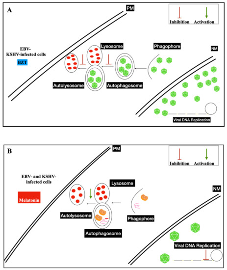
  43. Xia, J.; He, Y.; Meng, B.; Chen, S.; Zhang, J.; Wu, X.; Zhu, Y.; Shen, Y.; Feng, X.; Guan, Y.; et al. NEK2 induces autophagy-mediated bortezomib resistance by stabilizing Beclin-1 in multiple myeloma. Mol. Oncol. 2020, 14, 763–778.
  44. Granato, M.; Romeo, M.A.; Tiano, M.S.; Santarelli, R.; Gonnella, R.; Gilardini Montani, M.S.; Faggioni, A.; Cirone, M. Bortezomib promotes KHSV and EBV lytic cycle by activating JNK and autophagy. Sci. Rep. 2017, 7, 13052.
  45. Granato, M.; Santarelli, R.; Filardi, M.; Gonnella, R.; Farina, A.; Torrisi, M.R.; Faggioni, A.; Cirone, M. The activation of KSHV lytic cycle blocks autophagy in PEL cells. Autophagy 2015, 11, 1978–1986.
  46. Purushothaman, P.; Uppal, T.; Sarkar, R.; Verma, S.C. KSHV-Mediated Angiogenesis in Tumor Progression. Viruses 2016, 8, 198.
  47. Purushothaman, P.; Dabral, P.; Gupta, N.; Sarkar, R.; Verma, S.C. KSHV Genome Replication and Maintenance. Front. Microbiol. 2016, 7, 54.
  48. Ueda, K. KSHV Genome Replication and Maintenance in Latency. Adv. Exp. Med. Biol. 2018, 1045, 299–320.
  49. Uppal, T.; Banerjee, S.; Sun, Z.; Verma, S.C.; Robertson, E.S. KSHV LANA—The master regulator of KSHV latency. Viruses 2014, 6, 4961–4998.
  50. Wei, F.; Gan, J.; Wang, C.; Zhu, C.; Cai, Q. Cell Cycle Regulatory Functions of the KSHV Oncoprotein LANA. Front. Microbiol. 2016, 7, 334.
  51. Fajgenbaum, D.C. Novel insights and therapeutic approaches in idiopathic multicentric Castleman disease. Blood 2018, 132, 2323–2330.
  52. Gruffaz, M.; Vasan, K.; Tan, B.; Ramos da Silva, S.; Gao, S.J. TLR4-Mediated Inflammation Promotes KSHV-Induced Cellular Transformation and Tumorigenesis by Activating the STAT3 Pathway. Cancer Res. 2017, 77, 7094–7108.
  53. Sakakibara, S.; Tosato, G. Contribution of viral mimics of cellular genes to KSHV infection and disease. Viruses 2014, 6, 3472–3486.
  54. Rose-John, S. Interleukin-6 Family Cytokines. Cold Spring Harb. Perspect. Biol. 2018, 10, 951.
  55. Cengiz Seval, G.; Beksac, M. The safety of bortezomib for the treatment of multiple myeloma. Expert Opin. Drug Saf. 2018, 17, 953–962.
More
Information
Contributor MDPI registered users' name will be linked to their SciProfiles pages. To register with us, please refer to https://encyclopedia.pub/register :
View Times: 322
Revisions: 2 times (View History)
Update Date: 26 Oct 2021
1000/1000