Submitted Successfully!
To reward your contribution, here is a gift for you: A free trial for our video production service.
Thank you for your contribution! You can also upload a video entry or images related to this topic.
Version Summary Created by Modification Content Size Created at Operation
1 + 2228 word(s) 2228 2021-10-11 06:06:19 |
2 Format Change Meta information modification 2228 2021-10-22 09:42:10 |

Video Upload Options

Do you have a full video?

Confirm

Are you sure to Delete?
Cite
If you have any further questions, please contact Encyclopedia Editorial Office.
Usman, S. Vimentin at the Heart of Epithelial Mesenchymal Transition. Encyclopedia. Available online: https://encyclopedia.pub/entry/15230 (accessed on 29 March 2024).
Usman S. Vimentin at the Heart of Epithelial Mesenchymal Transition. Encyclopedia. Available at: https://encyclopedia.pub/entry/15230. Accessed March 29, 2024.
Usman, Saima. "Vimentin at the Heart of Epithelial Mesenchymal Transition" Encyclopedia, https://encyclopedia.pub/entry/15230 (accessed March 29, 2024).
Usman, S. (2021, October 21). Vimentin at the Heart of Epithelial Mesenchymal Transition. In Encyclopedia. https://encyclopedia.pub/entry/15230
Usman, Saima. "Vimentin at the Heart of Epithelial Mesenchymal Transition." Encyclopedia. Web. 21 October, 2021.
Vimentin at the Heart of Epithelial Mesenchymal Transition
Edit

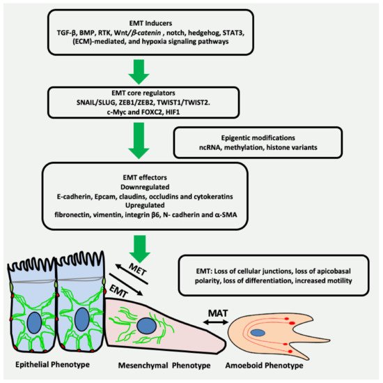
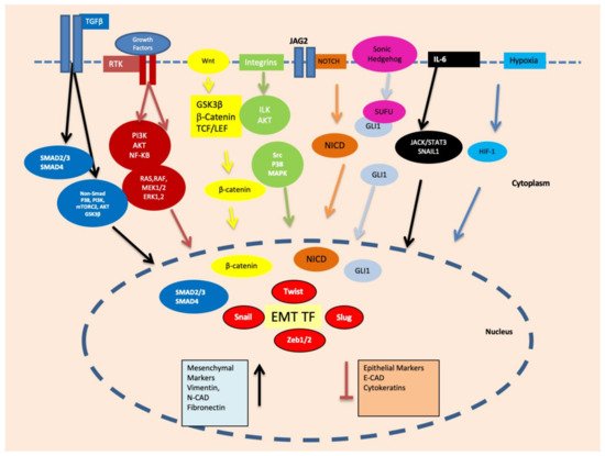
Epithelial-mesenchymal transition (EMT) is a reversible plethora of molecular events where epithelial cells gain the phenotype of mesenchymal cells to invade the surrounding tissues. EMT is a physiological event during embryogenesis (type I) but also happens during fibrosis (type II) and cancer metastasis (type III). It is a multifaceted phenomenon governed by the activation of genes associated with cell migration, extracellular matrix degradation, DNA repair, and angiogenesis. The cancer cells employ EMT to acquire the ability to migrate, resist therapeutic agents and escape immunity. One of the key biomarkers of EMT is vimentin, a type III intermediate filament that is normally expressed in mesenchymal cells but is upregulated during cancer metastasis. 

cancer invasion mesenchymal epithelial transition Stem Cells Micro RNA Vimentin EMT Inducers

1. Introduction

Epithelial to mesenchymal transition (EMT) is a reversible biological process in which epithelial cells lose their unique features of apicobasal polarity, epithelial markers, intercellular junctions, reorganization of the cytoskeletal architecture, immobility and differentiation and redirect to mesenchymal phenotype with the ability to migrate and invade [1]. EMT can be of three different types based on pathophysiological tissue context: type-1 EMT is an important physiological event during organogenesis and embryonic development, such as gastrulation or the outmigration of various cell types from the neural crest; type-2 EMT happens during wound healing for the induction of cell migration, growth and organ fibrosis [2] and type-3 EMT is described in the initiation and progression of multiple pathologies, including cancer and metastasis. In different carcinomas, EMT is characterized by the migration of epithelial cancer cells to invade the distant body sites by transforming into cells with the mesenchymal phenotype [3]. Cells previously activated by the EMT programme often revert to the epithelial state; this mechanism is called mesenchymal–epithelial transition (MET) [4]. In addition to the classical concept of EMT/MET in cancer cells, a recent concept of partial EMT (EM) was introduced, in which cells simultaneously express both epithelial and mesenchymal hybrid features [5][6]. This hybrid state makes them metastable, which is a dynamic state enabling cancer cells to induce or revert to EMT. Cancer cells may stably acquire one or more hybrid EM phenotypes expressing mixture of epithelial and mesenchymal traits. This multishaded EMT concept is known as epithelial–mesenchymal plasticity [7]. Researchers have categorized the hybrid EM into early and late types. The cells in early hybrid EM express both epithelial (cytokeratins) and mesenchymal (vimentin) markers but are less adhesive and rounded in shape. In the late hybrid stage, the mesenchymal markers become more pronounced and the epithelial phenotype is suppressed. Their shape becomes elongated, and adhesion is completely lost. Late hybrid EM stage can lead into a stable mesenchymal state [8].
The phenomenon of EMT is an intricate process with a timely interplay of a variety of complex network comprising inducers, core regulators and effectors [9]. EMT inducers include transforming growth factor-beta (TGF-β), bone morphogenetic protein (BMP), receptor tyrosine kinase (RTK), Wnt/β-catenin, NOTCH, hedgehog, signal transducer and activator of transcription 3 (STAT3), extracellular matrix (ECM)-mediated, and hypoxia signalling pathways (Figure 1) [10][11][12]. These EMT inducers lead to expression and functional activation of EMT core regulators, which among others include three major groups of EMT-activating transcription factors (EMT-TFs): the Snail family of the zinc-finger transcription factors Snail/Slug, the zinc-finger E-box binding homeobox (ZEB) family of transcription factors ZEB1/ZEB2, and the Twist family of basic helix-loop-helix (bHLH) transcription factors TWIST1/TWIST2 [13]. Other EMT-TFs are c-Myc, FOXC2 and HIF1. The activation of EMT-TFs is further fine-tuned by epigenetic modification leading to the induction of the expression of several EMT effectors that define the identity of the cell [14]. The epithelial biomarkers, such as E-cadherin, EpCAM, claudins, occludins and cytokeratins are downregulated, whereas the mesenchymal markers such as fibronectin, vimentin, integrin β6, N-cadherin and α-SMA are upregulated [15]. The key events during EMT are summarized in Figure 1.
Figure 1. Key events in EMT. Multiple growth factors, microenvironmental factors and other EMT inducers activate the transcription factors related to EMT. As a result, there is downregulation of the genes related to cell junctions and differentiation. Moreover, the genes specific to mesenchymal phenotype such as vimentin are upregulated, resulting in loss of intercellular junctions, apicobasal polarity, differentiation and increased cell motility, ultimately leading to cancer invasion. ncRNA = non-coding RNA.
Vimentin is an important type III intermediate filament (IF) protein alongside other cytoskeletal components, such as microfilaments and microtubules. Its dynamic role in different fundamental cellular processes such as structural support, attachment, migration and signalling is widely accepted [16]. Vimentin is consistently observed to be overexpressed during cancer metastasis and is therefore generally acknowledged as a canonical biomarker of type-3 EMT [17][18]. Several studies have highlighted its central role in the regulation of this complex process [19][20]. Vimentin filaments protect the cancer cells from mechanical stresses during the migration or squeezing through narrow spaces by providing a viscoelastic framework and support the positioning and integrity of organelles, especially the nucleus, during EMT and cancer progression [21]. In addition, it is reported that vimentin protects the cancer cells from the internal stress of misfolded proteins by directly binding to stress granules and aggresomes, supporting their subsequent destruction [22]. However, the exact mechanism used by vimentin to perform these functions is not known and requires further investigation.
In this review, we discuss various aspects of the pathophysiological mechanisms and regulation of type-3 EMT, and the driving role of vimentin as an upstream and/or downstream effector in signalling feedback loops in regulating and sustaining EMT, cancer invasion and metastasis.

2. EMT May Produce Cancer Stem Cells (CSCs) Expressing Vimentin

Cancer stem cells (CSC) are a small population of cells capable of self-renewal, which are known to resist therapeutic interventions and immune responses. Being pluripotent, these cells can provide cellular seeds to initiate new tumours at distant sites [23]. It was proposed that EMT can transform non-CSCs into cancer stem cells, which are invariably vimentin-positive [24]. In addition, it is believed that CSCs are generated as a result of adaptations and crosstalks with a tumour microenvironment, as well as in therapeutic interventions resulting in the generation of a heterogeneous subpopulation. Hypoxic conditions particularly contribute to the development of CSC characteristics including self-renewal, EMT, and drug resistance [25]. Hypoxia-inducible factors (HIFs) are the primary mediators of cellular responses, such as proliferation, EMT and metastasis, to hypoxic conditions [26]. Several other pathways implicated in the regulation of stemness phenotypes via HIFs include the TGF-β [27], Wnt/β-catenin [28], TNFα and NF-κB signalling [29]. These signalling cascades are also implicated in the induction of EMT via the transcriptional control of EMT-associated transcription factors, such as SNAI1, TWIST, ZEB1, SLUG and TCF3 leading to vimentin expression, as described above (Figure 2).
Figure 2. Important signalling pathways, such as Smad, PI3K/mTORC2/AKT/GSK3β, RAS/RAF/MEK1/2/ERK1, PI3K/AKT/NF-κB, ILK/AKT, Src/P38/MAPK, NOTCH, GLI1/SNAI2, JACK/STAT3 and HIF-1 are activated during EMT via growth factors, hypoxia and other microenvironmental factors. These signalling pathways ultimately upregulate the transcription factors related to EMT such as Snail, Slug, Twist, ZEB1 and ZEB2.
Cancer stem cells can exist in epithelial, mesenchymal or hybrid (mixed) states. They have the ability to switch between these different cellular states to maintain their survival, escape the immunity and grow at secondary sites. This switching is conducted by the complex interactions of various transcription factors, signalling pathways and microenvironmental factors [30]. The epithelial-like state of CSCs is characterized by the downregulation of mesenchymal markers, such as vimentin, and the upregulation of epithelial markers, such as CDH1. The expression of these markers is reversed in the mesenchymal state of CSCs. Both epithelial and mesenchymal markers are expressed in hybrid state [31]. CSCs express different cell surface proteins, such as CD34, CD44, CD24 and CD133, transcriptional factors, such as SOX2, NANOG, OCT ¾, SALL4 and other proteins that are not characterised as cell surface proteins, or transcription factors such as, ALDH, BMI1, Nestin and CXCR. These diverse markers are used to distinguish CSCs from the rest of the tumour population in different cancers [32]. These markers are expressed in a tissue-specific manner e.g., CD44, CD24 and ALDH are specific to breast cancer, CD34, CD8 to leukaemia, CD133 to colon cancer, CD44 to head neck cancer and CD90 to liver cancer [33]. These markers can be variably expressed according to the state of the CSCs. For example, in breast cancer stem cells, the CD24-CD44+ signature is related to a mesenchymal state with higher vimentin expression while the ALDH+ signature corresponds to an epithelial state of the cancer stem cells [31]. In normal cells, CD44 is a glycoprotein receptor for hyaluronic acid that is involved in cell adhesion, proliferation, differentiation, migration, angiogenesis and cell survival [34]. It is overexpressed in a variety of cancers, such as breast, colon, bladder, gastric, glioma, head and neck, prostate and leukaemia [32]. A soluble form of CD44 exists and is also overexpressed in certain cancers [35]. This soluble CD44 can bind to the vimentin head domain on the surface of endothelial cells, which is consistent with the fact that both CD44 and vimentin are overexpressed in oral squamous cell carcinoma (OSCC) and prostate cancer; however, the molecular basis of this association has not been fully elucidated [35]. CD24 is a cell adhesion sialoglycoprotein identified as a differentiation marker for hematopoietic and neuronal cells [36]. A higher CD44/CD24 ratio is positively correlated with vimentin in the breast CSC population [37]. ALDH1 (specifically isoform ALDH1A1) is another recently identified CSC marker in different tumours [38][39][40] and regulates the oxidation of retinal substrates into retinoic acid [40]. The increased expression of ALDH1 is related to the MET state of CSCS expressing lower levels of vimentin in breast cancer [41]. The sex-determining region Y-box 2 (SOX2) is an important transcription factor essential for the potential of stem cell multi-lineage. It can reprogram primary cells into stem cells [42], its levels are frequently upregulated in carcinomas such as HNSCC [43] with an inverse correlation between SOX2 and vimentin expression [44], and with a loss of SOX2 inducing tumour invasion through the upregulation of vimentin expression [45]. The possible role of miR-378 in the SOX2/Vim inverse functional relationship was also reported [31][46].

3. Vimentin Expression during Mesenchymal–Amoeboid Transition (MAT) of CSC

The concept of amoeboid movement was taken from amoeba Dictyostelium discoideum that rapidly move via contraction and expansion of the cell body without integrin interactions with the substrate. The belief that the amoeboid movement of cancer cells is linked to their invasive potential was first proposed in 1867 [47][48][49]. Different patterns of cell motility in cancer cells have been proposed by researchers and activated on an ad hoc basis during cancer invasion, which may also coexist within a population. This “switching of migration modes” is termed as “plasticity of cell motility”, which is considered imperative for cancer invasion [50]. One of the most familiar classifications of cell motility is individual versus combined cell migration that is further segregated based on mesenchymal or amoeboid phenotype. The single-cell motility-based “plasticity of migration” involves mesenchymal-to-amoeboid (MAT) and amoeboid-to-mesenchymal transitions (AMT) (Figure 1) [51]. A similar pattern could be observed in multicellular combined motility as the “plasticity of migration”, and involves collective-mesenchymal transition (CMT) and collective-amoeboid transition (CAT). As both CMT and CAT are reversible, therefore the terms, mesenchymal-collective transition (MCT) and amoeboid-collective transition (ACT), were also proposed [52]. During collective cell migration, a heterogeneous population of cells move together; the leader cells move by amoeboid movement while other members express mesenchymal phenotype and have intercellular connections [52].
Amoeboid cancer cells, unlike mesenchymal ones, migrate through the ECM barrier without proteolytic degradation of ECM and integrin clustering. As the amoeboid cells are rounded or ellipsoid, highly deformable, lacking focal adhesions and exhibit minimal cell-matrix contact, they move much faster than the mesenchymal cells [53][54]. Their nuclei are highly deformed, compressed and shifted towards the leading edge to allow movement through narrow spaces in ECM [55]. An amoeboid pattern of motility is reported when the cancer cells migrate through a soft medium such as blood or the lymphatic system [53]. Amoeboid movement is generally defined as a ‘path finding’ rather than the ‘path generating’ movement of mesenchymal cancer cells [52][56]. The detailed molecular basis of mobility shift from mesenchymal to amoeboid movement is not clear. Most studies are unable to describe the true molecular signature and transcriptional regulation of MAT in relation to tumour microenvironment and host immune response [51][57]. However, cellular stiffness, density, and other ECM factors along with the presence of chemotactic agents may be the determining factors in switching between mesenchymal and amoeboid states of cell motility [54]. A flexible vimentin network is reported to support the amoeboid mode of cell motility by conferring viscoelastic properties to the cell, protecting the nucleus and DNA from damage during propulsive squeezing movements [58]. Breast carcinoma cells devoid of vimentin are reported to be less contractile and less effective in migration [59]. In 3D cell cultures, vimentin is a prerequisite to the generation of propulsive pressure necessary to drive cell migration through confined spaces and vimentin knockdown leads to defective migration [59].
During metastasis, cancer cells transition from epithelial to mesenchymal phenotype or vice versa until they find suitable secondary host sites. Earlier reports suggested that stem cell-like features in cancer cells were induced by EMT; however, the latest research has linked the gain of stemness to cellular plasticity [24][60]. There are published reports proclaiming that EMT alone is not a prerequisite for cancer metastasis [61] and targeting EMT alone may lead to chemoresistance relapse, therefore both EMT and MET should be therapeutically targeted. Whether metastasis requires EMT or not is debatable; however, it is certain that EMT and metastasis both lead to the expression of vimentin [54][56][62].

References

  1. Kalluri, R.; Weinberg, R.A. The basics of epithelial-mesenchymal transition. J. Clin. Investig. 2009, 119, 1420–1428.
  2. Rout-Pitt, N.; Farrow, N.; Parsons, D.; Donnelley, M. Epithelial mesenchymal transition (EMT): A universal process in lung diseases with implications for cystic fibrosis pathophysiology. Respir. Res. 2018, 19, 1–10.
  3. Roche, J. The Epithelial-to-Mesenchymal Transition in Cancer. Cancers 2018, 10, 52.
  4. Bakir, B.; Chiarella, A.M.; Pitarresi, J.R.; Rustgi, A.K. EMT, MET, Plasticity, and Tumor Metastasis. Trends Cell Biol. 2020, 30, 764–776.
  5. Nieto, M.A.; Huang, R.Y.J.; Jackson, R.A.; Thiery, J.P. EMT: 2016. Cell 2016, 166, 21–45.
  6. Jordan, N.V.; Johnson, G.L.; Abell, A.N. Tracking the intermediate stages of epithelial-mesenchymal transition in epithelial stem cells and cancer. Cell Cycle 2011, 10, 2865–2873.
  7. Jolly, M.K.; Somarelli, J.A.; Sheth, M.; Biddle, A.; Tripathi, S.C.; Armstrong, A.J.; Hanash, S.M.; Bapat, S.A.; Rangarajan, A.; Levine, H. Hybrid epithelial/mesenchymal phenotypes promote metastasis and therapy resistance across carcinomas. Pharmacol. Ther. 2018, 194, 161–184.
  8. Pastushenko, I.; Blanpain, C. EMT Transition States during Tumor Progression and Metastasis. Trends Cell Biol. 2018, 29, 212–226.
  9. Tsai, J.H.; Yang, J. Epithelial-mesenchymal plasticity in carcinoma metastasis. Genes Dev. 2013, 27, 2192–2206.
  10. Yeo, C.D.; Kang, N.; Choi, S.Y.; Kim, B.N.; Park, C.K.; Kim, J.W.; Kim, S.J. The role of hypoxia on the acquisition of epithelial-mesenchymal transition and cancer stemness: A possible link to epigenetic regulation. Korean J. Intern. Med. 2017, 32, 589–599. Available online: https://www.ncbi.nlm.nih.gov/pmc/articles/PMC5511947/ (accessed on 2 October 2021).
  11. Gonzalez, D.; Medici, D. Signaling mechanisms of the epithelial-mesenchymal transition. Sci. Signal. 2014, 7, re8.
  12. Lindsey, S.; Langhans, S.A. Crosstalk of Oncogenic Signaling Pathways during Epithelial–Mesenchymal Transition. Front. Oncol. 2014, 4, 358.
  13. Georgakopoulos-Soares, I.; Chartoumpekis, D.V.; Kyriazopoulou, V.; Zaravinos, A. EMT Factors and Metabolic Pathways in Cancer. Front. Oncol. 2020, 10, 499.
  14. Lin, Y.-T.; Wu, K.-J. Epigenetic regulation of epithelial-mesenchymal transition: Focusing on hypoxia and TGF-β signaling. J. Biomed. Sci. 2020, 27, 1–10.
  15. Scanlon, C.; Van Tubergen, E.; Inglehart, R.; D’Silva, N. Biomarkers of Epithelial-Mesenchymal Transition in Squamous Cell Carcinoma. J. Dent. Res. 2012, 92, 114–121.
  16. Danielsson, F.; Peterson, M.K.; Araújo, H.C.; Lautenschläger, F.; Gad, A.K.B. Vimentin Diversity in Health and Disease. Cells 2018, 7, 147.
  17. Wu, S.; Du, Y.; Beckford, J.; Alachkar, H. Upregulation of the EMT marker vimentin is associated with poor clinical outcome in acute myeloid leukemia. J. Transl. Med. 2018, 16, 1–9.
  18. Liu, C.-Y.; Lin, H.-H.; Tang, M.-J.; Wang, Y.-K. Vimentin contributes to epithelial-mesenchymal transition cancer cell mechanics by mediating cytoskeletal organization and focal adhesion maturation. Oncotarget 2015, 6, 15966–15983.
  19. Ivaska, J. Vimentin. Small GTPases 2011, 2, 51–53.
  20. Vuoriluoto, K.; Haugen, H.; Kiviluoto, S.; Mpindi, J.-P.; Nevo, J.; Gjerdrum, C.; Tiron, C.; Lorens, J.B.; Ivaska, J. Vimentin regulates EMT induction by Slug and oncogenic H-Ras and migration by governing Axl expression in breast cancer. Oncogene 2010, 30, 1436–1448.
  21. Patteson, A.E.; Vahabikashi, A.; Pogoda, K.; Adam, S.A.; Mandal, K.; Kittisopikul, M.; Sivagurunathan, S.; Goldman, A.; Goldman, R.D.; Janmey, P.A. Vimentin protects cells against nuclear rupture and DNA damage during migration. J. Cell Biol. 2019, 218, 4079–4092.
  22. Pattabiraman, S.; Azad, G.K.; Amen, T.; Brielle, S.; Park, J.E.; Sze, S.K.; Meshorer, E.; Kaganovich, D. Vimentin protects differentiating stem cells from stress. Sci. Rep. 2020, 10, 1–15.
  23. Usman, S.; Jamal, A.; Teh, M.-T.; Waseem, A. Major Molecular Signaling Pathways in Oral Cancer Associated With Therapeutic Resistance. Front. Oral Heal. 2021, 1, 15.
  24. Thankamony, A.P.; Saxena, K.; Murali, R.; Jolly, M.K.; Nair, R. Cancer Stem Cell Plasticity – A Deadly Deal. Front. Mol. Biosci. 2020, 7, 79.
  25. Carnero, A.; Lleonart, M. The hypoxic microenvironment: A determinant of cancer stem cell evolution. BioEssays 2016, 38, S65–S74.
  26. Semenza, G.L. Oxygen Sensing, Hypoxia-Inducible Factors, and Disease Pathophysiology. Ann. Rev. Pathol. Mech. Dis. 2014, 9, 47–71.
  27. Anido, J.; Sáez-Borderías, A.; Gonzàlez-Juncà, A.; Rodón, L.; Folch, G.; Carmona, M.A.; Prieto-Sánchez, R.M.; Barba, I.; Martinez-Saez, E.; Prudkin, L.; et al. TGF-β Receptor Inhibitors Target the CD44high/Id1high Glioma-Initiating Cell Population in Human Glioblastoma. Cancer Cell 2010, 18, 655–668.
  28. Scheel, C.; Eaton, E.N.; Li, S.H.-J.; Chaffer, C.L.; Reinhardt, F.; Kah, K.-J.; Bell, G.; Guo, W.; Rubin, J.; Richardson, A.L.; et al. Paracrine and Autocrine Signals Induce and Maintain Mesenchymal and Stem Cell States in the Breast. Cell 2011, 145, 926–940.
  29. Scheel, C.; Weinberg, R.A. Phenotypic plasticity and epithelial-mesenchymal transitions in cancer and normal stem cells? Int. J. Cancer 2011, 129, 2310–2314.
  30. Walcher, L.; Kistenmacher, A.-K.; Suo, H.; Kitte, R.; Dluczek, S.; Strauß, A.; Blaudszun, A.-R.; Yevsa, T.; Fricke, S.; Kossatz-Boehlert, U. Cancer Stem Cells—Origins and Biomarkers: Perspectives for Targeted Personalized Therapies. Front. Immunol. 2020, 11, 1280.
  31. Zhang, R.; Tu, J.; Liu, S. Novel molecular regulators of breast cancer stem cell plasticity and heterogeneity. Semin. Cancer Biol. 2021.
  32. Zhang, X.; Zhao, W.; Li, Y. Stemness-related markers in cancer. Cancer Transl. Med. 2017, 3, 87–95.
  33. Yu, Z.; Pestell, T.G.; Lisanti, M.P.; Pestell, R.G. Cancer stem cells. Int. J. Biochem. Cell Biol. 2012, 44, 2144–2151.
  34. Thapa, R.; Wilson, G. The Importance of CD44 as a Stem Cell Biomarker and Therapeutic Target in Cancer. Stem Cells Int. 2016, 2016, 1–15.
  35. Patteson, A.E.; Vahabikashi, A.; Goldman, R.D.; Janmey, P.A. Mechanical and Non-Mechanical Functions of Filamentous and Non-Filamentous Vimentin. BioEssays 2020, 42, e2000078.
  36. Fang, X.; Zheng, P.; Tang, J.; Liu, Y. CD24: From A to Z. Cell. Mol. Immunol. 2010, 7, 100–103.
  37. Meyer, M.J.; Fleming, J.M.; Ali, M.A.; Pesesky, M.W.; Ginsburg, E.; Vonderhaar, B.K. Dynamic regulation of CD24 and the invasive, CD44posCD24negphenotype in breast cancer cell lines. Breast Cancer Res. 2009, 11, 14–82.
  38. Ginestier, C.; Hur, M.H.; Charafe-Jauffret, E.; Monville, F.; Dutcher, J.; Brown, M.; Jacquemier, J.; Viens, P.; Kleer, C.G.; Liu, S.; et al. ALDH1 Is a Marker of Normal and Malignant Human Mammary Stem Cells and a Predictor of Poor Clinical Outcome. Cell Stem Cell. 2007, 1, 555–567.
  39. Pearce, D.J.; Taussig, D.; Simpson, C.; Allen, K.; Rohatiner, A.Z.; Lister, T.A.; Bonnet, D. Characterization of Cells with a High Aldehyde Dehydrogenase Activity from Cord Blood and Acute Myeloid Leukemia Samples. Stem Cells 2005, 23, 752–760.
  40. Wu, A.; Luo, W.; Zhang, Q.; Yang, Z.; Zhang, G.; Li, S.; Yao, K. Aldehyde dehydrogenase 1, a functional marker for identifying cancer stem cells in human nasopharyngeal carcinoma. Cancer Lett. 2013, 330, 181–189.
  41. Liu, S.; Cong, Y.; Wang, D.; Sun, Y.; Deng, L.; Liu, Y.; Martin-Trevino, R.; Shang, L.; McDermott, S.P.; Landis, M.D.; et al. Breast Cancer Stem Cells Transition between Epithelial and Mesenchymal States Reflective of their Normal Counterparts. Stem Cell Rep. 2013, 2, 78–91.
  42. Wang, J.; Zeng, H.; Li, H.; Zhang, J.; Wang, S. Roles of sex-determining region Y-box 2 in cell pluripotency and tumor-related signaling pathways. Mol. Clin. Oncol. 2015, 3, 1203–1207.
  43. Li, B.; Chen, M.; Pan, M.-X. Sex determining region Y-box 2 is a prognostic factor for head and neck squamous cell carcinoma: Evidence from 11 published investigations. J. Cancer Res. Ther. 2020, 16, 434.
  44. Freier, K.; Knoepfle, K.; Flechtenmacher, C.; Pungs, S.; Devens, F.; Toedt, G.; Hofele, C.; Joos, S.; Lichter, P.; Radlwimmer, B. Recurrent copy number gain of transcription factorSOX2and corresponding high protein expression in oral squamous cell carcinoma. Genes Chromosom. Cancer 2010, 49, 9–16.
  45. Bayo, P.; Jou, A.; Stenzinger, A.; Shao, C.; Gross, M.; Jensen, A.D.; Grabe, N.; Mende, C.H.; Rados, P.V.; Debus, J.; et al. Loss of SOX2 expression induces cell motility via vimentin up-regulation and is an unfavorable risk factor for survival of head and neck squamous cell carcinoma. Mol. Oncol. 2015, 9, 1704–1719.
  46. Deng, Z.; Du, W.W.; Fang, L.; Shan, S.W.; Qian, J.; Lin, J.; Qian, W.; Ma, J.; Rutnam, Z.J.; Yang, B.B. The Intermediate Filament Vimentin Mediates MicroRNA miR-378 Function in Cellular Self-renewal by Regulating the Expression of the Sox2 Transcription Factor*. J. Biol. Chem. 2013, 288, 319–331.
  47. Waldeyer, R. Die Entwicklung der Carcinome. Virchows Arch. F. Path. Anat. 1872, 55, 67–159.
  48. Waldeyer., R. Die Entwicklung der Carcinome. Virchows Arch. F. Path. Anat. 1867, 41, 470–523.
  49. Enterline, H.T.; Coman, D.R. The ameboid motility of human and animal neoplastic cells. Cancer 1950, 3, 1033–1038.
  50. Taddei, M.L.; Giannoni, E.; Morandi, A.; Ippolito, L.; Ramazzotti, M.; Callari, M.; Gandellini, P.; Chiarugi, P. Mesenchymal to amoeboid transition is associated with stem-like features of melanoma cells. Cell Commun. Signal. 2014, 12, 24.
  51. Emad, A.; Ray, T.; Jensen, T.; Parat, M.; Natrajan, R.; Sinha, S.; Ray, S.P. An epithelial-mesenchymal-amoeboid transition gene signature reveals subtypes of breast cancer progression and metastasis. bioRxiv 2017. Available online: https://www.biorxiv.org/content/10.1101/219410v2.full (accessed on 13 September 2021).
  52. Wu, J.-S.; Jiang, J.; Chen, B.-J.; Wang, K.; Tang, Y.-L.; Liang, X.-H. Plasticity of cancer cell invasion: Patterns and mechanisms. Transl. Oncol. 2020, 14, 100899.
  53. Krakhmal, N.V.; Zavyalova, M.; Denisov, E.V.; Vtorushin, S.V.; Perelmuter, V. Cancer Invasion: Patterns and Mechanisms. Acta Naturae 2015, 7, 17–28.
  54. Talkenberger, K.; Cavalcanti-Adam, E.; Voss-Böhme, A.; Deutsch, A. Amoeboid-mesenchymal migration plasticity promotes invasion only in complex heterogeneous microenvironments. Sci. Rep. 2017, 7, 1–12.
  55. Yamada, K.M.; Sixt, M. Mechanisms of 3D cell migration. Nat. Rev. Mol. Cell Biol. 2019, 20, 738–752.
  56. Wolf, K.; Mazo, I.; Leung, H.; Engelke, K.; von Andrian, U.H.; Deryugina, E.I.; Strongin, A.Y.; Bröcker, E.-B.; Friedl, P. Compensation mechanism in tumor cell migration: Mesenchymal-amoeboid transition after blocking of pericellular proteolysis. J. Cell Biol. 2003, 160, 267–277.
  57. Holle, A.; Devi, N.G.K.; Clar, K.; Fan, A.; Saif, M.T.; Kemkemer, R.; Spatz, J.P. Cancer Cells Invade Confined Microchannels via a Self-Directed Mesenchymal-to-Amoeboid Transition. Nano Lett. 2019, 19, 2280–2290.
  58. Lavenus, S.B.; Tudor, S.M.; Ullo, M.F.; Vosatka, K.W.; Logue, J.S. A flexible network of vimentin intermediate filaments promotes migration of amoeboid cancer cells through confined environments. J. Biol. Chem. 2020, 295, 6700–6709.
  59. Strouhalova, K.; Přechová, M.; Gandalovičová, A.; Brábek, J.; Gregor, M.; Rosel, D. Vimentin Intermediate Filaments as Potential Target for Cancer Treatment. Cancers 2020, 12, 184.
  60. Qin, S.; Jiang, J.; Lu, Y.; Nice, E.C.; Huang, C.; Zhang, J.; He, W. Emerging role of tumor cell plasticity in modifying therapeutic response. Signal Transduct. Target. Ther. 2020, 5, 1–36.
  61. Jolly, M.K.; Ware, K.E.; Gilja, S.; Somarelli, J.A.; Levine, H. EMT and MET: Necessary or permissive for metastasis? Mol. Oncol. 2017, 11, 755–769.
  62. Melzer, C.; Von Der Ohe, J.; Hass, R. Breast Carcinoma: From Initial Tumor Cell Detachment to Settlement at Secondary Sites. BioMed Res. Int. 2017, 2017, 1–11.
More
Information
Subjects: Oncology
Contributor MDPI registered users' name will be linked to their SciProfiles pages. To register with us, please refer to https://encyclopedia.pub/register :
View Times: 658
Revisions: 2 times (View History)
Update Date: 22 Oct 2021
1000/1000