Submitted Successfully!
To reward your contribution, here is a gift for you: A free trial for our video production service.
Thank you for your contribution! You can also upload a video entry or images related to this topic.
Version Summary Created by Modification Content Size Created at Operation
1 + 1879 word(s) 1879 2021-08-16 10:42:57 |
2 The format is correct Meta information modification 1879 2021-08-17 04:49:40 |

Video Upload Options

Do you have a full video?

Confirm

Are you sure to Delete?
Cite
If you have any further questions, please contact Encyclopedia Editorial Office.
Lee, S. Exercise and Parkinson’s Neural Mitochondria. Encyclopedia. Available online: https://encyclopedia.pub/entry/13215 (accessed on 25 April 2024).
Lee S. Exercise and Parkinson’s Neural Mitochondria. Encyclopedia. Available at: https://encyclopedia.pub/entry/13215. Accessed April 25, 2024.
Lee, Shin-Da. "Exercise and Parkinson’s Neural Mitochondria" Encyclopedia, https://encyclopedia.pub/entry/13215 (accessed April 25, 2024).
Lee, S. (2021, August 16). Exercise and Parkinson’s Neural Mitochondria. In Encyclopedia. https://encyclopedia.pub/entry/13215
Lee, Shin-Da. "Exercise and Parkinson’s Neural Mitochondria." Encyclopedia. Web. 16 August, 2021.
Exercise and Parkinson’s Neural Mitochondria
Edit

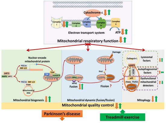
Treadmill training attenuated complex I deficits, cytochrome c release, ATP depletion, and complexes II–V abnormalities in Parkinson’s disease. Studies analyzed the neural mitochondrial quality-control, reporting that treadmill exercise improved mitochondrial biogenesis, mitochondrial fusion, and mitophagy in Parkinson’s disease. The hypothesis that treadmill training could attenuate both neural mitochondrial respiratory deficiency and neural mitochondrial quality-control dysregulation in Parkinson’s disease, suggesting that treadmill training might slow down the progression of Parkinson’s disease.

treadmill exercise Parkinson’s disease neural mitochondrial functions

1. Introduction

Parkinson’s disease (PD) is the second most common neurodegenerative disorder, causing a considerable number of disabilities globally [1][2]. The underlying mechanisms of PD are unclear, making it difficult to find efficient targeted therapies [3]. The current treatment of PD addresses symptomatic improvement only because none of the available treatment strategies have been confirmed to slow down PD progression [2].
Recently, neural mitochondrial respiratory deficiency has emerged as a central hallmark of Parkinsonian etiology [4][5]. In PD, the electron transport system of the neural mitochondria is impaired, mainly characterized by mitochondrial complex I deficit, cytochrome c release, and ATP depletion [6]. Impaired mitochondrial respiration in the brain increases oxidative stress and neuron loss, thereby augmenting PD progression [4].
Studies have also linked neural mitochondrial respiratory deficiency to neural mitochondrial quality-control dysregulation in PD [7][8]. In the physiological condition, neural mitochondrial quality-control involves a balance among biogenesis, dynamics (fusion/fission), and mitophagy (autophagy of mitochondria) [7]. Mitochondrial biogenesis produces the new mitochondria and mitochondrial content, accompanied by mitochondrial fusion to maintain a healthy mitochondrial network [9]. Meanwhile, mitochondrial fission segregates the damaged mitochondria and provides those for the mitophagy process, preventing the accumulation of dysfunctional mitochondria in the brain [10].
In PD, the biogenesis regulators and import machinery of neural mitochondria are reduced, leading to inhibition of mitochondrial biogenesis [11]. Additionally, PD has been confirmed to induce an imbalance of neural mitochondrial dynamics (fusion/fission) [4]. Moreover, the mitophagy process has been proven to be impaired with reductions of lysosomal activities in PD [12]. Disorders of biogenesis, fusion/fission, and mitophagy reduce the quantity and quality of neural mitochondria, leading to a neural mitochondrial respiratory deficiency in PD [4][7].
Treadmill exercise (TE) has been widely applied in PD rehabilitation [13][14]. Previous evidence indicated that TE training improved both the symptoms and the quality of life in PD patients [13]. In addition, a previous study showed that TE training improved gait functions by modulating neural mitochondrial dynamics in a PD rats model [15]. Another study reported that TE training reduced both neuron loss and behavioral disorders by improving mitochondrial respiration in a PD mouse model [16]. Those data suggested that TE training not only improves symptoms but also delays PD progression by attenuating PD-induced neural mitochondrial damage. However, the various approaches of the individual studies make it difficult to comprehensively understand the effects of TE on neural mitochondria in PD. Although several systematic reviews have been carried out to summarize the neuroprotective effects of TE training on PD, none of them specifically analyzed neural mitochondrial respiratory deficiency and neural mitochondrial quality-control dysregulation.

2. Treadmill Exercise on Neural Mitochondrial Functions in Parkinson’s Disease

2.1. Effects of TE Training on Neural Mitochondrial Respiratory Deficiency in PD

Three studies analyzed the effects of TE training on mitochondrial complex I in PD [15][17][18]. Two of those showed that the protein levels of complex I were reduced in PD compared to normal, whereas TE training enhanced their levels in PD [15][18]. However, the other study observed that the protein levels of complex I were similar among the normal control group, the PD group, and the TE-trained PD group [17].
Two studies observed that the protein levels of cytochrome c in neural mitochondria were reduced in PD compared to normal, whereas TE training increased those levels in PD [19][20]. One study reported that ATP production was reduced in PD compared to normal, whereas TE training enhanced ATP production in PD [21].
Five authors accessed the expression of complexes II, III, IV, and V, reporting different results [15][19][22][17][18]. One study reported that the protein levels of complexes II, III, IV, and V were unchanged among the normal control group, the sedentary PD group, and the TE-trained PD group [18]. The other study showed that the protein levels of complex II and complex V were reduced in PD compared to normal and those levels were restored by TE training in PD, whereas the protein levels of complex III and complex IV were unchanged among the normal control group, the PD group, and the TE-trained PD group [17]. Another study observed that TE training reduced the overexpression of complex II, complex III, and complex IV protein levels in PD, whereas complex V protein levels were unchanged among the normal control group, the PD group, and the TE trained-PD group [15]. Two studies showed that the protein levels of complex IV were reduced in PD compared to normal, whereas TE training increased those levels in PD [19][22].

2.2. Effects of TE Training on Neural Mitochondrial Biogenesis in PD

Six publications analyzed TE effects on biogenesis regulators of neural mitochondria in PD [16][19][23][17][20][18]. Four of those showed that the protein levels of biogenesis regulators, including SIRT3 [17], SIRT1 [16][19], PGC-1α [19][18], NRF-1,2 [19][17][18], and TFAM [19][17][18] were reduced in PD compared to normal, whereas TE training increased those levels in PD. In the other study that analyzed mRNA and protein levels of biogenesis regulators, they observed reduced levels of two biogenesis regulators (AMPK and PGC-1α) along with increased levels of two others (SIRT1 and TFAM) in PD compared to normal [23]. However, in this study, all of those levels were enhanced by TE training in PD [23]. On the contrary, another study showed that the mRNA levels of biogenesis regulators (PGC-1α and TFAM) were increased in PD compared to normal, and those levels were reduced by TE training in PD [20].
Two studies analyzed the effects of TE on translocase factors of neural mitochondria in PD [15][22]. One study reported that the protein levels of translocase proteins (TOM-20, TOM-40, TIM-23, and mtHSP70) were reduced in PD compared to normal, whereas TE training increased those levels in PD [22]. The other study showed that the level of translocase protein (TOM-20) in the substantia nigra was reduced in PD and recovered by TE training, but its level in the striatum was similar among the normal control group, the PD group and the TE-trained PD group [15].

2.3. Effects of TE Training on Neural Mitochondrial Dynamics in PD

Two studies analyzed the effects of TE training on mitochondrial fusion and fission proteins [15][17]. They reported that the protein levels of fusion proteins (OPA1, MFN2) were reduced in PD compared to normal, whereas TE training enhanced those levels in PD [15][17]. Regarding neural mitochondrial fission, one of two studies showed that the fission protein (Drp-1) was reduced in PD compared to normal, whereas TE training enhanced those levels in PD [15]. However, the other study showed that the anti-fission protein level (p-Drp1Ser637) was reduced in PD compared to normal, whereas TE training enhanced those levels in PD [17].

2.4. Effects of TE Training on Neural Mitophagy in PD

Three studies analyzed the effects of TE training on neural mitophagy in PD [15][19][24]. Those studies showed that the levels of mitophagy detector proteins, including PINK1 [15][24], parkin [24], and p62 [19][24] were increased in PD compared to normal, whereas TE training reduced those levels in PD. Two of those studies showed that the levels of autophagosomal proteins, including beclin-1 [19] and LC3 II/I [19][24] were increased in PD compared to normal, whereas TE training had no effect on their levels in PD. One of those studies reported that the levels of lysosomal proteins (LAMP2 and cathepsin L) were reduced in PD compared to normal, whereas TE training enhanced those levels in PD [24].

3. Summary

(1) Treadmill training attenuated neural mitochondrial respiratory deficiency in Parkinson’s disease, supported by the evidence that treadmill training normalized the levels of complexes I–V, cytochrome c, and ATP production in the Parkinsonian brain. (2) Treadmill training optimized neural mitochondrial biogenesis in Parkinson’s disease, supported by the evidence that treadmill training increased or normalized the levels of biogenesis regulators (SIRT3, SIRT1, AMPK, PGC-1α, NRF-1,2, and TFAM) and import machinery (TOM-20, TOM-40, TIM-23, and mtHSP70) in the Parkinsonian brain. (3) Treadmill training enhanced the neural mitochondrial fusion in Parkinson’s disease, supported by the evidence that treadmill training increased mitochondrial fusion factors (OPA-1 and MFN-2) in the Parkinsonian brain. (4) Treadmill training repaired the impairment of mitophagy in Parkinson’s disease, supported by the evidence that treadmill training reduced the levels of dysfunctional mitochondria detectors (PINK1, parkin, and p62) and increased the levels of lysosomal factors (LAMP2 and cathepsin L) in the Parkinsonian brain. Taking these findings with the previously hypothesized pathophysiology of Parkinson’s disease together, we drew a hypothesized figure (Figure 1), which suggests that treadmill training could counteract the neurodegeneration of Parkinson’s disease in both the neural mitochondrial respiratory system and neural mitochondrial quality-control.

Figure 1. The hypothesized figure. The figure shows the structure of the electron transport system and the cycle of mitochondrial quality-control. The electron transport system includes complexes I–V and cytochrome c, taking the primary responsibility for producing ATP (adenosine triphosphate) in neurons. Neural mitochondrial quality-control is the balance among mitochondrial biogenesis, mitochondrial dynamics (fusion/fission), and mitophagy (autophagy of mitochondria). Mitochondrial biogenesis produces the new mitochondria and mitochondrial content, controlled by biogenesis regulators (e.g., SIRT3, SIRT1, AMPK, PGC-1α, NRF-1,2, and TFAM). SIRT3/SIRT1/AMPK activates PGC-1α, then PGC-1α binds with NRF-1,2 in both the nucleus and mitochondria. In the nucleus, PGC-1α and NRF-1,2 promote the production of nuclear-encoded mitochondrial proteins. Nuclear-encoded mitochondrial proteins are imported into mitochondria, which involves the import machinery (e.g., TOM, TIM, and mtHSP70). In the mitochondria, PGC-1α and NRF-1,2 bind with TFAM to activate replication, transcription, and translation of mitochondrial DNA. Mitochondrial fusion merged mitochondria to the large mitochondrion, regulated by OPA-1 in the inner membrane and MFN-2 in the outer membrane. When the mitochondria are damaged, Drp-1 promotes mitochondrial fission to segregate and provide dysfunctional mitochondria for the mitophagy process to destroy. In the mitophagy process, the overexpression of detectors (e.g., PINK1, parkin, p62) on the dysfunctional mitochondrial membrane recruits autophagosomal factors (e.g., beclin-1 and LC3II) to form an autophagosome. Supported by LAMP2, the autophagosome fuses with lysosomes, destroying the dysfunctional mitochondria by enzymes (e.g., cathepsin L). In Parkinson’s disease, the evidence shows that complex I, cytochrome c, and ATP production in the mitochondria are reduced. Moreover, the mitochondria quality-control is dysregulated in Parkinson’s diseases, characterized by biogenesis reduction, fusion/fission imbalance, and mitophagy reduction. The included studies suggested that treadmill training activated complex I, cytochrome c, and ATP production. Additionally, treadmill training was shown to optimize the levels of mitochondrial biogenesis regulators (SIRT3, SIRT1, AMPK, PGC-1α, NRF-1,2, and TFAM), translocase factors (TOM-20, TOM-40, TIM-23, and mtHSP70), fusion proteins (OPA-1 and MFN-2), and lysosomal factors (LAMP2 and cathepsin L) as well as reducing dysfunctional mitochondrial detectors (PINK1, parkin, and p62). These data imply that treadmill training could attenuate neural mitochondrial respiratory deficiencies and neural mitochondrial quality-control dysregulation in Parkinson’s disease.

4. The Implications for Future Research

Further interdisciplinary studies are required to investigate the effects of treadmill training on the neural mitochondrial respiratory system, biogenesis, dynamics, and mitophagy in both genetic models and toxin models of Parkinson’s disease. Additionally, clinical studies should clarify the possible therapeutic applications through different exercise interventions into neural mitochondrial dysfunction in Parkinson’s disease.

References

  1. Sveinbjornsdottir, S. The clinical symptoms of Parkinson’s disease. J. Neurochem. 2016, 139, 318–324.
  2. Kalia, L.V.; Lang, A.E. Parkinson’s disease. Lancet 2015, 386, 896–912.
  3. Cacabelos, R. Parkinson’s disease: From pathogenesis to pharmacogenomics. Int. J. Mol. Sci. 2017, 18, 551.
  4. Chen, C.; Turnbull, D.M.; Reeve, A.K. Mitochondrial dysfunction in Parkinson’s disease—Cause or consequence? Biology 2019, 8, 38.
  5. Dossi, G.; Squarcina, L.; Rango, M. In Vivo Mitochondrial Function in Idiopathic and Genetic Parkinson’s Disease. Metabolites 2020, 10, 19.
  6. Moon, H.E.; Paek, S.H. Mitochondrial dysfunction in Parkinson’s disease. Exp. Neurobiol. 2015, 24, 103–116.
  7. Banerjee, R.; Starkov, A.A.; Beal, M.F.; Thomas, B. Mitochondrial dysfunction in the limelight of Parkinson’s disease pathogenesis. Biochim. Biophys. Acta 2009, 1792, 651–663.
  8. Meng, H.; Yan, W.-Y.; Lei, Y.-H.; Wan, Z.; Hou, Y.-Y.; Sun, L.-K.; Zhou, J.-P. SIRT3 regulation of mitochondrial quality control in neurodegenerative diseases. Front. Aging Neurosci. 2019, 11, 313.
  9. Diaz, F.; Moraes, C.T. Mitochondrial biogenesis and turnover. Cell Calcium. 2008, 44, 24–35.
  10. Lodish, H.; Berk, A.; Kaiser, C.A.; Krieger, M.; Scott, M.P.; Bretscher, A.; Ploegh, H.; Matsudaira, P. Molecular Cell Biology, 6th ed.; W. H. Freeman and Company: New York, NY, USA, 2008.
  11. Perier, C.; Vila, M. Mitochondrial biology and Parkinson’s disease. Cold Spring Harb. Perspect. Med. 2012, 4, a009332.
  12. Liu, J.; Liu, W.; Li, R.; Yang, H. Mitophagy in Parkinson’s disease: From pathogenesis to treatment. Cells 2019, 8, 712.
  13. Mehrholz, J.; Kugler, J.; Storch, A.; Pohl, M.; Elsner, B.; Hirsch, K. Treadmill Training for Patients with Parkinson’s Disease; John Wiley & Sons: Hoboken, NJ, USA, 2015.
  14. Wang, R.; Tian, H.; Guo, D.; Tian, Q.; Yao, T.; Kong, X. Impacts of exercise intervention on various diseases in rats. J. Sport Health Sci. 2020, 9, 211–227.
  15. Chuang, C.-S.; Chang, J.-C.; Cheng, F.-C.; Liu, K.-H.; Suc, H.-L.; Liu, C.-S. Modulation of mitochondrial dynamics by treadmill training to improve gait and mitochondrial deficiency in a rat model of Parkinson’s disease. Life Sci. 2017, 191, 236–244.
  16. Tuon, T.; Souza, P.S.; Santos, M.F.; Pereira, F.T.; Pedroso, G.S.; Luciano, T.F.; Souza, C.T.D.; Dutra, R.C.; Silveira, P.C.L.; Pinho, R.A. Physical training regulates mitochondrial parameters and neuroinflammatory mechanisms in an experimental model of Parkinson’s disease. Oxidative Med. Cell. Longev. 2015, 2015, 261809.
  17. Jang, Y.; Kwon, I.; Song, W.; Cosio-Lima, L.M.; Taylor, S.; Lee, Y. Modulation of mitochondrial phenotypes by endurance exercise contributes to neuroprotection against a MPTP-induced animal model of PD. Life Sci. 2018, 209, 455–465.
  18. Ferreira, A.F.F.; Binda, K.H.; Singulani, M.P.; Pereira, C.P.M.; Ferrari, G.D.; Alberici, L.C.; Real, C.C.; Britto, L.R. Physical exercise protects against mitochondria alterations in the 6-hidroxydopamine rat model of Parkinson’s disease. Behav. Brain Res. 2020, 387, 11260.
  19. Koo, J.-H.; Cho, J.-Y. Treadmill exercise attenuates α-synuclein levels by promoting mitochondrial function and autophagy possibly via SIRT1 in the chronic MPTP/P-induced mouse model of Parkinson’s disease. Neurotox Res. 2017, 32, 473–486.
  20. Patki, G.; Lau, Y.-S. Impact of exercise on mitochondrial transcription factor expression and damage in the striatum of a chronic mouse model of Parkinson’s disease. Neurosci. Lett. 2011, 505, 268–272.
  21. Lau, Y.-S.; Patki, G.; Das-Panja, K.; Le, W.-D.; Ahmad, S.O. Neuroprotective effects and mechanisms of exercise in a chronic mouse model of Parkinson’s disease with moderate neurodegeneration. Eur. J. Neurosci. 2011, 33, 1264–1274.
  22. Koo, J.-H.; Cho, J.-Y.; Lee, U.-B. Treadmill exercise alleviates motor deficits and improves mitochondrial import machinery in an MPTP-induced mouse model of Parkinson’s disease. Exp. Gerontol. 2017, 89, 20–29.
  23. Rezaee, Z.; Marandi, S.M.; Alaei, H.; Esfarjani, F.; Feyzollahzadeh, S. Effects of preventive treadmill exercise on the recovery of metabolic and mitochondrial factors in the 6-hydroxydopamine rat model of Parkinson’s disease. Neurotox. Res. 2019, 35, 908–917.
  24. Hwang, D.; Koo, J.; Kwon, K.; Choi, D.; Shin, S.; Jeong, J.; Um, H.; Cho, J. Neuroprotective effect of treadmill exercise possibly via regulation of lysosomal degradation molecules in mice with pharmacologically induced Parkinson’s disease. J. Physiol. Sci. 2018, 68, 707–716.
More
Information
Subjects: Neurosciences
Contributor MDPI registered users' name will be linked to their SciProfiles pages. To register with us, please refer to https://encyclopedia.pub/register :
View Times: 385
Entry Collection: Neurodegeneration
Revisions: 2 times (View History)
Update Date: 17 Aug 2021
1000/1000