Submitted Successfully!
To reward your contribution, here is a gift for you: A free trial for our video production service.
Thank you for your contribution! You can also upload a video entry or images related to this topic.
Version Summary Created by Modification Content Size Created at Operation
1 + 2787 word(s) 2787 2021-08-05 04:42:31

Video Upload Options

Do you have a full video?

Confirm

Are you sure to Delete?
Cite
If you have any further questions, please contact Encyclopedia Editorial Office.
Beaulieu, J. Integrin α6β4 in Colorectal Cancer. Encyclopedia. Available online: https://encyclopedia.pub/entry/12962 (accessed on 20 April 2024).
Beaulieu J. Integrin α6β4 in Colorectal Cancer. Encyclopedia. Available at: https://encyclopedia.pub/entry/12962. Accessed April 20, 2024.
Beaulieu, Jean-François. "Integrin α6β4 in Colorectal Cancer" Encyclopedia, https://encyclopedia.pub/entry/12962 (accessed April 20, 2024).
Beaulieu, J. (2021, August 09). Integrin α6β4 in Colorectal Cancer. In Encyclopedia. https://encyclopedia.pub/entry/12962
Beaulieu, Jean-François. "Integrin α6β4 in Colorectal Cancer." Encyclopedia. Web. 09 August, 2021.
Integrin α6β4 in Colorectal Cancer
Edit

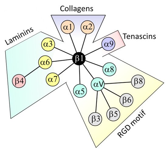
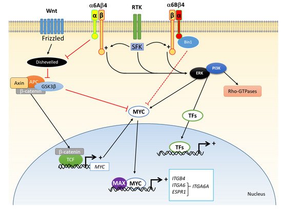
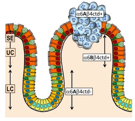
Integrin α6β4 is one of the main laminin receptors and is primarily expressed by epithelial cells as an active component of hemidesmosomes. In this article, after a brief summary about integrins in the gut epithelium in general, I review the knowledge and clinical potential of this receptor in human colorectal cancer (CRC) cells. Most CRC cells overexpress both α6 and β4 subunits, in situ in primary tumours as well as in established CRC cell lines. The mechanisms that lead to overexpression have not yet been elucidated but clearly involve specific transcription factors such as MYC. From a functional point of view, one key element affecting CRC cell behaviour is the relocalization of α6β4 to the actin cytoskeleton, favouring a more migratory and anoikis-resistant phenotype. 

colorectal cancer integrin α6β4 ITGA6 ITGB4 alternative splicing expression function biomarker

1. Introduction

Epithelial cells lie and attach to the basement membrane, a specialized network of extracellular matrix molecules that comprise specific collagens, proteoglycans and exclusive glycoproteins such as laminins as well as non-exclusive ones such as fibronectins. Cell adhesion is mediated by membrane receptors specific for these glycoproteins. In the intestine, there are a number of these receptors that have been identified for epithelial cells including dystroglycan and the 37/67 kDa laminin receptor [1] as well as many that belong to the integrin family [2][3]. Integrins are transmembrane αβ heterodimers that function as bidirectional signal transduction mediators acting as mechanosensors and participating in the regulation of main cell functions such as adhesion, migration, proliferation, survival and differentiation [4][5]. The gut epithelium expresses a wide variety of integrins as well as their corresponding ligands. Figure 1 illustrates the various heterodimers identified and their extracellular ligands.
Figure 1. Integrins expressed in intestinal epithelial cells under normal or pathologic conditions. Integrins are classified according to their preferential ligands: collagens for α1/α2 coupled to β1, laminins for α3/α6/α7 coupled to β1 or β4, tenascin for α9β1 and RGD-containing ligands (fibronectin, osteopontin and vitronectin) for α5/α8 coupled to β1 and αV coupled to β1/β3/β5/β6/β8.
It is noteworthy that while some of these integrins are mostly associated with the differentiated state in the intestinal epithelium cells such as α3β1 and α7β1, many are predominantly expressed by the proliferative cells at the base of the epithelial cells of the crypts in both the small and large intestines [2][3][6] and, more importantly in the context of this review, in primary and/or metastatic colorectal cancer (CRC) cells. This has been well documented recently for the α1β1 integrin, a collagen receptor. While exclusively expressed in the proliferative compartment in the normal colon, α1β1 was found to be overexpressed in CRC primary tumour and cell lines [7][8] and knockdown of α1 subunit expression resulted in the reduction of cell proliferation and tumour growth [9]. Integrin α9β1 is another interesting case. Expressed in the proliferative cells in the normal gut at the fetal stage but not in the adult, its resurgence in CRC cells both in primary tumours and in vitro is indicative of an oncofetal pattern of expression [10][11]. Two of the RGD-dependent integrins, α5β1 and αVβ6, appear to contribute to the metastatic process of CRCs [12][13]. Interestingly, the β6 subunit is expressed at very low levels in epithelia under normal conditions but is detected in both CRC primary tumours and metastasis where it acts through various mechanisms and systems to promote cancer progression [12]. However, there are exceptions. For instance, α8β1 which has also been shown to promote intestinal cell migration and proliferation in normal cells [14] appears to be no longer expressed by CRC cells [15]. Further investigation using normal intestinal cells revealed that this integrin acts as an anoikis-sensitizing trigger factor while the repression of its expression abolished this checkpoint [15]. Conversely, its reintroduction into CRC cells restores sensitivity to anoikis [15]. The αVβ3 integrin was also reported to act as an anoikis-sensitizing integrin for CRC cells [16][17]. These examples illustrate the crucial importance of the integrins as extracellular matrix molecule receptors involved in CRC cell progression, some susceptible to directly act in its promotion such as α1β1, α5β1 and αVβ6 while others act as preventive checkpoints such as α8β1 and αVβ3 or favour quiescence and differentiation rather than proliferation such as α3β1 and α7β1. These effects are also context-dependent over the entire process, some acting at early stages of cancer development while, as mentioned above, others are more likely to act at a later stage such as in the metastatic process. Some integrins likely exert dual promoting/suppressive influence on CRC development and progression [18]. In this context, integrin α6β4 is one of the most intriguing cases since its different role in CRC may in part derive from it ability to assemble hemidesmosomes, a very stable cell-extracellular matrix adhesion structure, and its potential to modulate oncogenic cell signalling events when released from hemidesmosomes as well as its α6A/α6B variant composition. While it belongs to the laminin-binding integrin subset, which also includes α3β1, α6β1 and α7β1 [2][19][20], α6β4 is characterized by a large β subunit and its predominant expression is in parenchymal cells such as the epithelial cells that line the luminal surface of the colonic crypts.

2. β4 Integrin Subunit (ITGB4) in CRC

2.1. Expression

The expression of the integrin β4 subunit in CRC has been the subject of a number of studies. While most of these have shown that β4 is uniformly distributed at the basal side of the colonic epithelium, its level of expression in primary CRC tumours was initially found to be quite variable depending on the study and antibody used, being found to be reduced or lost [21], maintained [22] or increased [23][24]. Increased expression was finally confirmed using a panel of distinct anti-β4 antibodies and validated at the transcript level [25].
The use of this panel of anti-β4 antibodies including two directed against extracellular epitopes and two targeting the cytoplasmic COOH-domain revealed interesting features. Indeed, in the normal small intestine and colon, cells of the crypts expressed a co-translationally processed form of β4 lacking the cytoplasmic terminal domain (β4ctd-) [26] so that only antibodies directed to the extracellular domain stained epithelial cells of the colonic crypts [25]. In contrast, the differentiated cells of the small intestinal villi and colonic surface epithelium expressed normal β4ctd+ suggesting that the β4ctd- form is related to the proliferative and less differentiated cells of the renewing intestinal epithelium [25][26]. Unexpectedly, colonic cells from both primary CRC and established adenocarcinoma cell lines expressed the β4ctd+ form, suggesting that this processing of β4 is lost or at least downregulated in neoplastic cells [25]. The potential impact of this is addressed below.

2.2. Regulation of Expression

Integrin β4 subunit expression appears to be mainly regulated at the transcript level but the mechanism of its overexpression in CRC cells is still incompletely understood. There have been a number of studies that have reported specific potential interactions of transcription factors with the ITGB4 promoter since its cloning [27] in a variety of cell types including RUNX1 [28], JUN [29] and KLF4 [30]. In CRC cells, MYC was the first transcription factor identified that promotes ITGB4 transcription [25] but recent studies have found others such as ZKSCAN3 [31] and FOSL1 [32] that need to be considered. Epigenetic regulation of ITGB4 expression has also been suggested after identification of microRNAs that may target the ITGB4 transcript such as miR-21 [33] and miR-335-5p [32] as well as hypomethylation of the ITGB4 promoter [32].

2.3. Change in Functionality

In normal epithelial cells, the β4 subunit is one of the key components of hemidesmosomes, a specialized adhesive structure found in epithelia. Most integrin β subunits bear short cytoplasmic domains smaller than 50 amino acids able to interact with actin filaments through cytoplasmic linker proteins such as talin and vinculin that are also used as scaffolding for the recruitment and activation of intracellular signalling pathways [34]. In contrast, β4 has a 1000 amino acid cytoplasmic domain composed of distinct sequences not found in other β integrins: a Calxβ motif adjacent to the plasma membrane, two pairs of fibronectin type III domains, a connecting segment and a COOH-terminal end domain [35]. In hemidesmosomes, α6β4 mediates the intracellular interaction with cytoskeletal keratins through various plakins such as plectin and the extracellular interaction with laminins, preferentially laminin-332 [35]. However, hemidesmosomes are dynamic structures that need to be dismantled to allow cell migration and other cell dynamic functions. There are a number of mechanisms that have been suggested for the release of α6β4 from the hemidesmosomes [18] mainly the phosphorylation of the β4 cytoplasmic tail in response to receptor tyrosine kinase activation by growth factors [18][36]. Upon release, α6β4 switches its association with cytokeratin to relocalize with actin filaments favouring the formation of motility structures [36]. In this context, it is worth mentioning that the processing of β4 into β4ctd- in normal proliferating intestinal cells impairs the ability of α6β4 to bind to laminin [26], which by increasing susceptibility to anoikis may represent an additional checkpoint mechanism for preventing aberrations in the permanent cell population responsible for epithelial renewal in the intestine [37].
In carcinoma cells, numerous studies have demonstrated that α6β4 promotes cell motility and invasive behaviour rather than stable anchoring to the extracellular matrix via a mechanism related to the one used for cell migration in wound healing in normal cells [36]. Since the discovery that α6β4 promotes colonic carcinoma cell invasiveness by activating the PI3K pathway [38], there have been a number of studies that have contributed to better documenting the potential of integrin α6β4 to regulate multiple signal transduction cascades involved in the promotion of cell proliferation, migration, invasion and suppression of anoikis [18][36][39]. While I invite the reader to refer to these seminal reviews [18][36][39] for further details about the multiple signalling transduction cascades that can be activated by α6β4 upon binding to its ligand, a few crucial elements are worth mentioning herein in the context of colorectal cancer (Figure 2). First, is the ability of α6β4 to synergistically cooperate with oncogenic receptor tyrosine kinases such as those of the epidermal growth factor receptor family and c-Met, which via their downstream effectors, the Src family of kinases, trigger the phosphorylation of tyrosine residues in the cytoplasmic domain of the β4 subunit to enhance the signal [34]. Second are the mechanisms by which α6β4 promote migration and invasion, which include the activation of the small GTPase RhoA and the up-regulation of the metastasis-associated protein S-100A4 [36]. Third is the apparent dual influence of α6β4 on cell survival. In most cell types, cell survival requires an α6β4-dependent PI3K activation and blocking α6β4-mediated adhesion trigger apoptosis [39]. However, using the RKO colon carcinoma cell line, Mercurio’s team showed that the ability of α6β4 to promote or inhibit apoptosis depends on the p53 cell status. Furthermore, the loss of the β4ctd- form in carcinoma cells, which strengthens α6β4-laminin interactions, and thus outside-in signalling, may also account for these effects.
Figure 2. Some of the signaling pathways regulated by integrin α6β4 in colorectal cancer cells. First is the cooperation between receptor tyrosine kinases (RTK), which via Src family of kinases (SFK) trigger phosphorylation of the cytoplasmic domain of β4 to activate the ERK and PI3K signaling pathways, which in turn regulate various cellular functions such as migration, proliferation and survival by the modulation of specific transcription factors (TFs) and activation of the Rho-GTPases. Second are the regulatory effects of the α6 subunit on cell proliferation where α6A promotes the Wnt/β-catenin pathway and the expression of downstream effectors such as MYC while α6B appears to inhibit MYC activity by a possible interaction with the protein bridging integrator 1 (Bin1). MYC appears also as one key regulator of integrin expression since both ITGB4 and ITGA6 have MYC-responsive elements in their promoters as for ESPR1 that encodes a splicing factor that regulate ITGA6A expression. Adapted from [18][36][39][40].

3. α6 Integrin Subunit (ITGA6) in CRC

3.1. Expression

As mentioned above, the integrin α6 subunit is the only possible dimerization partner for β4. ITGA6, as for ITGA3 and ITGA7 thought to have been derived from a common ancestor [20], is subject to alternative splicing of its exon 25, leading to the generation of two variants of the α6 subunits, α6A and α6B, that are distinct in their unique cytoplasmic sequences [20]. Although the α6 variants were reported and characterized more than 25 years ago and are likely to participate in distinct signalling cascades [41], the relationship between variant expression and distinctive cell functions has remained elusive especially in tumorigenesis [18].
As for many other epithelial cell types, intestinal cells can express both variants [41]. Closer analysis of α6A and α6B distribution in the intact normal human gut with a panel of anti-α6 antibodies revealed that α6A was predominantly detected in the proliferative cells of the small and large intestine while the α6B subunit was more restricted to the quiescent/differentiated cells lying on the villus and surface epithelium of the two intestinal segments [42][43]. Predominant expression of α6A in proliferative/undifferentiated cells and its gradual replacement by the α6B subunit in differentiating cells has also been confirmed in intestinal cell models [43].
Expression of the α6 variants was investigated in CRC in situ and in adenocarcinoma cell lines. In CRC primary tumours, although most carcinoma cells expressing both α6A and α6B a clear loss of α6A/α6B segregation was observed [43]. Quantitatively, a clear increase in total ITGA6 transcripts was noted in CRC as compared to the corresponding resection margins and this increase was attributed to an up-regulation in ITGA6A expression, with total levels of ITGA6B remaining comparable to those of the resection margins [44]. Consistently, all colorectal adenocarcinoma cell lines tested under proliferative conditions showed predominant ITGA6A expression.

3.2. Regulation of Expression

Information about transcriptional regulation of ITGA6 expression is still limited. Among the consensus binding sites for the transcription factors SP1, NF-kB, AP1 and MYC identified on the ITGA6 promoter [45], only SP1/SP3 have been confirmed [46]. Considering the fact that MYC expression is up-regulated in a large proportion of CRC [47][48] and that data indicating that MYC controls the expression of many genes in CRC cells including several key integrin subunits [40], which comprise ITGB4 [25], MYC represents a potential regulator of ITGA6 transcription. Pharmacological inhibition of MYC activity as well as molecular manipulations of intracellular MYC levels has shown a direct concordance between MYC levels and ITGA6 expression in intestinal cells while chromatin immunoprecipitation assays have confirmed the functionality of the MYC binding site on the ITGA6 promoter in the intestinal context [49].
Since neo ITGA6 is expressed under the form of ITGA6A in CRC cells [24], the mechanism responsible for the up-regulation of this spliced form was further investigated. Alternative splicing of ITGA6 has been relatively well studied and at least 10 splicing factors have been identified under various contexts but not in CRC cells. Among the various factors tested, the epithelial splicing regulatory protein 2 (ESPR2) was identified to be the main splicing factor responsible for ITGA6A expression in CRC cells [49]. It is noteworthy that ESPR1 was found to be responsible for ITGA6 splicing in breast cancer cells [50] but in contrast to ESPR2 that was found to be up-regulated by MYC, ESPR1 was not modulated in CRC cells by this factor [49]. Further identification of MYC as a direct activator of ESPR2 active transcription indicates that MYC regulates both ITGA6 and ESPR2 expression resulting in the specific up regulation of ITGA6A in CRC cells [30].

3.3. Change in Functionality

It is interesting to note that the cells of the crypt proliferative compartment, which predominantly express α6A/ITGA6A [42][43], are also the cells that express MYC in the normal colon [9]. Experimental studies using a normal intestinal epithelial crypt cell line showed that MYC regulates ITGA6A expression [49]. These data suggest that the MYC-dependent regulation of ITGA6A is a phenomenon occurring in the normal intestine, which appears to be exploited throughout the neoplastic process.
Indeed, colorectal cancer cell lines express 5–10 times more ITGA6A and MYC that their normal counterparts [49] while the integrin α6Aβ4 was found to promote proliferation in colorectal cancer cells [44]. Specific knockdown expression of ITGA6A in various CRC cell lines was also accompanied by a reduction in the capacity of these cells to develop tumours [44]. Incidentally, the ability of α6Aβ4 to promote proliferation in cancer cells was found to be mediated by activation of the Wnt/β-catenin pathway [44], a central signalling cascade required for intestinal crypt stem cell homoeostasis that is also exploited by CRC cells [51]. As MYC is a Wnt/B-catenin target gene [40], this suggests a feed forward loop for α6Aβ4 and MYC expressions responsible for the overexpression of the two molecules in cancer cells. In this context, it is worth mentioning that experimental overexpression of α6B results in the inhibition of MYC activity [43], an effect potentially mediated by the selective interaction of the MYC inhibitor nucleoshuttling scaffold protein bridging integrator 1 with the α6B cytoplasmic domain [40].

4. The Integrin α6β4 in CRC

In summary, while there is a clear segregation of α6β4 forms in the normal intestinal mucosa where α6Aβ4cdt- is expressed by the proliferative cells of the lower crypt and α6Bβ4ctd+ is mainly expressed by the quiescent and differentiated cells of the upper crypt and surface epithelium, cancer cells predominantly overexpress α6β4 under the α6Aβ4ctd+ hybrid form (Figure 3), which appears to mediate both cell proliferation and suppression of anoikis [39][52].
Figure 3. Integrin α6β4 expressed in the human colonic mucosa. In the lower crypt (LC), which contains stem cells (yellow) and proliferative absorptive and goblet cell precursors (gold and light green), α6β4 is expressed under the form of α6Aβ4ctd- while in the upper crypt (UC) and surface epithelium (SE), which contains quiescent absorptive and goblet cells (orange and green), it is under the form of α6Bβ4ctd+. In carcinoma (blue), this segregation is lost and α6β4 is predominantly expressed under the pro-proliferative form α6Aβ4ctd+.

References

  1. Cloutier, G.; Sallenbach-Morrissette, A.; Beaulieu, J.F. Non-integrin laminin receptors in epithelia. Tissue Cell 2019, 56, 71–78.
  2. Beaulieu, J.F. Integrins and human intestinal cell functions. Front. Biosci. 1999, 4, 310–321.
  3. Beaulieu, J.F. Extracellular matrix components and integrins in relationship to human intestinal epithelial cell differentiation. Prog. Histochem. Cytochem. 1997, 31, 1–76.
  4. Humphries, J.D.; Chastney, M.R.; Askari, J.A.; Humphries, M.J. Signal transduction via integrin adhesion complexes. Curr. Opin. Cell Biol. 2019, 56, 14–21.
  5. Kechagia, J.Z.; Ivaska, J.; Roca-Cusachs, P. Integrins as biomechanical sensors of the microenvironment. Nat. Rev. Mol. Cell Biol. 2019, 20, 457–473.
  6. Hussey, G.S.; Keane, T.J.; Badylak, S.F. The extracellular matrix of the gastrointestinal tract: A regenerative medicine platform. Nat. Rev. Gastroenterol. Hepatol. 2017, 14, 540–552.
  7. Boudjadi, S.; Carrier, J.C.; Beaulieu, J.F. Integrin alpha1 subunit is up-regulated in colorectal cancer. Biomark. Res. 2013, 1, 16.
  8. Boudjadi, S.; Carrier, J.C.; Groulx, J.F.; Beaulieu, J.F. Integrin alpha1beta1 expression is controlled by c-MYC in colorectal cancer cells. Oncogene 2016, 35, 1671–1678.
  9. Boudjadi, S.; Bernatchez, G.; Senicourt, B.; Beausejour, M.; Vachon, P.H.; Carrier, J.C.; Beaulieu, J.F. Involvement of the Integrin alpha1beta1 in the Progression of Colorectal Cancer. Cancers 2017, 9, 96.
  10. Basora, N.; Desloges, N.; Chang, Q.; Bouatrouss, Y.; Gosselin, J.; Poisson, J.; Sheppard, D.; Beaulieu, J.F. Expression of the alpha9beta1 integrin in human colonic epithelial cells: Resurgence of the fetal phenotype in a subset of colon cancers and adenocarcinoma cell lines. Int. J. Cancer 1998, 75, 738–743.
  11. Desloges, N.; Basora, N.; Perreault, N.; Bouatrouss, Y.; Sheppard, D.; Beaulieu, J.F. Regulated expression of the integrin alpha9beta1 in the epithelium of the developing human gut and in intestinal cell lines: Relation with cell proliferation. J. Cell. Biochem. 1998, 71, 536–545.
  12. Cantor, D.I.; Cheruku, H.R.; Nice, E.C.; Baker, M.S. Integrin alphavbeta6 sets the stage for colorectal cancer metastasis. Cancer Metastasis Rev. 2015, 34, 715–734.
  13. Pelillo, C.; Bergamo, A.; Mollica, H.; Bestagno, M.; Sava, G. Colorectal Cancer Metastases Settle in the Hepatic Microenvironment Through alpha5beta1 Integrin. J. Cell. Biochem. 2015, 116, 2385–2396.
  14. Benoit, Y.D.; Lussier, C.; Ducharme, P.A.; Sivret, S.; Schnapp, L.M.; Basora, N.; Beaulieu, J.F. Integrin alpha8beta1 regulates adhesion, migration and proliferation of human intestinal crypt cells via a predominant RhoA/ROCK-dependent mechanism. Biol. Cell 2009, 101, 695–708.
  15. Benoit, Y.D.; Larrivee, J.F.; Groulx, J.F.; Stankova, J.; Vachon, P.H.; Beaulieu, J.F. Integrin alpha8beta1 confers anoikis susceptibility to human intestinal epithelial crypt cells. Biochem. Biophys. Res. Commun. 2010, 399, 434–439.
  16. Kozlova, N.I.; Morozevich, G.E.; Chubukina, A.N.; Berman, A.E. Integrin alphavbeta3 promotes anchorage-dependent apoptosis in human intestinal carcinoma cells. Oncogene 2001, 20, 4710–4717.
  17. Morozevich, G.E.; Kozlova, N.I.; Chubukina, A.N.; Berman, A.E. Role of integrin alphavbeta3 in substrate-dependent apoptosis of human intestinal carcinoma cells. Biochemistry (Moscow) 2003, 68, 416–423.
  18. Ramovs, V.; Te Molder, L.; Sonnenberg, A. The opposing roles of laminin-binding integrins in cancer. Matrix Biol. 2017, 57–58, 213–243.
  19. Pouliot, N.; Kusuma, N. Laminin-511: A multi-functional adhesion protein regulating cell migration, tumor invasion and metastasis. Cell Adhes. Migr. 2013, 7, 142–149.
  20. de Melker, A.A.; Sonnenberg, A. Integrins: Alternative splicing as a mechanism to regulate ligand binding and integrin signaling events. Bioessays 1999, 21, 499–509.
  21. Stallmach, A.; von Lampe, B.; Matthes, H.; Bornhoft, G.; Riecken, E.O. Diminished expression of integrin adhesion molecules on human colonic epithelial cells during the benign to malign tumour transformation. Gut 1992, 33, 342–346.
  22. Lohi, J.; Oivula, J.; Kivilaakso, E.; Kiviluoto, T.; Frojdman, K.; Yamada, Y.; Burgeson, R.E.; Leivo, I.; Virtanen, I. Basement membrane laminin-5 is deposited in colorectal adenomas and carcinomas and serves as a ligand for alpha3beta1 integrin. Apmis 2000, 108, 161–172.
  23. Falcioni, R.; Turchi, V.; Vitullo, P.; Navarra, G.; Ficari, F.; Cavaliere, F.; Sacchi, A.; Marianicostantini, R. Integrin beta-4 expression in colorectal-cancer. Int. J. Oncol. 1994, 5, 573–578.
  24. Sordat, I.; Bosman, F.T.; Dorta, G.; Rousselle, P.; Aberdam, D.; Blum, A.L.; Sordat, B. Differential expression of laminin-5 subunits and integrin receptors in human colorectal neoplasia. J. Pathol. 1998, 185, 44–52.
  25. Ni, H.; Dydensborg, A.B.; Herring, F.E.; Basora, N.; Gagne, D.; Vachon, P.H.; Beaulieu, J.F. Upregulation of a functional form of the beta4 integrin subunit in colorectal cancers correlates with c-Myc expression. Oncogene 2005, 24, 6820–6829.
  26. Basora, N.; Herring-Gillam, F.E.; Boudreau, F.; Perreault, N.; Pageot, L.P.; Simoneau, M.; Bouatrouss, Y.; Beaulieu, J.F. Expression of functionally distinct variants of the beta4A integrin subunit in relation to the differentiation state in human intestinal cells. J. Biol. Chem. 1999, 274, 29819–29825.
  27. Takaoka, A.S.; Yamada, T.; Gotoh, M.; Kanai, Y.; Imai, K.; Hirohashi, S. Cloning and characterization of the human beta4-integrin gene promoter and enhancers. J. Biol. Chem. 1998, 273, 33848–33855.
  28. Phillips, J.L.; Taberlay, P.C.; Woodworth, A.M.; Hardy, K.; Brettingham-Moore, K.H.; Dickinson, J.L.; Holloway, A.F. Distinct mechanisms of regulation of the ITGA6 and ITGB4 genes by RUNX1 in myeloid cells. J. Cell. Physiol. 2018, 233, 3439–3453.
  29. An, X.Z.; Zhao, Z.G.; Luo, Y.X.; Zhang, R.; Tang, X.Q.; Hao, D.; Zhao, X.; Lv, X.; Liu, D. Netrin-1 suppresses the MEK/ERK pathway and ITGB4 in pancreatic cancer. Oncotarget 2016, 7, 24719–24733.
  30. Ma, B.; Zhang, L.; Zou, Y.; He, R.; Wu, Q.; Han, C.; Zhang, B. Reciprocal regulation of integrin beta4 and KLF4 promotes gliomagenesis through maintaining cancer stem cell traits. J. Exp. Clin. Cancer Res. 2019, 38, 23.
  31. Yang, L.; Zhang, L.; Wu, Q.; Boyd, D.D. Unbiased screening for transcriptional targets of ZKSCAN3 identifies integrin beta 4 and vascular endothelial growth factor as downstream targets. J. Biol. Chem. 2008, 283, 35295–35304.
  32. Li, M.; Jiang, X.; Wang, G.; Zhai, C.; Liu, Y.; Li, H.; Zhang, Y.; Yu, W.; Zhao, Z. ITGB4 is a novel prognostic factor in colon cancer. J. Cancer 2019, 10, 5223–5233.
  33. Ferraro, A.; Kontos, C.K.; Boni, T.; Bantounas, I.; Siakouli, D.; Kosmidou, V.; Vlassi, M.; Spyridakis, Y.; Tsipras, I.; Zografos, G.; et al. Epigenetic regulation of miR-21 in colorectal cancer: ITGB4 as a novel miR-21 target and a three-gene network (miR-21-ITGBeta4-PDCD4) as predictor of metastatic tumor potential. Epigenetics 2014, 9, 129–141.
  34. Cooper, J.; Giancotti, F.G. Integrin Signaling in Cancer: Mechanotransduction, Stemness, Epithelial Plasticity, and Therapeutic Resistance. Cancer Cell 2019, 35, 347–367.
  35. Walko, G.; Castanon, M.J.; Wiche, G. Molecular architecture and function of the hemidesmosome. Cell Tissue Res. 2015, 360, 529–544.
  36. Stewart, R.L.; O’Connor, K.L. Clinical significance of the integrin alpha6beta4 in human malignancies. Lab. Investig. 2015, 95, 976–986.
  37. Benoit, Y.D.; Groulx, J.F.; Gagne, D.; Beaulieu, J.F. RGD-Dependent Epithelial Cell-Matrix Interactions in the Human Intestinal Crypt. J. Signal Transduct. 2012, 2012.
  38. Shaw, L.M.; Rabinovitz, I.; Wang, H.H.; Toker, A.; Mercurio, A.M. Activation of phosphoinositide 3-OH kinase by the alpha6beta4 integrin promotes carcinoma invasion. Cell 1997, 91, 949–960.
  39. Beausejour, M.; Boutin, A.; Vachon, P.H. Anoikis regulation—Complexities, distinction, and cell differentiation. In Apoptosis and Beyond: The Many Ways Cells Die; Radosevich, J.A., Ed.; Wiley: Hoboken, NJ, USA, 2017; pp. 145–182.
  40. Boudjadi, S.; Beaulieu, J.F. MYC and integrins interplay in colorectal cancer. Oncoscience 2016, 3, 50–51.
  41. Hogervorst, F.; Admiraal, L.G.; Niessen, C.; Kuikman, I.; Janssen, H.; Daams, H.; Sonnenberg, A. Biochemical characterization and tissue distribution of the A and B variants of the integrin alpha 6 subunit. J. Cell. Biol. 1993, 121, 179–191.
  42. Dydensborg, A.B.; Teller, I.C.; Basora, N.; Groulx, J.F.; Auclair, J.; Francoeur, C.; Escaffit, F.; Pare, F.; Herring, E.; Menard, D.; et al. Differential expression of the integrins alpha6Abeta4 and alpha6Bbeta4 along the crypt-villus axis in the human small intestine. Histochem. Cell Biol. 2009, 131, 531–536.
  43. Dydensborg, A.B.; Teller, I.C.; Groulx, J.F.; Basora, N.; Pare, F.; Herring, E.; Gauthier, R.; Jean, D.; Beaulieu, J.F. Integrin alpha6Bbeta4 inhibits colon cancer cell proliferation and c-Myc activity. BMC Cancer 2009, 9, 223.
  44. Groulx, J.F.; Giroux, V.; Beausejour, M.; Boudjadi, S.; Basora, N.; Carrier, J.C.; Beaulieu, J.F. Integrin alpha6A splice variant regulates proliferation and the Wnt/beta-catenin pathway in human colorectal cancer cells. Carcinogenesis 2014, 35, 1217–1227.
  45. Nishida, K.; Kitazawa, R.; Mizuno, K.; Maeda, S.; Kitazawa, S. Identification of regulatory elements of human alpha 6 integrin subunit gene. Biochem. Biophys. Res. Commun. 1997, 241, 258–263.
  46. Gaudreault, M.; Vigneault, F.; Leclerc, S.; Guerin, S.L. Laminin reduces expression of the human alpha6 integrin subunit gene by altering the level of the transcription factors Sp1 and Sp3. Investig. Ophthalmol. Vis. Sci. 2007, 48, 3490–3505.
  47. Sikora, K.; Chan, S.; Evan, G.; Gabra, H.; Markham, N.; Stewart, J.; Watson, J. c-myc oncogene expression in colorectal cancer. Cancer 1987, 59, 1289–1295.
  48. Erisman, M.D.; Rothberg, P.G.; Diehl, R.E.; Morse, C.C.; Spandorfer, J.M.; Astrin, S.M. Deregulation of c-myc gene expression in human colon carcinoma is not accompanied by amplification or rearrangement of the gene. Mol. Cell. Biol. 1985, 5, 1969–1976.
  49. Groulx, J.F.; Boudjadi, S.; Beaulieu, J.F. MYC Regulates alpha6 Integrin Subunit Expression and Splicing Under Its Pro-Proliferative ITGA6A form in Colorectal Cancer Cells. Cancers 2018, 10, 42.
  50. Goel, H.L.; Gritsko, T.; Pursell, B.; Chang, C.; Shultz, L.D.; Greiner, D.L.; Norum, J.H.; Toftgard, R.; Shaw, L.M.; Mercurio, A.M. Regulated splicing of the alpha6 integrin cytoplasmic domain determines the fate of breast cancer stem cells. Cell Rep. 2014, 7, 747–761.
  51. Kretzschmar, K.; Clevers, H. Wnt/beta-catenin signaling in adult mammalian epithelial stem cells. Dev. Biol. 2017, 428, 273–282.
  52. Beaulieu, J.F. Integrin alpha6beta4 in colorectal cancer. World J. Gastrointest. Pathophysiol. 2010, 1, 3–11.
More
Information
Subjects: Oncology
Contributor MDPI registered users' name will be linked to their SciProfiles pages. To register with us, please refer to https://encyclopedia.pub/register :
View Times: 562
Entry Collection: Gastrointestinal Disease
Revision: 1 time (View History)
Update Date: 09 Aug 2021
1000/1000