Submitted Successfully!
To reward your contribution, here is a gift for you: A free trial for our video production service.
Thank you for your contribution! You can also upload a video entry or images related to this topic.
Version Summary Created by Modification Content Size Created at Operation
1 + 2303 word(s) 2303 2021-07-19 03:34:16 |
2 format correct -21 word(s) 2282 2021-08-05 11:15:07 |

Video Upload Options

Do you have a full video?

Confirm

Are you sure to Delete?
Cite
If you have any further questions, please contact Encyclopedia Editorial Office.
Brunetti, D. PITRM1 in Neurodegeneration. Encyclopedia. Available online: https://encyclopedia.pub/entry/12820 (accessed on 25 April 2024).
Brunetti D. PITRM1 in Neurodegeneration. Encyclopedia. Available at: https://encyclopedia.pub/entry/12820. Accessed April 25, 2024.
Brunetti, Dario. "PITRM1 in Neurodegeneration" Encyclopedia, https://encyclopedia.pub/entry/12820 (accessed April 25, 2024).
Brunetti, D. (2021, August 05). PITRM1 in Neurodegeneration. In Encyclopedia. https://encyclopedia.pub/entry/12820
Brunetti, Dario. "PITRM1 in Neurodegeneration." Encyclopedia. Web. 05 August, 2021.
PITRM1 in Neurodegeneration
Edit

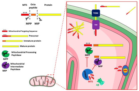
Mounting evidence shows a link between mitochondrial dysfunction and neurodegenerative disorders, including Alzheimer Disease. Increased oxidative stress, defective mitodynamics, and impaired oxidative phosphorylation leading to decreased ATP production, can determine synaptic dysfunction, apoptosis, and neurodegeneration. Furthermore, mitochondrial proteostasis and the protease-mediated quality control system, carrying out degradation of potentially toxic peptides and misfolded or damaged proteins inside mitochondria, are emerging as potential pathogenetic mechanisms. The enzyme pitrilysin metallopeptidase 1 (PITRM1) is a key player in these processes; it is responsible for degrading mitochondrial targeting sequences that are cleaved off from the imported precursor proteins and for digesting a mitochondrial fraction of amyloid beta (Aβ).

PITRM1 pitrilysin metallopeptidase 1 mitochondrial proteostasis Alzheimer Disease neurodegeneration neurodegenerative diseases neurodegenerative dementia spinocerebellar ataxia mitochondrial protein quality control protein aggregation mitochondrial dysfun

1. Introduction

Mitochondrial dysfunction, whether primary or secondary, is increasingly recognized as a hallmark of neurodegeneration [1] and a wide body of literature provides evidence of impaired mitochondrial function as a cause rather than a consequence of neurodegeneration [2][3][4]. Mitochondrial demise can be observed even before the appearance of pathognomonic histopathological hallmarks of the disease [5]. Neurons are obligatorily dependent on mitochondrial energy production, which sustains a myriad of functions, including membrane remodeling, synaptic spine formation, and the generation of transmembrane resting and action potentials [6][7].
Mitochondria also play a pivotal role in cell survival and death by regulating apoptotic pathways and contributing to different cellular functions including intracellular calcium homeostasis, maintenance of the cellular redox potential, and cell cycle regulation. Recent evidence demonstrated that mitochondria are also important in regulating cell fate towards stemness or neurogenesis [8].
Thus, it is not surprising that mitochondrial dysfunction can have devastating effects on neuronal differentiation and survival. The brain is a major target in primary, genetically determined mitochondrial disease, but mitochondrial dysfunction is also a prominent feature in many of the most prevalent neurodegenerative diseases, including Parkinson’s Disease (PD) [9], Huntington’s Disease [10], neurodegeneration associated with stroke [11], Amyotrophic Lateral Sclerosis [12], neurodegenerative ataxias [13] and different types of psychiatric and cognitive disorders such as Dementia with Lewy Bodies [14], Frontotemporal Dementia [15], and Alzheimer’s disease (AD) [16]. In AD, for example, the “mitochondrial cascade” hypothesis proposes that organellar dysfunction is the primary event in AD pathology [17]. Moreover, although the extracellular deposition of amyloid-beta (Aβ) peptides (Aβ1–40, Aβ1–42) is the key histopathological hallmark of AD, Aβ accumulation is proposed to also occur in mitochondria, through the mitochondrial import machinery [18][19], causing impairment of different mitochondrial pathways such as respiration, reactive oxygen species (ROS) detoxification, and organelle morphology [20][21][22][23][24][25][26].
The protease-mediated quality control system is a first-line homeostatic defense against mitochondrial damage, and includes degradation of non-assembled, misfolded or damaged proteins as a result of oxidative stress, elimination of cleaved products during protein processing, and overall regulation of protein turnover and homeostasis (referred to as proteostasis) [27]. The proteolytic system in mitochondria is crucial for the maintenance of protein turnover and the integrity of mitochondria. The majority of mitochondrial proteins are synthesized on cytosolic ribosomes with an N-terminal peptide (the presequence or mitochondrial targeting sequence—MTS) which is recognized by—and binds to—specific receptors in the mitochondrial outer membrane. Following this event, these precursor mitochondrial proteins are translocated through the mitochondrial entry gate, the TOM (Translocase of Outer Membrane) complex and then, via a specific TIM (Translocase of Inner Membrane) system, TIM23, into the matrix [28], where they undergo proteolytic processes, including the cleavage of MTS, and structural modifications that lead to mature, functional proteins [29].
Several proteases and peptidases have been identified in different mitochondrial sub compartments. Most are ATP dependent proteases, such as the matrix-located Lon protease 1 (LONP1) and the membrane-bound AAA (ATPases Associated with diverse cellular Activities) proteases. The latter include i-AAA (Yme1), active in the intermembrane space (IMS), and m-AAA (Yta10/Yta12), exposed to the matrix. These enzymes catalyse the initial step of degradation, cleaving proteins into peptides, thus contributing to mitochondrial quality control [30]. Other ATP-independent proteases such as the mitochondrial processing peptidase (MPP), the mitochondrial intermediate protease (MIP), and the inner membrane peptidase (IMP), also generate short peptides. Upon import of the mitochondrial precursor proteins, MPP in the matrix cleaves the MTSs, releasing the mature proteins [31][32][33]. MPP activity can produce import intermediates, which are further processed by MIP (the octapeptidyl peptidase Oct1 in yeast) or IMP (the intermediate cleaving peptidase Icp55 in yeast) [33][34][35][36]. Therefore, all these proteases are involved in the processing of precursor protein and release free MTS peptides after proteolytic cleavage (Figure 1) [37].
Figure 1. Schematic representation of PITRM1 function and interaction. Mitochondrial Precursor proteins are imported from cytosol to the matrix through the Translocase of the Outer Membrane (TOM) and Translocase of the Inner Membrane (TIM) complexes. Dysfunctional activity of PITRM1 results in the accumulation of Amyloid beta (Aβ), of Mitochondrial Targeting Sequences (MTS) and of octapeptides that trigger feedback inhibition of Mitochondrial Processing Peptidase (MPP) and Mitochondrial Intermediate Peptidase (MIP), leading to the accumulation and aggregation of unprocessed precursor. Figure 1 was modified from SMART (Servier Medical Art), licensed under a Creative Common Attribution 3.0 Generic License. http://smart.servier.com/.
Typically, the MTSs are amphiphilic species, with a polar, positively charged, arginine-rich side, opposite to an apolar side (Figure 1). Therefore, if these MTS peptides fail to be cleared from the mitochondrial matrix, they may act as detergent-like, toxic agents, forming pores in the membranes and resulting in uncoupling of oxidative phosphorylation and dissipation of the mitochondrial membrane potential [36].

2. PITRM1/PreP

PITRM1, also termed Presequence protease (PreP), is localized in the mitochondrial matrix; it is the only known protein responsible for the degradation of the MTS, thus completing the last step of the protein import process. PreP was initially identified in Arabidopsis thaliana (AtPreP), and shown to degrade the MTS of both mitochondria and chloroplasts [38]. A yeast mutant lacking the PITRM1 homolog Cym1 [39] displayed mitochondrial accumulation of precursor proteins and processing intermediates, as well as decreased levels of cleaved, mature proteins [32][40]. Impaired preprotein maturation leads to accelerated protein degradation and an unbalanced mitochondrial proteome, resulting in mitochondrial dysfunction manifested by reduced respiration, altered membrane potential, and high ROS levels [28][37].
The human gene PITRM1 is located in chromosomal region 10p15.2, contains 27 exons, and is transcribed from the antisense strand. Three different isoforms of the protein correspond to transcript variants derived from alternative splicing. In isoforms 1 and 2, all 27 exons of the gene are retained and translated, with a total of 1038 and 1037 aminoacids (aa), respectively, due to the use of two different splice donor sites at exon 17. The third isoform is the smallest one and contains 939 aa derived from 24 exons of the gene.
Human PITRM1 was initially identified as metalloprotease 1 [41], and shows 31% sequence identity to AtPreP, performing a similar function in human mitochondria. PITRM1 belongs to the M16 metalloproteases family, which are Zn2+-dependent and ATP-independent enzymes that share a conserved architecture of two ~ 50 kDa homologous domains enclosing a large catalytic chamber. Three families, M16-A, -B and -C, have been characterized depending on the connection between these two domains [42]. PITRM1 is a 117 kDa M16C enzyme arranged in four domains, forming two enzyme halves, hPreP-N (amino acids 33–509) and hPreP-C (aa 576–1037) domains, connected by a hinge region (aa 510–575), which is possibly involved in the opening and closing of the two enzyme halves. Active site residues are located in both halves, which interact to form a large peptidasome chamber [43]. In addition to targeting peptides generated during the import processing, the large catalytic chamber of PITRM1 is also able to degrade a wide range of unstructured peptides, ranging from 10 to 65 aa, but not larger folded proteins [44].
In vitro studies demonstrated that human PITRM1 can degrade Aβ1–40, Aβ1–42 and the Aβ arctic (Aβ1–40 E22G), a peptide that causes increased fibril formation and early onset of a familial variant of AD. Its proteolytic activity generates several fragments, which are unique to PITRM1 (in comparison to other proteases), and recognizes the cleavage sites in the very hydrophobic C-terminal portion of Aβ that is prone to aggregation [38]. Interestingly, the 3D structures of PITRM1 are highly similar to IDE (Insulin degrading enzyme), a zinc metallopeptidase that degrades intracellular insulin, as well as glucagon, amylin, beta-amyloid, bradykinin, and kallidin. Moreover, IDE has been suggested to have a role in the degradation of cleaved MTSs [37][45]. The preferential affinity of this enzyme for insulin results in the insulin-mediated inhibition of degradation of other peptides, such as beta-amyloid [46]. Variants in the IDE gene and deficiencies in the protein function have previously been associated with type 2 diabetes mellitus and Alzheimer’s disease [47][48].

3. PITRM1/PreP and Alzheimer’s Disease

3.1. PITRM1/PreP Is Downregulated in AD Patients and Mouse Models

It was reported that mitochondria isolated from the temporal lobe, an area of the brain that is highly susceptible to Aβ accumulation, showed significantly lower PITRM1 activity in AD patients compared to age-matched control samples. In contrast, activity in mitochondria isolated from the cerebellum showed no differences [49]. Decreased expression of a transcript antisense to PITRM1 was found in the posterior cingulate cortex extracted from AD patients, suggesting changes in the regulation of PITRM1 expression [50].
Further, global proteomic studies revealed a mitochondrial proteome imbalance in AD samples, with presequence-containing mitochondrial proteins being particularly affected [23][51]. Similar experiments were performed in transgenic (Tg) mouse models of AD, including a Tg for APP (Amyloid-β Protein Precursor), which is the most studied AD model, and ABAD (Aβ-binding alcohol dehydrogenase), which is an accelerated AD model. Indeed, Aβ peptides are produced by regulated proteolysis of the APP through the action of β- and γ- secretases. ABAD or HSD17B10 interacts with Aβ and mediates its mitochondrial toxicity [20]. The proteolytic activity of mouse PreP (mPreP) was significantly reduced in the matrix of cortical mitochondria isolated from 5-month-old Tg mAPP mice compared to non-Tg littermates. Similarly, the mPreP activity in Tg mAPP/ABAD mice was significantly lower than that in both non-Tg mice and Tg mAβPP mice [49]. Transgenic mice overexpressing APP also showed lower mPreP proteolytic activity compared to age-matched, non-transgenic mice and a progressive, age dependent reduction in the same activity [49].
These results suggest that the loss of PITRM1 activity may result not only in the accumulation of Aβ, but also of free presequence peptides [36]. While the relevance of Aβ accumulation still remains to be determined, the accumulation of presequence peptides is likely to be toxic.
The lower PITRM1 activity observed in AD brain mitochondria appears not to be due to lowered protein levels but to a functional alteration of the enzyme, possibly through post-translational modifications such as protein oxidation. Oxidative stress is well-documented in AD brains and confirmed by the increase in several reactive oxygen species (ROS) markers [52]. For example, levels of 4-hydroxynonenal derived from lipid peroxidation, a biomarker of oxidation, were found to be higher in the temporal lobe of AD patients than controls, while were unchanged in the cerebellum [49]. These results suggest that lower temporal lobe PITRM1 activity reflects protein oxidation, and this is confirmed by several biochemical analyses using purified PITRM1 [53]. In addition, it has been shown that PITRM1 can be inactivated by the oxidative-dependent formation of disulfide bridges [43], which can be reverted by the antioxidant enzyme methionine sulfoxide reductase A (MsrA) [53].
Interestingly, it appears that PITRM1 activity is not only important for degrading mitochondrial Aβ; it also appears to influence total brain Aβ levels, suggesting that mitochondrial Aβ somehow plays an important overall regulatory role. Since exogenous or intra-cellular Aβ can enter mitochondria via the aforementioned pathways, the mitochondrial pool of Aβ may undergo dynamic changes, contributing to the balance of intra-cellular/extra-cellular Aβ accumulation.
Increased neuronal mPreP in double transgenic mAPP/PreP mice not only reduced Aβ accumulation in the brain but also remarkably suppressed the expression of the Receptor for Advanced Glycation End-products (RAGE) as compared with mutant APP mice. RAGE are important cell-surface receptor-mediating chemotactic and inflammatory reactions that occur in response to Aβ and other proinflammatory ligands [54][55]. RAGE signalling is known to promote the activation of microglia (as shown by increased expression of microglial markers e.g., CD4 and CD11) in the Aβ-enriched brain, and the production of proinflammatory mediators including cytokines and chemokines. These results suggest the involvement of RAGE in PreP/Aβ-mediated cytokine and chemokine production along with microglial activation [56].

3.2. Targeting PITRM1/PreP as a New Therapeutic Strategy for Treatment of AD

Besides AD patients, reduced levels of mPreP were found in both the hippocampus and cortex of the APPswe/PS1dE9 (APP/PS1) transgenic mouse, an AD model that develops spatial memory impairment, increased Aβ deposition in the brain, synaptic loss, and mitochondrial dysfunction, features that are similar to those observed in AD patients. Importantly, 12 weeks of treatment with Ligustilide (LIG, 3-butylidene-4, 5-dihydrophthalide) in 7 month old APP/PS1 mice increased PreP protein levels and reduced mitochondrial Aβ1–42 concentration in both the hippocampus and neocortex [57]. Recently, an age-dependent decrease in mPreP levels was reported by our group [58] in the hippocampus of the Senescence Accelerated Mouse Prone 8 (SAMP8), a model characterized by high oxidative stress and cognitive decline, with the onset of an AD–like phenotype between 9 and 15 months of age. Stimulation of the Sirt1-PGC-1α axis by a metabolic activator increased mPreP protein levels and improved the cognitive dysfunction [59].
A potential role for PITRM1 in AD is also fostered by studies showing that increased expression and activity of neuronal mPreP significantly reduced the mitochondrial Aβ load, and improved mitochondrial function, and synaptic plasticity and strength, in AD mouse models, and prevented the development of impaired spatial learning and memory. This enhancement of PITRM1 activity provided a new therapeutic option for the treatment of AD [56][60]. Accordingly, there has been an increasing interest in the resolution of PITRM1 structure to develop specific agonists; benzimidazole derivatives (3c and 4c) were reported to act as agonists that boost PITRM1 proteolytic activity [61]. Unfortunately, pharmacological attempts using this approach have, so far, been unsuccessful [62] and therapeutic modulators of PITRM1 remain to be identified.

References

  1. Morán, M.; Moreno-Lastres, D.; Marín-Buera, L.; Arenas, J.; Martín, M.A.; Ugalde, C. Mitochondrial Respiratory Chain Dysfunction: Implications in Neurodegeneration. Free. Radic. Biol. Med. 2012, 53, 595–609.
  2. Monzio Compagnoni, G.; Di Fonzo, A.; Corti, S.; Comi, G.P.; Bresolin, N.; Masliah, E. The Role of Mitochondria in Neurodegenerative Diseases: The Lesson from Alzheimer’s Disease and Parkinson’s Disease. Mol. Neurobiol. 2020, 57, 2959–2980.
  3. Wu, Y.; Chen, M.; Jiang, J. Mitochondrial Dysfunction in Neurodegenerative Diseases and Drug Targets via Apoptotic Signaling. Mitochondrion 2019, 49, 35–45.
  4. Tapias, V. Editorial: Mitochondrial Dysfunction and Neurodegeneration. Front. Neurosci. 2019, 13, 1372.
  5. Mosconi, L.; Pupi, A.; De Leon, M.J. Brain Glucose Hypometabolism and Oxidative Stress in Preclinical Alzheimer’s Disease. Ann. N. Y. Acad. Sci. 2008, 1147, 180–195.
  6. Bifari, F.; Dolci, S.; Bottani, E.; Pino, A.; Di Chio, M.; Zorzin, S.; Ragni, M.; Zamfir, R.G.; Brunetti, D.; Bardelli, D.; et al. Complete Neural Stem Cell (NSC) Neuronal Differentiation Requires a Branched Chain Amino Acids-Induced Persistent Metabolic Shift towards Energy Metabolism. Pharmacol. Res. 2020, 158, 104863.
  7. Son, G.; Han, J. Roles of Mitochondria in Neuronal Development. BMB Rep. 2018, 51, 549–556.
  8. Inak, G.; Rybak-Wolf, A.; Lisowski, P.; Pentimalli, T.M.; Jüttner, R.; Glažar, P.; Uppal, K.; Bottani, E.; Brunetti, D.; Secker, C.; et al. Defective Metabolic Programming Impairs Early Neuronal Morphogenesis in Neural Cultures and an Organoid Model of Leigh Syndrome. Nat. Commun. 2021, 12, 1929.
  9. Winklhofer, K.F.; Haass, C. Mitochondrial Dysfunction in Parkinson’s Disease. Biochim. Biophys. Acta (BBA)-Mol. Basis Dis. 2010, 1802, 29–44.
  10. Sharma, A.; Behl, T.; Sharma, L.; Aelya, L.; Bungau, S. Mitochondrial Dysfunction in Huntington’s Disease: Pathogenesis and Therapeutic Opportunities. CDT 2021, 22.
  11. Carinci, M.; Vezzani, B.; Patergnani, S.; Ludewig, P.; Lessmann, K.; Magnus, T.; Casetta, I.; Pugliatti, M.; Pinton, P.; Giorgi, C. Different Roles of Mitochondria in Cell Death and Inflammation: Focusing on Mitochondrial Quality Control in Ischemic Stroke and Reperfusion. Biomedicines 2021, 9, 169.
  12. Mehta, A.R.; Gregory, J.M.; Dando, O.; Carter, R.N.; Burr, K.; Nanda, J.; Story, D.; McDade, K.; Smith, C.; Morton, N.M.; et al. Mitochondrial Bioenergetic Deficits in C9orf72 Amyotrophic Lateral Sclerosis Motor Neurons Cause Dysfunctional Axonal Homeostasis. Acta Neuropathol. 2021, 141, 257–279.
  13. Gohel, D.; Berguerand, N.C.; Tassone, F.; Singh, R. The Emerging Molecular Mechanisms for Mitochondrial Dysfunctions in FXTAS. Biochim. Biophys. Acta (BBA)-Mol. Basis Dis. 2020, 1866, 165918.
  14. Spano, M. The Possible Involvement of Mitochondrial Dysfunctions in Lewy Body Dementia: A Systematic Review. Funct. Neurol. 2015, 30, 151–158.
  15. Huang, C.; Yan, S.; Zhang, Z. Maintaining the Balance of TDP-43, Mitochondria, and Autophagy: A Promising Therapeutic Strategy for Neurodegenerative Diseases. Transl. Neurodegener. 2020, 9, 40.
  16. Moreira, P.I.; Carvalho, C.; Zhu, X.; Smith, M.A.; Perry, G. Mitochondrial Dysfunction Is a Trigger of Alzheimer’s Disease Pathophysiology. Biochim. Biophys. Acta (BBA)-Mol. Basis Dis. 2010, 1802, 2–10.
  17. Swerdlow, R.H. Mitochondria and Mitochondrial Cascades in Alzheimer’s Disease. JAD 2018, 62, 1403–1416.
  18. Hansson Petersen, C.A.; Alikhani, N.; Behbahani, H.; Wiehager, B.; Pavlov, P.F.; Alafuzoff, I.; Leinonen, V.; Ito, A.; Winblad, B.; Glaser, E.; et al. The Amyloid -Peptide Is Imported into Mitochondria via the TOM Import Machinery and Localized to Mitochondrial Cristae. Proc. Natl. Acad. Sci. USA 2008, 105, 13145–13150.
  19. Pagani, L.; Eckert, A. Amyloid-Beta Interaction with Mitochondria. Int. J. Alzheimer’s Dis. 2011, 2011, 1–12.
  20. Lustbader, J.W.; Cirilli, M.; Lin, C.; Xu, H.W.; Takuma, K.; Wang, N.; Caspersen, C.; Chen, X.; Pollak, S.; Chaney, M.; et al. ABAD Directly Links Abeta to Mitochondrial Toxicity in Alzheimer’s Disease. Science 2004, 304, 448–452.
  21. Manczak, M.; Calkins, M.J.; Reddy, P.H. Impaired Mitochondrial Dynamics and Abnormal Interaction of Amyloid Beta with Mitochondrial Protein Drp1 in Neurons from Patients with Alzheimer’s Disease: Implications for Neuronal Damage. Hum. Mol. Genet. 2011, 20, 2495–2509.
  22. Yao, J.; Irwin, R.W.; Zhao, L.; Nilsen, J.; Hamilton, R.T.; Brinton, R.D. Mitochondrial Bioenergetic Deficit Precedes Alzheimer’s Pathology in Female Mouse Model of Alzheimer’s Disease. Proc. Natl. Acad. Sci. USA 2009, 106, 14670–14675.
  23. Rhein, V.; Song, X.; Wiesner, A.; Ittner, L.M.; Baysang, G.; Meier, F.; Ozmen, L.; Bluethmann, H.; Drose, S.; Brandt, U.; et al. Amyloid- and Tau Synergistically Impair the Oxidative Phosphorylation System in Triple Transgenic Alzheimer’s Disease Mice. Proc. Natl. Acad. Sci. USA 2009, 106, 20057–20062.
  24. Morais, V.A.; De Strooper, B. Mitochondria Dysfunction and Neurodegenerative Disorders: Cause or Consequence. JAD 2010, 20, S255–S263.
  25. Selfridge, J.E.; Lezi, E.; Lu, J.; Swerdlow, R.H. Role of Mitochondrial Homeostasis and Dynamics in Alzheimer’s Disease. Neurobiol. Dis. 2013, 51, 3–12.
  26. Pinho, C.M.; Teixeira, P.F.; Glaser, E. Mitochondrial Import and Degradation of Amyloid-β Peptide. Biochim. Biophys. Acta (BBA)-Bioenerg. 2014, 1837, 1069–1074.
  27. Swerdlow, R.H.; Khan, S.M. A “Mitochondrial Cascade Hypothesis” for Sporadic Alzheimer’s Disease. Med. Hypotheses 2004, 63, 8–20.
  28. Rugarli, E.I.; Langer, T. Mitochondrial Quality Control: A Matter of Life and Death for Neurons: Mitochondrial Quality Control and Neurodegeneration. EMBO J. 2012, 31, 1336–1349.
  29. Schmidt, O.; Pfanner, N.; Meisinger, C. Mitochondrial Protein Import: From Proteomics to Functional Mechanisms. Nat. Rev. Mol. Cell Biol. 2010, 11, 655–667.
  30. Bolender, N.; Sickmann, A.; Wagner, R.; Meisinger, C.; Pfanner, N. Multiple Pathways for Sorting Mitochondrial Precursor Proteins. EMBO Rep. 2008, 9, 42–49.
  31. Hawlitschek, G.; Schneider, H.; Schmidt, B.; Tropschug, M.; Hartl, F.-U.; Neupert, W. Mitochondrial Protein Import: Identification of Processing Peptidase and of PEP, a Processing Enhancing Protein. Cell 1988, 53, 795–806.
  32. Yang, M.J.; Geli, V.; Oppliger, W.; Suda, K.; James, P.; Schatz, G. The MAS-Encoded Processing Protease of Yeast Mitochondria. Interaction of the Purified Enzyme with Signal Peptides and a Purified Precursor Protein. J. Biol. Chem. 1991, 266, 6416–6423.
  33. Vögtle, F.-N.; Wortelkamp, S.; Zahedi, R.P.; Becker, D.; Leidhold, C.; Gevaert, K.; Kellermann, J.; Voos, W.; Sickmann, A.; Pfanner, N.; et al. Global Analysis of the Mitochondrial N-Proteome Identifies a Processing Peptidase Critical for Protein Stability. Cell 2009, 139, 428–439.
  34. Vögtle, F.-N.; Prinz, C.; Kellermann, J.; Lottspeich, F.; Pfanner, N.; Meisinger, C. Mitochondrial Protein Turnover: Role of the Precursor Intermediate Peptidase Oct1 in Protein Stabilization. MBoC 2011, 22, 2135–2143.
  35. Mossmann, D.; Meisinger, C.; Vögtle, F.-N. Processing of Mitochondrial Presequences. Biochim. Biophys. Acta (BBA)-Gene Regul. Mech. 2012, 1819, 1098–1106.
  36. Teixeira, P.F.; Glaser, E. Processing Peptidases in Mitochondria and Chloroplasts. Biochim. Biophys. Acta (BBA)-Mol. Cell Res. 2013, 1833, 360–370.
  37. Kücükköse, C.; Taskin, A.A.; Marada, A.; Brummer, T.; Dennerlein, S.; Vögtle, F. Functional Coupling of Presequence Processing and Degradation in Human Mitochondria. FEBS J. 2021, 288, 600–613.
  38. Ståhl, A.; Nilsson, S.; Lundberg, P.; Bhushan, S.; Biverståhl, H.; Moberg, P.; Morisset, M.; Vener, A.; Mäler, L.; Langel, U.; et al. Two Novel Targeting Peptide Degrading Proteases, PrePs, in Mitochondria and Chloroplasts, so Similar and Still Different. J. Mol. Biol. 2005, 349, 847–860.
  39. Kambacheld, M.; Augustin, S.; Tatsuta, T.; Müller, S.; Langer, T. Role of the Novel Metallopeptidase MoP112 and Saccharolysin for the Complete Degradation of Proteins Residing in Different Subcompartments of Mitochondria. J. Biol. Chem. 2005, 280, 20132–20139.
  40. Taylor, A.B.; Smith, B.S.; Kitada, S.; Kojima, K.; Miyaura, H.; Otwinowski, Z.; Ito, A.; Deisenhofer, J. Crystal Structures of Mitochondrial Processing Peptidase Reveal the Mode for Specific Cleavage of Import Signal Sequences. Structure 2001, 9, 615–625.
  41. Mzhavia, N.; Berman, Y.L.; Qian, Y.; Yan, L.; Devi, L.A. Cloning, Expression, and Characterization of Human Metalloprotease 1: A Novel Member of the Pitrilysin Family of Metalloendoproteases. DNA Cell Biol. 1999, 18, 369–380.
  42. King, J.V.; Liang, W.G.; Scherpelz, K.P.; Schilling, A.B.; Meredith, S.C.; Tang, W.-J. Molecular Basis of Substrate Recognition and Degradation by Human Presequence Protease. Structure 2014, 22, 996–1007.
  43. Falkevall, A.; Alikhani, N.; Bhushan, S.; Pavlov, P.F.; Busch, K.; Johnson, K.A.; Eneqvist, T.; Tjernberg, L.; Ankarcrona, M.; Glaser, E. Degradation of the Amyloid β-Protein by the Novel Mitochondrial Peptidasome, PreP. J. Biol. Chem. 2006, 281, 29096–29104.
  44. Johnson, K.A.; Bhushan, S.; Ståhl, A.; Hallberg, B.M.; Frohn, A.; Glaser, E.; Eneqvist, T. The Closed Structure of Presequence Protease PreP Forms a Unique 10 000 Å3 Chamber for Proteolysis. EMBO J. 2006, 25, 1977–1986.
  45. Taskin, A.A.; Kücükköse, C.; Burger, N.; Mossmann, D.; Meisinger, C.; Vögtle, F.-N. The Novel Mitochondrial Matrix Protease Ste23 Is Required for Efficient Presequence Degradation and Processing. MBoC 2017, 28, 997–1002.
  46. Im, H.; Manolopoulou, M.; Malito, E.; Shen, Y.; Zhao, J.; Neant-Fery, M.; Sun, C.-Y.; Meredith, S.C.; Sisodia, S.S.; Leissring, M.A.; et al. Structure of Substrate-Free Human Insulin-Degrading Enzyme (IDE) and Biophysical Analysis of ATP-Induced Conformational Switch of IDE. J. Biol. Chem. 2007, 282, 25453–25463.
  47. Björk, B.F.; Katzov, H.; Kehoe, P.; Fratiglioni, L.; Winblad, B.; Prince, J.A.; Graff, C. Positive Association between Risk for Late-Onset Alzheimer Disease and Genetic Variation in IDE. Neurobiol. Aging 2007, 28, 1374–1380.
  48. González-Casimiro, C.M.; Merino, B.; Casanueva-Álvarez, E.; Postigo-Casado, T.; Cámara-Torres, P.; Fernández-Díaz, C.M.; Leissring, M.A.; Cózar-Castellano, I.; Perdomo, G. Modulation of Insulin Sensitivity by Insulin-Degrading Enzyme. Biomedicines 2021, 9, 86.
  49. Alikhani, N.; Guo, L.; Yan, S.; Du, H.; Pinho, C.M.; Chen, J.X.; Glaser, E.; Yan, S.S. Decreased Proteolytic Activity of the Mitochondrial Amyloid-β Degrading Enzyme, PreP Peptidasome, in Alzheimer’s Disease Brain Mitochondria. JAD 2011, 27, 75–87.
  50. Sekar, S.; McDonald, J.; Cuyugan, L.; Aldrich, J.; Kurdoglu, A.; Adkins, J.; Serrano, G.; Beach, T.G.; Craig, D.W.; Valla, J.; et al. Alzheimer’s Disease Is Associated with Altered Expression of Genes Involved in Immune Response and Mitochondrial Processes in Astrocytes. Neurobiol. Aging 2015, 36, 583–591.
  51. Begcevic, I.; Kosanam, H.; Martínez-Morillo, E.; Dimitromanolakis, A.; Diamandis, P.; Kuzmanov, U.; Hazrati, L.-N.; Diamandis, E.P. Semiquantitative Proteomic Analysis of Human Hippocampal Tissues from Alzheimer’s Disease and Age-Matched Control Brains. Clin. Proteom. 2013, 10, 5.
  52. Niedzielska, E.; Smaga, I.; Gawlik, M.; Moniczewski, A.; Stankowicz, P.; Pera, J.; Filip, M. Oxidative Stress in Neurodegenerative Diseases. Mol. Neurobiol. 2016, 53, 4094–4125.
  53. Chen, J.; Teixeira, P.F.; Glaser, E.; Levine, R.L. Mechanism of Oxidative Inactivation of Human Presequence Protease by Hydrogen Peroxide. Free. Radic. Biol. Med. 2014, 77, 57–63.
  54. Kierdorf, K.; Fritz, G. RAGE Regulation and Signaling in Inflammation and Beyond. J. Leukoc. Biol. 2013, 94, 55–68.
  55. Sparvero, L.J.; Asafu-Adjei, D.; Kang, R.; Tang, D.; Amin, N.; Im, J.; Rutledge, R.; Lin, B.; Amoscato, A.A.; Zeh, H.J.; et al. RAGE (Receptor for Advanced Glycation Endproducts), RAGE Ligands, and Their Role in Cancer and Inflammation. J. Transl. Med. 2009, 7, 17.
  56. Fang, D.; Wang, Y.; Zhang, Z.; Du, H.; Yan, S.; Sun, Q.; Zhong, C.; Wu, L.; Vangavaragu, J.R.; Yan, S.; et al. Increased Neuronal PreP Activity Reduces Aβ Accumulation, Attenuates Neuroinflammation and Improves Mitochondrial and Synaptic Function in Alzheimer Disease’s Mouse Model. Hum. Mol. Genet. 2015, 24, 5198–5210.
  57. Xu, Y.-J.; Mei, Y.; Qu, Z.-L.; Zhang, S.-J.; Zhao, W.; Fang, J.-S.; Wu, J.; Yang, C.; Liu, S.-J.; Fang, Y.-Q.; et al. Ligustilide Ameliorates Memory Deficiency in APP/PS1 Transgenic Mice via Restoring Mitochondrial Dysfunction. BioMed Res. Int. 2018, 2018, 1–15.
  58. Brunetti, D.; Bottani, E.; Segala, A.; Marchet, S.; Rossi, F.; Orlando, F.; Malavolta, M.; Carruba, M.O.; Lamperti, C.; Provinciali, M.; et al. Targeting Multiple Mitochondrial Processes by a Metabolic Modulator Prevents Sarcopenia and Cognitive Decline in SAMP8 Mice. Front. Pharmacol. 2020, 11, 1171.
  59. Morley, J.E.; Armbrecht, H.J.; Farr, S.A.; Kumar, V.B. The Senescence Accelerated Mouse (SAMP8) as a Model for Oxidative Stress and Alzheimer’s Disease. Biochim. Biophys. Acta (BBA)-Mol. Basis Dis. 2012, 1822, 650–656.
  60. Du, F.; Yu, Q.; Yan, S.; Zhang, Z.; Vangavaragu, J.R.; Chen, D.; Yan, S.F.; Yan, S.S. Gain of PITRM1 Peptidase in Cortical Neurons Affords Protection of Mitochondrial and Synaptic Function in an Advanced Age Mouse Model of Alzheimer’s Disease. Aging Cell 2021, 20.
  61. Vangavaragu, J.R.; Valasani, K.R.; Gan, X.; Yan, S.S. Identification of Human Presequence Protease (HPreP) Agonists for the Treatment of Alzheimer’s Disease. Eur. J. Med. Chem. 2014, 76, 506–516.
  62. Li, N.-S.; Liang, W.; Piccirilli, J.A.; Tang, W.-J. Reinvestigating the Synthesis and Efficacy of Small Benzimidazole Derivatives as Presequence Protease Enhancers. Eur. J. Med. Chem. 2019, 184, 111746.
More
Information
Subjects: Neurosciences
Contributor MDPI registered users' name will be linked to their SciProfiles pages. To register with us, please refer to https://encyclopedia.pub/register :
View Times: 561
Revisions: 2 times (View History)
Update Date: 05 Aug 2021
1000/1000