Submitted Successfully!
To reward your contribution, here is a gift for you: A free trial for our video production service.
Thank you for your contribution! You can also upload a video entry or images related to this topic.
Version Summary Created by Modification Content Size Created at Operation
1 + 3956 word(s) 3956 2021-06-11 08:53:53 |
2 format correct Meta information modification 3956 2021-06-16 08:28:16 |

Video Upload Options

Do you have a full video?

Confirm

Are you sure to Delete?
Cite
If you have any further questions, please contact Encyclopedia Editorial Office.
Johansen, P. Photochemical Internalization. Encyclopedia. Available online: https://encyclopedia.pub/entry/10892 (accessed on 16 April 2024).
Johansen P. Photochemical Internalization. Encyclopedia. Available at: https://encyclopedia.pub/entry/10892. Accessed April 16, 2024.
Johansen, Pål. "Photochemical Internalization" Encyclopedia, https://encyclopedia.pub/entry/10892 (accessed April 16, 2024).
Johansen, P. (2021, June 16). Photochemical Internalization. In Encyclopedia. https://encyclopedia.pub/entry/10892
Johansen, Pål. "Photochemical Internalization." Encyclopedia. Web. 16 June, 2021.
Photochemical Internalization
Edit

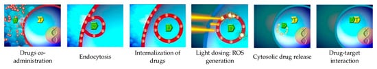
Photochemical internalization (PCI) is a further development of photodynamic therapy (PDT). In this report, we describe PCI as a potential tool for cellular internalization of chemotherapeutic agents or antigens and systematically review the ongoing research. One Phase-I clinical trial has been conducted, and it demonstrated that PCI-mediated bleomycin treatment was safe and identified tolerable doses of the photosensitizer disulfonated tetraphenyl chlorin (TPCS2a). Likewise, PCI was pre-clinically shown to mediate major histocompatibility complex (MHC) class I antigen presentation and generation of tumor-specific cytotoxic CD8+ T-lymphocytes (CTL) and cancer remission. A first clinical Phase I trial with the photosensitizer TPCS2a combined with human papilloma virus antigen (HPV) was recently completed and results are expected in 2020. Hence, photosensitizers and light can be used to mediate cytosolic delivery of endocytosed chemotherapeutics or antigens. While the therapeutic potential in cancer has been clearly demonstrated pre-clinically, further clinical trials are needed to reveal the true translational potential of PCI in humans.

photochemical internalization photodynamic therapy cytosolic delivery cancer vaccination cancer immunotherapy cross-presentation CTL

1. Introduction

1.1. Cancer Therapy Development

Cancer therapy has evolved since Coley’s toxins and the birth of radiotherapy in the early twentieth century. The 1940s saw the development of chemotherapeutics, the first monoclonal antibodies for targeted therapies appeared in the 1980s, and photodynamic therapy (PDT), which is the topic of the current special issue of Cancers, was approved for treatment of cancer in the 1990s [1][2]. However, the first reports on photochemical treatment of cancer lesions date back to the early twentieth century [3]. Moreover, the complex relationship and crosstalk between tumors and the immune system has become gradually untangled and changed our understanding of oncology and cancer therapy. Therefore, the development of cancer immunotherapies has recently gained scientific and clinical momentum, with a giant leap made in 2011 as the FDA approved Ipilimumab ®for treatment of advanced melanoma. Ipilimumab is a monoclonal antibody targeting cytotoxic T-lymphocyte-associated protein 4 (CTLA-4) [4][5]. Second generation checkpoint inhibitors, such as programmed cell death protein 1 (PD-1) and programmed cell death 1 ligand 1 (PD-L1) inhibitors, have followed [6][7], and, today, more than 10 different immunotherapeutic agents, including checkpoint inhibitors, vaccine-based therapies, oncolytic viruses, and T-cell directed therapies for nearly 20 different indications across countless tumor types are available [1].
Despite these success stories, cancer therapy remains challenging, which is a major goal that is efficient and provides site-specific delivery of the therapeutic agents, the improvement of therapeutic outcomes, and the reduction of damage to healthy tissue. These problems are, in part, being addressed by defining and targeting cancer-specific molecules. In this case, personalized treatment can aid the design of a patient-tailored specific targeted therapy, which allows administration of the right treatment to the right person at the right time [8]. Therefore, the utilization of macromolecules is becoming increasingly relevant. However, since many of the therapeutic targets or receptors are intracellular, internalization to the cell cytosol is often crucial to achieve the expected biological effect [9].

1.2. Methods for Cytosolic Targeting

During the last three decades, several approaches have been suggested for the delivery of antigens and drugs to the cytosol. The mechanism by which a virus fuses with the cell membrane and hijacks the host protein production machinery has been one inspiration. Viral vectors and viral transcellular transduction proteins such as the twin-arginin translocation (TAT) sequence from the human immunodeficiency virus (HIV) or VP22 from the herpes simplex virus (HSV) have been widely used [10][11][12][13][14]. Since most viruses are immunogenic, tumor antigens have become more immunogenic when delivered with a viral vector [15][16]. In addition, recombinant viruses can be easily produced, administered, and quality-controlled [10]. As some tumor cells, e.g., ovarian cancer or lung adenocarcinoma, express folate receptors on their surface, folic acid was applied to facilitate delivery of chemotherapeutic agents [17] in patients with platinum-refractory epithelial ovarian, primary peritoneal, or endometrial cancer [18] and progressive lung adenocarcinoma [19]. Other pharmaceutical strategies for cytosolic targeting of drugs include cationic particles to shuffle antigens across the negatively charged cellular membrane [20][21], pH-sensitive and fusogenic liposomes that break up acidified phagolysosomes [22][23], and micelle-based immune-stimulating complexes (ISCOMs) that may facilitate antigen cross-presentation [22][24]. Very recently, reports on the use of photochemical internalization (PCI), which is a further development of PDT, has been suggested as a method to internalize cytotoxic therapeutics in tumor cells [25][26][27][28][29][30][31][32] or vaccine antigens in antigen-presenting cells (APC) [20][22][33][34][35][36][37].

1.3. Photodynamic Therapy (PDT)

Photodynamic therapy (PDT) is a well-established technique for the clinical treatment of several neoplasms such as non-melanoma skin cancer, esophageal cancer, non-small-cell lung cancer (NSCLC), bladder cancer, cervical cancer, head and neck cancer, breast cancer, pancreatic cancer, or prostate cancer [38], as described elsewhere in this issue of Cancers. Briefly, a photosensitizer is administered to the tumor lesion, and subsequent light activation induces photochemical energy, the generation of reactive oxygen species (ROS), which damage the cell membrane to cause cell death [39][40][41].
The physicochemical cytotoxicity mediated by PDT can further trigger inflammatory reactions and even tumor-specific adaptive immune reactions [42][43]. This immunological effect of PDT typically follows the production of damage-associated molecular patterns (DAMPs) by necrotic or apoptotic tumor cells, which are then recognized by APCs [44]. Activated APCs can present tumor antigens to T cells for stimulation of tumor-specific immune responses. Although the exact mechanism for such PDT-mediated immune effects is unclear, it has been demonstrated that PDT can elicit production of pro-inflammatory cytokines and anti-tumor immune responses [45][46].

1.4. PCI—A Photosensitizer—And Light-Driven Technology for Cellular Internalization of Molecules

Photochemical internalization (PCI) was developed as a method for light-enhanced cytosolic release of membrane-impermeable molecules that have been taken up by cells and entrapped in endocytic vesicles [47] (Figure 1). The molecules, e.g., chemicals, proteins, or nucleic acid DNA or RNA, together with the photosensitizer, get internalized by the cell via endocytosis or phagocytosis. This process leaves the internalized molecules entrapped within the lumen of the endosome [48][49]. PCI is based on amphiphilic photosensitizers that allow time-dependent dissociation from the outer plasma membrane, but not from the endosome. While the plasma membrane is light-insensitive after a certain time, the photosensitizer cannot escape the endosomal lumen or membrane, which, therefore, remains light sensitive. Light-induced ROS formation causes endosomal leakage with translocation of the internalized molecule into the cytosol for interacting with its designated target [37][50]. By these means, PCI has been demonstrated to enhance the therapeutic effects of a large number of molecules, including many types of macromolecules and some chemotherapeutic agents, that are subject to endosome-lysosome entrapment [48][50]. Hence, PCI is a method for intracellular delivery of molecules, but also a technology to enhance therapeutic specificity and the efficacy of drugs [9].
Figure 1. Photochemical internalization. The drug is co-administered with the photosensitizer. The photosensitizer accumulates in cell membranes and the drug is taken up through endocytosis. ROS are generated during illumination, which leads to disruption of the endocytic membrane and release of the drug into the cytosol (modified with courtesy from PCI Biotech: http://pcibiotech.no/what-is-pci/).

1.5. Photosensitizers in Use

There are several characteristics a photosensitizer drug should meet, e.g., specific tumor uptake, low toxicity in the absence of light, and long absorption wavelength. Longer wavelengths allow deeper tissue penetration [44]. Some of the most investigated photosensitizing drugs include hypericin [39], porfimer [38], and 5-aminolevulinic acid (ALA) [50]. The lifetime and diffusion distance of ROS are very limited, and the location of damage upon light activation is, therefore, highly dependent on the localization of the photosensitizer [25]. Porfimer sodium (Photofrin) was the first approved PDT agent (in Canada in 1993), and was FDA approved in 1995 for the palliative treatment of obstructive esophageal cancer [38]. The prodrug ALA or ALA esters (Metvix and Hexvix/Cysview) are the most widely used porphyrin-based photosensitizers for PDT. The topical application of these prodrugs leads to the production of protoporphyrin IX, which is very effective for photodynamic detection (PDD) and fluorescence guided resection of non-muscle invasive bladder cancer or for the treatment of thin and superficial skin lesions [50].
On the other hand, photosensitizers for PCI should be amphiphilic in order to enable non-receptor mediated endocytosis and dissociation of excess photosensitizers from the plasma membrane [29]. Examples of specific amphiphilic photosensitizers are disulfonated aluminum phthalocyanine (AlPcS2a) and disulfonated tetraphenyl porphyrin (TPPS2a) [48]. Disulfonated tetraphenyl chlorin (TPCS2a) is a second generation photosensitizer developed for clinical use in PCI [51]. In a recent clinical Phase-I trial, TPCS2a was administered to patients with solid cutaneous or sub-cutaneous malignancies for internalization of the cytotoxic agent bleomycin [52]. Furthermore, a Phase-I dose-escalation study to assess the safety of fimaporfin-induced PCI of gemcitabine in patients with inoperable extrahepatic bile duct cancer (cholangiocarcinoma) and, based on the positive data, a pivotal Phase-II trial on extrahepatic biliary tract cancer has started recruiting patients [53]Table 1 gives an overview of the photosensitizers currently in use.
Table 1. Photosensitizers approved or under clinical trials.
Name Ex Wave-Length (nm) Manufacturer Application
FIRST GENERATION PHOTOSENSITIZERS
Porfimer sodium 630 Axcan Pharma PDT of esophageal cancer, lung adenocarcinoma, and endobronchial cancer
SECOND GENERATION PHOTOSENSITIZERS/Prodrugs
5-aminolaevulinic acid 635 DUSA
Stabiopharma
PDT of mild to moderate actinic keratosis Fluorescence guided resection of glioma
Methyl-aminolevulinic acid 579–670 Galderma PDT of non-hyperkeratotic actinic keratosis and basal cell carcinoma
Temoporfin 652 Biolitec PDT of advanced head and neck cancer
Talaporfin 664 Meiji Seika
Novartis
PDT of early centrally located lung cancer
Verteporfin 690 Novartis PDT of age-related macular degeneration
Redaporfin 749 Luzitin PDT of biliary tract cancer
PHOTOSENSITIZERS UNDER CLINICAL INVESTIGATIONS
Fotolon 665 Apocare Pharma PDT of nasopharyngeal, sarcoma
Hexylaminolevulinate 635 Photocure PDT of HPV-induced cervical precancerous lesions and non-muscle invasive bladder cancer
Radachlorin 662 Rada-pharma PDT of skin cancer
Photochlor (HTTP) 664 Rosewell Park PDT of head and neck cancer
Padeliporfin 762 Negma-Lerads PDT of prostate cancer
Motexafin lutetium 732 Pharmacyclics PDT of coronary artery disease
Rostaprofin 664 Miravant PDT of age-related macular degeneration
Talaporfin 664 Meiji Seika PDT of colorectal neoplasms, liver metastasis
Fimaporfin 435 PCI Biotech PCI of cutaneous or sub-cutaneous malignancies, cholangiocarcinoma and PCI of vaccine antigens

1.6. PCI in Immunotherapy

Immunotherapies directed against cancer cells can broadly be divided into active, passive, or immunomodulatory. Passive immunotherapy involves the administration of tumor-specific lymphocytes or antibodies, whereas adjuvants or other immunologically active compounds can be immunomodulatory [4]. Checkpoint inhibitors operate by modulating the immune system’s endogenous mechanisms of T-cell regulation. They block co-inhibitory molecules on cytotoxic T lymphocytes (CTLs) and, consequently, debunk the inhibitory signals tumor cells elicit on T cells with promising results in clinical trials [54][55][56][57].
Cancer vaccines intend to elicit an active immune response by stimulating the body’s own immune system to target tumor-specific antigens. For recognition, such antigens can be presented by APCs to T cells by either intracellular or extracellular pathways. Extracellular pathogens and vaccines enter the APCs via the endosome or phagosome formation (Figure 2). The antigen uptake and the maturation of the antigen-containing endosome depend on the activation of pathogen recognition receptors such as Toll-like receptors (TLRs) and nucleotide-binding oligomerization domain-like or NOD-like receptors (NLRs). The endo- or phagosomes then fuse with lysosomes to form endo- or phagolysosomes in which the antigen gets loaded on major histocompatibility complex (MHC) class II molecules for presentation to CD4 T-helper cells. These cells typically provide help to B-cells for production of antibodies [20][58]. In that manner, the antigen never reaches the cytosol of the APC. Intracellular antigens, such as proteins from worn-out endogenous proteins, have direct access to the cytosol of the APCs. These antigens are processed by the proteasome to produce small peptide-fragments that can bind MHC class-I molecules in the endoplasmic reticulum. The peptide-MHC complex is transported to the cell surface for presentation to cognate T-cell receptors on CD8 T cells, which may then differentiate into CTLs. Since CTLs have been found to be one of the key players in fighting cancer [20][37][59], vaccination with tumor-associated antigens aims at inducing strong CTL immune responses. Hence, one major challenge is accessing the MHC class I pathway of antigen presentation. For this cross-presentation, antigens need to reach the cytosol of APCs, and this can be achieved by direct diffusion through the cell membrane or through endosomal escape after uptake.
Figure 2. Antigen uptake, processing, and T-cell presentation in PCI-based vaccination. Photosensitizer and antigen are endocytosed into an antigen-presenting cell (APC). The photosensitizer is attached to the endosomal membrane and the antigen is contained in the endosomal lumen. After a wash-out period, where excess photosensitizer dissociates from the outer plasma membrane, light exposure causes endosomal eruption and cytosolic release of antigen for proteasomal degradation and MHC class-I presentation to CD8 T cells. In the absence of the photosensitizer and light, endosomes mature and fuse with lysosomes for MHC class-II presentation of digested antigens to CD4 T cells.
Once it was demonstrated, PCI enabled internalization of chemotherapeutics into cells. The idea to deliver antigens to the cytosol of APCs for stimulation of MHC class I-restricted CD8 T-cell responses was born. Antigen and photosensitizer are co-administered, and subsequent endocytosis of antigen into photosensitized APCs enables ROS-mediated disruption of endosomes upon light treatment. Time is given for the amphiphilic photosensitizer to dissociate from the outer cellular membrane before light-activation. Lastly, the antigen is released from the endosomes into the cytosol where it can enter the MHC class I pathway of antigen presentation and stimulation of CTLs. Figure 2 illustrates PCI-mediated endosomal escape, proteasomal degradation, and CTL induction.

1.7. Cancer Vaccines

While prophylactic cancer vaccines can decrease the probability of late tumor development and have already been proven effective, the development of therapeutic vaccines against an established tumor is more difficult. Prophylactic vaccines against the human papilloma virus (HPV) and the hepatitis B virus (HBV) are clinically established to prevent cervical and oropharyngeal head and head and neck squamous cell carcinoma (HNSCC) or hepatocellular carcinoma (HCC), respectively [60][61]. The first and, so far, only approved therapeutic cancer vaccine is Sipuleucel-T, which is a dendritic cell (DC)-based cancer vaccine for the treatment of metastatic castration-resistant prostate cancer that increased overall survival by four to five months [4][62].

2. PCI of Cytotoxic Therapeutics

PDT kills tumorous tissue by means of photosensitizer and light. In contrast, photosensitizer and light in PCI is not primarily used to kill tumor cells but as a vehicle for specific and intracellular delivery of anti-cancer drugs. By existing data, PCI has proven to be a promising method for targeting therapeutic molecules to tumor cells for the purpose of specific killing. A wide range of drugs have been tested, e.g., macromolecular proteins, peptides, nucleic acids, and synthetic polymers, but also low-molecular weight chemotherapeutic drugs [47][49][50][63]. The method of PCI of cytotoxic therapeutics is especially applicable to drugs where the therapeutic target is intracellular and the PCI mediating cytosolic delivery of drugs has poor access to their cytosolic target.
One potential application of the PCI of cytotoxic therapeutics is to overcome drug resistance, which is one of the major challenges to reach effective cancer treatment. Up to 50% of malignant tumors are intrinsically resistant to chemotherapy [64], with the additional problem of attained resistance after repetitive drug administration. In this case, PDT represents an alternative treatment method that is usually not associated with resistance. However, PDT is often tissue and cell unspecific and, therefore, mostly applicable for superficial and solid tumors (Table 1). By contrast, the combination of PDT and chemotherapy in PCI has been suggested to enable PDT-guided delivery of chemotherapeutic drugs to specific tumor cells, and, thereby, overcome the problem of resistance [50]. For example, the chemotherapeutic drug bleomycin is approved for treatment of testicular carcinomas, lymphomas, head and neck cancers, and other non-melanoma skin cancers, but is known to become trapped in intracellular compartments after administration, which consequently leads to the need for higher therapeutic doses [65]. This is associated with an increased risk of pneumonitis and subsequent lung fibrosis [66]. However, PCI of bleomycin enhanced cytotoxicity compared to bleomycin alone both in vitro [26] and in vivo [28], which suggests improved bio-distribution and organ specificity with reduced resistance. By consequence, PCI can enable reduction of the dose needed to achieve a therapeutic effect and thereby reducing non-specific, adverse events [67].
The use of PDT for cell-targeted delivery of immunotoxins, such as the ribosome-inactivating proteins saporin and gelonin, have also been investigated with PCI. Immunotoxins were coupled with specific cell-targeting proteins that can bind to CSPG4 [32], to CD133-expressing cancer stem cells [29][30], to EGFR [27][31], or to VEGFR [68]. Targeting of CD133-expressing cancer stem cells is based on the knowledge that a small population of stem-like cancer cells are often resistant to traditional chemotherapies, where the consequence is tumor relapse and metastasis [69]. Unfortunately, the clinical success of CD133-directed immunotoxins has been compromised by the potential harm on normal stem cells that also express CD133 [70][71][72][73]. However, the PCI of CD133-directed immunotoxins seems to pose a potential solution to normal stem cell toxicity by increasing tumor-cell selectivity [29][30]. Systemically administered CD133-directed immunotoxins were found to localize predominantly in the tumor tissue, with no detection in normal tissue except in the kidney and the liver [30]. Hence, PCI may reduce the frequency of drug administrations, which by consequence may reduce treatment-associated AEs and resistance. Since the efficacy of conventional cancer therapies are often limited by a dose-dependent toxicity [50][63][74], PCI may represent a rational and promising approach for chemotherapeutic targeting and killing of drug-therapy or multi-therapy-resistant cancer cells. PCI of bleomycin was safe in patients with squamous cell carcinoma or other advanced or recurrent malignancies of the head and neck, torso, and upper limbs [52][75]. A pivotal Phase-II study is currently recruiting patients with inoperable bile duct cancer to assess effectiveness of PCI of gemcitabine complemented by systemic gemcitabine/cisplatin chemotherapy compared to gemcitabine/cisplatin alone [53].

3. PCI in Immunotherapy

The immune system can play an important role in fighting cancer cells. Immunotherapy, such as checkpoint-inhibition, cytokine therapy, adoptive cell transfer therapy, and therapeutic vaccines all have the potential to induce immune responses that can surveil tumor, suppress growth of or kill cancerous cells, and give the patient a long-lasting immunity that may prevent remissions. However, cancer cells can interfere with the immune system in many ways. In this case, the potential immune-suppressive tumor micro environment (TME) may represent a significant challenge for effective anti-tumor therapies. The TME can be seen as an environment generated by various interactions between cancer cells and immune cells. Cancer cells, as they develop and grow, exploit immune-regulatory mechanisms by interacting with immune cells such as regulatory T and B cells, DCs, and myeloid-derived suppressor cells. Tumors can downregulate protein p53 or other tumor suppressors, downregulate MHC class I or co-stimulatory molecules on APCs, attract immunosuppressive leucocytes, activate CTLA4, PD1, or other co-inhibitory receptors on T cells [4][59][76][77]. The tumor-cell mediated activation of co-inhibitory receptors on T cells directly interferes with T-cell mediated tumor destruction [78], whereas the lack of co-stimulation can lead to T-cell anergy [79]. Additionally, immune escape due to self-tolerance of tumor antigens makes it difficult to target the immune system, notably T cells [80].
While adjuvant immunotherapy with checkpoint inhibitors have found wide application during the last decade, therapeutic cancer vaccination has proven more laborious and less effective. Cancer vaccination aims to stimulate tumor-specific immune responses against delivered antigens. In order to achieve this, one has to overcome hurdles, such as the correct selection of antigens among the plethora of heterogeneously expressed and genetically unstable tumor antigens [4][81] and the use of appropriate adjuvants. To avoid mechanisms of central tolerance in the thymus, it is important to choose immunogenic antigens for vaccination [59]. In this case, neoantigens and viral antigens are not subject to self-tolerance mechanisms and could be used for stronger anti-tumor T-cell responses than regular tumor antigens. Since tumor neoantigens are usually patient-specific, they typically require personalized vaccines, whereas tumor-specific viral antigens could be used for off-the-shelf vaccines.
Cytotoxic T cells and natural killer (NK) cells have shown to make important immunological contributions in fighting tumors. In order for therapeutic vaccines to trigger the generation of tumor-specific CTLs, the MHC class I pathway of antigen presentation needs to be accessed. However, vaccine antigens end up in phagolysosomes of APCs and are presented in the context of MHC class II, which leads to stimulation of CD4 rather than CD8 T cells. One possible approach for CTL activation is to shuffle the antigen across the plasma membrane and, thereby, avoid endocytosis altogether. Another way has been obtained by triggering endosomal escape of the internalized antigen subsequent to the endocytosis or phagocytosis of antigens into APCs. The combination of antigens with a photosensitizer and light can facilitate cytosolic release of the endocytosed antigen by disruption of the endosomal membrane. The now cytosolic antigen can be processed by proteasomes and presented via MHC class I pathway for stimulation of CD8 T-cell responses, which, therefore, overcame the problem of the CD8 deficit after vaccination (Figure 2).
Successful stimulation of tumor-specific immunity by PCI has been demonstrated in several mouse models of cancer. PCI mediated induction of antigen-specific CD8 T-cell proliferation and IFN-γ production [20][22][33][34][35][36], prevention of tumor grafting [34][35], suppression of tumor growth, and improved progression-free survival in mice [35][37]. Studies have demonstrated the mechanism of antigen and photosensitizer uptake in APCs and that, upon application of light, the antigen is released from endosomes or even phagolysosomes into the cytosol [34][35][36]. While it has been recognized that the generation of primary CD8 T-cell responses to non-inflammatory antigens typically require MHC class II-restricted CD4 T helper cells, Varypataki et al. demonstrated that CD8 T-cell responses and their ability to control tumor growth after PCI-based vaccination were not impaired in MHC class-II and CD4 T-cell deficient mice [37]. In order to verify the significance of the data, further tumor models will be needed, and future findings may have clinical importance with regard to the fact that many tumor patients are treated with CD4 T-cell-sensitive immunosuppressive agents [82]. Antigen-specific CD8 T-cell responses could also be generated autologously in mice after prior PCI-mediated loading of the antigen to DCs in vitro [33]. In light of the autologous vaccine Sipuleucel-T, which is the only FDA-approved and therapeutic cancer vaccine, it would be interesting to follow up on this technique with other models, e.g., DNA or mRNA treated DCs or DCs treated with tumor antigens. This would enable us to conclude on the true potential of PCI-based autologous vaccination. In the above mentioned report [33], the DCs were treated with the model antigen OVA, which is a strong antigen, while tumor antigens are typically weak.
A Phase-I clinical trial was completed on 27 August, 2019 on the safety of photochemical internalization of a large immunogenic protein (KLH) and two smaller and less immunogenic peptides (HPV) in healthy volunteers [83]. The primary objective was to study the incidence of AEs after a single administration of the photosensitizer and light. The first results thereof were presented at the ESMO Immuno-Oncology Congress in December 2019 [84]. The induction of HPV-specific immune response in blood showed an increase in the number of healthy donors with HPV-specific CD4+ and CD8+ T-cell responses to PCI-based vaccination compared to baseline levels. Further details and results of the study are expected to be released. However, additional Phase-II and III trials will be needed to investigate the translational potential of current pre-clinical and anecdotal clinical reports.
Therapeutic benefits of anti-cancer vaccines in development are inconclusive. Even with optimized antigen selection and delivery, tumor-intrinsic evasive actions, as well as the lack of understanding of the tumor-microenvironment, pose unforeseeable obstacles. A deeper understanding of the interactions between the immune system and cancer cells will be inevitable for treatment optimization. One possible approach to overcome the immunosuppressant TME has been suggested to be the combination of cancer vaccines and other immunotherapies such as checkpoint inhibition, cytotoxic agents, or classical chemotherapies [59]. Therapeutic vaccination could help prime the immune system to recognize tumor antigens or individualized neoantigens, and the effect of established cancer therapies could, therefore, be improved [36]. Another obstacle for future clinical trials is expected to be the possible shift from self-tolerance to autoimmunity, triggered by immunotherapy combined with local inflammation following treatment. So far, no such effects have been observed with PCI, but, since autoimmunity typically develops slowly, a further assessment will be required. However, the risk of autoimmune reactions is not limited to cancer vaccination, but to all immune-stimulating procedures. Photochemical internalization, as a technology with the potential to enhance therapeutic cancer vaccines, can be seen as a promising tool to optimize anti-cancer immunotherapy.

References

  1. Osipov, A.; Murphy, A.; Zheng, L. From immune checkpoints to vaccines: The past, present and future of cancer immunotherapy. Adv. Cancer Res. 2019, 143, 63–144.
  2. Falzone, L.; Salomone, S.; Libra, M. Evolution of cancer pharmacological treatments at the turn of the third millennium. Front. Pharmacol. 2018, 9, 1300.
  3. Agostinis, P.; Berg, K.; Cengel, K.A. Photodynamic Therapy of Cancer: An Update. CA Cancer J. Clin. 2011, 61, 250–281.
  4. Melero, I.; Gaudernack, G.; Gerritsen, W.; Huber, C.; Parmiani, G.; Scholl, S.; Thatcher, N.; Wagstaff, J.; Zielinski, C.; Faulkner, I.; et al. Therapeutic vaccines for cancer: An overview of clinical trials. Nat. Rev. Clin. Oncol. 2014, 11, 509–524.
  5. Kvistborg, P.; Philips, D.; Kelderman, S.; Hageman, L.; Ottensmeier, C.; Joseph-Pietras, D.; Welters, M.J.P.; Van Der Burg, S.; Kapiteijn, E.; Michielin, O.; et al. Anti-CTLA-4 therapy broadens the melanoma-reactive CD8+ T cell response. Sci. Transl. Med. 2014, 6, 1–10.
  6. Topalian, S.L.; Hodi, S.F.; Brahmer, J.R.; Gettinger, S.N.; Smith, D.C.; McDermott, D.F.; Powderly, J.D.; Carvajal, R.D.; Sosman, J.A.; Atkins, M.B.; et al. Safety, Activity, and Immune Correlates of Anti–PD-1 Antibody in Cancer. N. Engl. J. Med. 2012, 366, 2443–2454.
  7. Brahmer, J.R.; Tykodi, S.S.; Chow, L.Q.M.; Hwu, W.-J.; Topalian, S.L.; Hwu, P.; Drake, C.G.; Camacho, L.H.; Kauh, J. Safety and Activity of Anti–PD-L1 Antibody in Patients with Advanced Cancer. N. Engl. J. Med. 2012, 366, 2455–2465.
  8. Palumbo, M.O.; Kavan, P.; Miller, W.H.; Panasci, L.; Assouline, S.; Johnson, N.; Cohen, V.; Patenaude, F.; Pollak, M.; Jagoe, R.T.; et al. Systemic cancer therapy: Achievements and challenges that lie ahead. Front. Pharmacol. 2013, 4, 1–9.
  9. Berg, K.; Weyergang, A.; Prasmickaite, L.; Bonsted, A.; Hogset, A.; Strand, M.-T.R.; Wagner, E.; Selbo, P.K. Photochemical Internalization (PCI): A Technology for Drug Delivery. In Photodynamic Therapy—Methods and Protocols, Methods in Molecular Biology; Gomer, C.J., Ed.; Springer: Berlin/Heidelberg, Germany, 2010; pp. 133–145. ISBN 9781607616962.
  10. Larocca, C.; Schlom, J. Viral Vector-Based Therapeutic Cancer Vaccines. Cancer J. 2011, 17, 359–371.
  11. Choi, Y.; Chang, J. Viral vectors for vaccine applications. Clin. Exp. Vaccine Res. 2013, 2, 97.
  12. Fittipaldi, A.; Giacca, M. Transcellular protein transduction using the Tat protein of HIV-1. Adv. Drug Deliv. Rev. 2005, 57, 597–608.
  13. Tanaka, M.; Kato, A.; Satoh, Y.; Ide, T.; Sagou, K.; Kimura, K.; Hasegawa, H.; Kawaguchi, Y. Herpes Simplex Virus 1 VP22 Regulates Translocation of Multiple Viral and Cellular Proteins and Promotes Neurovirulence. J. Virol. 2012, 86, 5264–5277.
  14. Lindgren, M.; Hällbrink, M.; Prochiantz, A.; Langel, Ü. Cell-penetrating peptides. Trends Pharmacol. Sci. 2000, 21, 99–103.
  15. Arlen, P.M.; Kaufman, H.L.; DiPaola, R.S. Pox viral vaccine approaches. Semin. Oncol. 2005, 32, 549–555.
  16. Kass, E.; Schlom, J.; Thompson, J.; Guadagni, F.; Graziano, P.; Greiner, J.W. Induction of protective host immunity to carcinoembryonic antigen (CEA), a self-antigen in CEA transgenic mice, by immunizing with a recombinant vaccinia-CEA virus. Cancer Res. 1999, 59, 676–683.
  17. Ledermann, J.A.; Canevari, S.; Thigpen, T. Targeting the folate receptor: Diagnostic and therapeutic approaches to personalize cancer treatments. Ann. Oncol. 2015, 26, 2034–2043.
  18. US National Library of Medicine Study of Vintafolide (MK-8109, EC145) in Participants with Advanced Ovarian and Endometrial Cancers (MK-8109-007, EC-FV-02). Available online: (accessed on 13 October 2019).
  19. US National Library of Medicine Study of Vintafolide (MK-8109, EC145) in Participants with Progressive Adenocarcinoma of the Lung (MK-8109-008, EC-FV-03). Available online: (accessed on 13 October 2019).
  20. Hjálmsdóttir, Á.; Bühler, C.; Vonwil, V.; Roveri, M.; Håkerud, M.; Wäckerle-Men, Y.; Gander, B.; Johansen, P. Cytosolic Delivery of Liposomal Vaccines by Means of the Concomitant Photosensitization of Phagosomes. Mol. Pharm. 2016, 13, 320–329.
  21. Karlsen, K.; Korsholm, K.S.; Mortensen, R.; Ghiasi, S.M.; Andersen, P.; Foged, C.; Christensen, D. A stable nanoparticulate DDA/MMG formulation acts synergistically with CpG ODN 1826 to enhance the CD4+ T-cell response. Nanomedicine 2014, 9, 2625–2638.
  22. Bruno, C.; Waeckerle-Men, Y.; Håkerud, M.; Kündig, T.M.; Gander, B.; Johansen, P. Photosensitizer and Light Pave the Way for Cytosolic Targeting and Generation of Cytosolic CD8 T Cells Using PLGA Vaccine Particles. J. Immunol. 2015, 195, 166–173.
  23. Watarai, S.; Iwase, T.; Tajima, T.; Yuba, E.; Kono, K. Efficiency of pH-Sensitive Fusogenic Polymer-Modified Liposomes as a Vaccine Carrier. Sci. World J. 2013.
  24. Sanders, M.T.; Brown, L.E.; Deliyannis, G.; Pearse, M.J. ISCOMTM-based vaccines: The second decade. Immunol. Cell Biol. 2005, 83, 119–128.
  25. Olsen, C.E.; Berg, K.; Selbo, P.K.; Weyergang, A. Circumvention of resistance to photodynamic therapy in doxorubicin- resistant sarcoma by photochemical internalization of gelonin. Free Radic. Biol. Med. 2013, 65, 1300–1309.
  26. O’Rourke, C.; Hopper, C.; MacRobert, A.J.; Phillips, J.B.; Woodhams, J.H. Could clinical photochemical internalisation be optimised to avoid neuronal toxicity? Int. J. Pharm. 2017, 528, 133–143.
  27. Martínez-Jothar, L.; Beztsinna, N.; Van Nostrum, C.F.; Hennink, W.E.; Oliveira, S. Selective Cytotoxicity to HER2 Positive Breast Cancer Cells by Saporin-Loaded Nanobody-Targeted Polymeric Nanoparticles in Combination with Photochemical Internalization. Mol. Pharm. 2019, 16, 1633–1647.
  28. Norum, O.J.; Fremstedal, A.S.V.; Weyergang, A.; Golab, J.; Berg, K. Photochemical delivery of bleomycin induces T-cell activation of importance for curative effect and systemic anti-tumor immunity. J. Control. Release 2017, 268, 120–127.
  29. Stratford, E.W.; Bostad, M.; Castro, R.; Skarpen, E.; Berg, K.; Høgset, A.; Myklebost, O.; Selbo, P.K. Photochemical internalization of CD133-targeting immunotoxins efficiently depletes sarcoma cells with stem-like properties and reduces tumorigenicity. Biochim. Biophys. Acta Gen. Subj. 2013, 1830, 4235–4243.
  30. Bostad, M.; Olsen, C.E.; Peng, Q.; Berg, K.; Høgset, A.; Selbo, P.K. Light-controlled endosomal escape of the novel CD133-targeting immunotoxin AC133-saporin by photochemical internalization—A minimally invasive cancer stem cell-targeting strategy. J. Control. Release 2015, 206, 37–48.
  31. Berstad, M.B.; Cheung, L.H.; Berg, K.; Peng, Q.; Fremstedal, A.S.V.; Patzke, S.; Rosenblum, M.G.; Weyergang, A. Design of an EGFR-targeting toxin for photochemical delivery: In vitro and in vivo selectivity and efficacy. Oncogene 2015, 34, 5582–5592.
  32. Eng, M.S.; Kaur, J.; Prasmickaite, L.; Engesæter, B.; Weyergang, A.; Skarpen, E.; Berg, K.; Rosenblum, M.G.; Mælandsmo, G.M.; Høgset, A.; et al. Enhanced targeting of triple-negative breast carcinoma and malignant melanoma by photochemical internalization of CSPG4-targeting immunotoxins. Photochem. Photobiol. Sci. 2018, 17, 539–551.
  33. Waeckerle-Men, Y.; Mauracher, A.; Håkerud, M.; Mohanan, D.; Kündig, T.M.; Høgset, A.; Johansen, P. Photochemical targeting of antigens to the cytosol for stimulation of MHC class-I-restricted T-cell responses. Eur. J. Pharm. Biopharm. 2013, 85, 34–41.
  34. Håkerud, M.; Waeckerle-Men, Y.; Selbo, P.K.; Kündig, T.M.; Høgset, A.; Johansen, P. Intradermal photosensitisation facilitates stimulation of MHC class-I restricted CD8 T-cell responses of co-administered antigen. J. Control. Release 2014, 174, 143–150.
  35. Håkerud, M.; Selbo, P.K.; Waeckerle-Men, Y.; Contassot, E.; Dziunycz, P.; Kündig, T.M.; Høgset, A.; Johansen, P. Photosensitisation facilitates cross-priming of adjuvant-free protein vaccines and stimulation of tumour-suppressing CD8 T cells. J. Control. Release 2015, 198, 10–17.
  36. Haug, M.; Brede, G.; Håkerud, M.; Nedberg, A.G.; Gederaas, O.A.; Flo, T.H.; Edwards, V.T.; Selbo, P.K.; Høgset, A.; Halaas, Ø. Photochemical internalization of peptide antigens provides a novel strategy to realize therapeutic cancer vaccination. Front. Immunol. 2018, 9, 1–14.
  37. Varypataki, E.M.; Hasler, F.; Waeckerle-Men, Y.; Vogel-Kindgen, S.; Høgset, A.; Kündig, T.M.; Gander, B.; Halin, C.; Johansen, P. Combined Photosensitization and Vaccination Enable CD8 T-Cell Immunity and Tumor Suppression Independent of CD4 T-Cell Help. Front. Immunol. 2019, 10, 1–12.
  38. Baskaran, R.; Lee, J.; Yang, S.-G. Clinical development of photodynamic agents and therapeutic applications. Biomater. Res. 2018, 22, 1–8.
  39. Agostinis, P.; Vantieghem, A.; Merlevede, W.; De Witte, P.A.M. Hypericin in cancer treatment: More light on the way. Int. J. Biochem. Cell Biol. 2002, 34, 221–241.
  40. Juarranz, Á.; Jaén, P.; Sanz-Rodríguez, F.; Cuevas, J.; González, S. Photodynamic therapy of cancer. Basic principles and applications. Clin. Transl. Oncol. 2008, 10, 148–154.
  41. Van Straten, D.; Mashayekhi, V.; de Bruijn, H.S.; Oliveira, S.; Robinson, D.J. Oncologic photodynamic therapy: Basic principles, current clinical status and future directions. Cancers 2017, 9, 19.
  42. Castano, A.P.; Mroz, P.; Wu, M.X.; Hamblin, M.R. Photodynamic therapy plus low-dose cyclophosphamide generates antitumor immunity in a mouse model. Proc. Natl. Acad. Sci. USA 2008, 105, 5495–5500.
  43. Mroz, P.; Szokalska, A.; Wu, M.X.; Hamblin, M.R. Photodynamic therapy of tumors can lead to development of systemic antigen-specific immune response. PLoS ONE 2010, 5, 15194.
  44. Brodin, N.P.; Guha, C.; Tomé, W.A. Photodynamic therapy and its role in combined modality anticancer treatment. Technol. Cancer Res. Treat. 2015, 14, 355–368.
  45. Mroz, P.; Hashmi, J.T.; Huang, Y.Y.; Lange, N.; Hamblin, M.R. Stimulation of anti-tumor immunity by photodynamic therapy. Expert Rev. Clin. Immunol. 2011, 7, 75–91.
  46. Garg, A.D.; Nowis, D.; Golab, J.; Agostinis, P. Photodynamic therapy: Illuminating the road from cell death towards anti-tumour immunity. Apoptosis 2010, 15, 1050–1071.
  47. Berg, K.; Selbo, P.K.; Prasmickaite, L.; Tjelle, T.E.; Sandvig, K.; Moan, J.; Gaudernack, G.; Fodstad, Ø.; Kjølsrud, S.; Anholt, H.; et al. Photochemical internalization: A novel technology for delivery of macromolecules into cytosol. Cancer Res. 1999, 59, 1180–1183.
  48. Shin, D.; Christie, C.; Ju, D.; Nair, R.K.; Molina, S.; Berg, K.; Krasieva, T.B.; Madsen, S.J.; Hirschberg, H. Photochemical internalization enhanced macrophage delivered chemotherapy. Photodiagnosis Photodyn. Ther. 2018, 21, 156–162.
  49. Selbo, P.K.; Weyergang, A.; Høgset, A.; Norum, O.J.; Berstad, M.B.; Vikdal, M.; Berg, K. Photochemical internalization provides time- and space-controlled endolysosomal escape of therapeutic molecules. J. Control. Release 2010, 148, 2–12.
  50. Berg, K.; Folini, M.; Prasmickaite, L.; Selbo, P.; Bonsted, A.; Engesaeter, B.; Zaffaroni, N.; Weyergang, A.; Dietzea, A.; Maelandsmo, G.; et al. Photochemical Internalization: A New Tool for Drug Delivery. Curr. Pharm. Biotechnol. 2007, 8, 362–372.
  51. Berg, K.; Nordstrand, S.; Selbo, P.K.; Tran, D.T.T.; Angell-Petersen, E.; Høgset, A. Disulfonated tetraphenyl chlorin (TPCS 2a), a novel photosensitizer developed for clinical utilization of photochemical internalization. Photochem. Photobiol. Sci. 2011, 10, 1637–1651.
  52. Sultan, A.A.; Jerjes, W.; Berg, K.; Høgset, A.; Mosse, C.A.; Hamoudi, R.; Hamdoon, Z.; Simeon, C.; Carnell, D.; Forster, M.; et al. Disulfonated tetraphenyl chlorin (TPCS2a)-induced photochemical internalisation of bleomycin in patients with solid malignancies: A phase 1, dose-escalation, first-in-man trial. Lancet Oncol. 2016, 17, 1217–1229.
  53. US National Library of Medicine PCI Treatment/Gemcitabine & Chemotherapy vs. Chemotherapy Alone in Patients with Inoperable Extrahepatic Bile Duct Cancer (RELEASE). Available online: (accessed on 11 October 2019).
  54. Page, D.B.; Postow, M.A.; Callahan, M.K.; Allison, J.P.; Wolchok, J.D. Immune Modulation in Cancer with Antibodies. Annu. Rev. Med. 2014, 65, 185–202.
  55. Naidoo, J.; Page, D.B.; Wolchok, J.D. Immune modulation for cancer therapy. Br. J. Cancer 2014, 111, 2214–2219.
  56. Larkin, J.; Chiarion-Sileni, V.; Gonzalez, R.; Grob, J.-J.; Rutkowski, P.; Lao, C.D.; Cowey, C.L.; Schadendorf, D.; Wagstaff, J.; Dummer, R.; et al. Five-Year Survival with Combined Nivolumab and Ipilimumab in Advanced Melanoma. N. Engl. J. Med. 2019, 381, 1535–1546.
  57. US National Library of Medicine Phase 3 Study of Nivolumab or Nivolumab Plus Ipilimumab Versus Ipilimumab Alone in Previously Untreated Advanced Melanoma (CheckMate 067). Available online: (accessed on 14 October 2019).
  58. Zinkernagel, R.M. Immunological memory protective immunity. Cell. Mol. Life Sci. 2012, 69, 1635–1640.
  59. Van Der Burg, S.H.; Arens, R.; Ossendorp, F.; Van Hall, T.; Melief, C.J.M. Vaccines for established cancer: Overcoming the challenges posed by immune evasion. Nat. Rev. Cancer 2016, 16, 219–233.
  60. Mammas, I.N.; Sourvinos, G.; Zaravinos, A.; Spandidos, D.A. Vaccination against human papilloma virus (HPV): Epidemiological evidence of HPV in non-genital cancers. Pathol. Oncol. Res. 2011, 17, 103–119.
  61. Chemin, I. Evaluation of a hepatitis B vaccination program in Taiwan: Impact on hepatocellular carcinoma development. Futur. Oncol. 2010, 6, 21–23.
  62. Graff, J.N.; Chamberlain, E.D. Sipuleucel-T in the treatment of prostate cancer: An evidence-based review of its place in therapy. Core Evid. 2014, 10, 1–11.
  63. Høgset, A.; Prasmickaite, L.; Selbo, P.K.; Hellum, M.; Engesæter, B.; Bonsted, A.; Berg, K. Photochemical internalisation in drug and gene delivery. Adv. Drug Deliv. Rev. 2004, 56, 95–115.
  64. Perez, R.P.; Hamilton, T.C.; Ozols, R.F.; Young, R.C. Mechanisms and modulation of resistance to chemotherapy in ovarian cancer. Cancer 1993, 71, 1571–1580.
  65. Pron, G.; Mahrour, N.; Orlowski, S.; Tounekti, O.; Poddevin, B.; Belehradek, J.; Mir, L.M. Internalisation of the bleomycin molecules responsible for bleomycin toxicity: A receptor-mediated endocytosis mechanism. Biochem. Pharmacol. 1999, 57, 45–56.
  66. Sleijfer, S. Bleomycin-induced pneumonitis. Chest 2001, 120, 617–624.
  67. Berg, K.; Dietze, A.; Kaalhus, O.; Høgset, A. Site-specific drug delivery by photochemical internalization enhances the antitumor effect of bleomycin. Clin. Cancer Res. 2005, 11, 8476–8485.
  68. Weyergang, A.; Fremstedal, A.S.; Skarpen, E.; Peng, Q.; Mohamedali, K.A.; Eng, M.S.; Cheung, L.H.; Rosenblum, M.G.; Waltenberger, J.; Berg, K. Light-enhanced VEGF121/rGel: A tumor targeted modality with vascular and immune-mediated efficacy. J. Control. Release 2018, 288, 161–172.
  69. Liu, B.; Ma, W.; Jha, R.K.; Gurung, K. Cancer stem cells in osteosarcoma: Recent progress and perspective. Acta Oncol. 2011, 50, 1142–1150.
  70. Waldron, N.N.; Kaufman, D.S.; Oh, S.; Inde, Z.; Hexum, M.K.; Ohlfest, J.R.; Vallera, D.A. Targeting tumor-initiating cancer cells with dCD133KDEL shows impressive tumor reductions in a xenotransplant model of human head and neck cancer Nate. Mol. Cancer Ther. 2011, 10, 1829–1838.
  71. Smith, L.M.; Nesterova, A.; Ryan, M.C.; Duniho, S.; Jonas, M.; Anderson, M.; Zabinski, R.F.; Sutherland, M.K.; Gerber, H.P.; Van Orden, K.L.; et al. CD133/prominin-1 is a potential therapeutic target for antibody-drug conjugates in hepatocellular and gastric cancers. Br. J. Cancer 2008, 99, 100–109.
  72. Alewine, C.; Hassan, R.; Pastan, I. Advances in Anticancer Immunotoxin Therapy. Oncologist 2015, 20, 176–185.
  73. Li, M.; Liu, Z.S.; Liu, X.L.; Hui, Q.; Lu, S.Y.; Qu, L.L.; Li, Y.S.; Zhou, Y.; Ren, H.L.; Hu, P. Clinical targeting recombinant immunotoxins for cancer therapy. Onco. Targets. Ther. 2017, 10, 3645–3665.
  74. Selbo, P.K.; Sivam, G.; Fodstad, Y.; Sandvig, K.; Berg, K. In vivo documentation of photochemical internalization, a novel approach to site specific cancer therapy. Int. J. Cancer 2001, 92, 761–766.
  75. University College London Safety Study of Amphinex Based Photochemical Internalisation (PCI) of Bleomycin in Patients with Cutaneous Cancer. Available online: (accessed on 27 September 2019).
  76. Zanetti, M. Tapping CD4 T Cells for Cancer Immunotherapy: The Choice of Personalized Genomics. J. Immunol. 2015, 194, 2049–2056.
  77. Gajewski, T.F.; Meng, Y.; Blank, C.; Brown, I.; Kacha, A.; Kline, J.; Harlin, H. Immune resistance orchestrated by the tumor microenvironment. Immunol. Rev. 2006, 213, 131–145.
  78. Dong, H.; Strome, S.E.; Salomao, D.R.; Tamura, H.; Hirano, F.; Flies, D.B.; Roche, P.C.; Lu, J.; Zhu, G.; Tamada, K.; et al. Tumor-associated B7-H1 promotes T-cell apoptosis: A potential mechanism of immune evasion. Nat. Med. 2002, 8, 793–800.
  79. Schwartz, R.H. T Cell Anergy. Annu. Rev. Immunol. 2003, 21, 305–334.
  80. Ochsenbein, A.F. Immunological ignorance of solid tumors. In Springer Seminars in Immunopathology; Springer: Berlin, Germany, 2005; Volume 27, pp. 19–35.
  81. Cebon, J. Perspective: Cancer vaccines in the era of immune checkpoint blockade. Mamm. Genome 2018, 29, 703–713.
  82. Xia, A.; Zhang, Y.; Xu, J.; Yin, T.; Lu, X.J. T Cell Dysfunction in Cancer Immunity and Immunotherapy. Front. Immunol. 2019, 10, 1719.
  83. US National Library of Medicine Study to Assess Safety, Tolerability and Immune Response of Fimaporfin-induced Photochemical Internalisation of Antigen/Adjuvant. Available online: (accessed on 28 October 2019).
  84. Selbo, P.K.; Janetzki, S.; Welters, M.J.P.; Håkerud, M.; Nedberg, A.G.; Edwards, V.T.; Olivecrona, H.; van der Burg, S.H.; Otterhaug, T.; Hogset, A. 109P Phase I clinical study for validation of fimaporfin-based photochemical internalisation: A novel technology for enhancing cellular immune responses important for therapeutic effect of peptide-and protein-based vaccines. Ann. Oncol. 2019, 30.
More
Information
Subjects: Oncology
Contributor MDPI registered users' name will be linked to their SciProfiles pages. To register with us, please refer to https://encyclopedia.pub/register :
View Times: 650
Revisions: 2 times (View History)
Update Date: 16 Jun 2021
1000/1000