Submitted Successfully!
To reward your contribution, here is a gift for you: A free trial for our video production service.
Thank you for your contribution! You can also upload a video entry or images related to this topic.
Version Summary Created by Modification Content Size Created at Operation
1 + 2140 word(s) 2140 2021-09-09 06:18:44 |
2 Format change Meta information modification 2140 2021-09-24 09:52:59 | |
3 Format change Meta information modification 2140 2021-09-24 09:59:47 | |
4 Format Change Meta information modification 2140 2021-09-24 10:01:20 |

Video Upload Options

Do you have a full video?

Confirm

Are you sure to Delete?
Cite
If you have any further questions, please contact Encyclopedia Editorial Office.
Hayden, M. Post-Acute Sequela of SARS-CoV-2. Encyclopedia. Available online: https://encyclopedia.pub/entry/14516 (accessed on 19 April 2024).
Hayden M. Post-Acute Sequela of SARS-CoV-2. Encyclopedia. Available at: https://encyclopedia.pub/entry/14516. Accessed April 19, 2024.
Hayden, Melvin. "Post-Acute Sequela of SARS-CoV-2" Encyclopedia, https://encyclopedia.pub/entry/14516 (accessed April 19, 2024).
Hayden, M. (2021, September 24). Post-Acute Sequela of SARS-CoV-2. In Encyclopedia. https://encyclopedia.pub/entry/14516
Hayden, Melvin. "Post-Acute Sequela of SARS-CoV-2." Encyclopedia. Web. 24 September, 2021.
Post-Acute Sequela of SARS-CoV-2
Edit

The COVID-19 pandemic has paralleled the great Spanish flu pandemic of 1918–1919 in the United States. Previous historical accounts have strongly suggested a post-viral syndrome and, currently, a post-COVID-19 viral syndrome is unquestionable, which shares many of the characteristics of myalgic encephalomyelitis/chronic fatigue syndrome that is present globally. The original term for this post-acute sequela of SARS-CoV-2 (PASC) was termed long haulers by those who were affected with this syndrome and it is now termed long COVID (LC) or PASC. International researchers and clinicians are desperately trying to better understand the pathobiological mechanisms possibly involved in this syndrome. This review aims to summarize many of the cumulated findings associated with LC/PASC and provides supportive and representative illustrations and transmission electron micrographic remodeling changes within brain tissues associated with a stress type of injury as occurs in the classic db/db and novel BTBR ob/ob obesity and diabetes mellitus mice models. These models are utilized to merely provide a response to metabolic stress injury wound healing mechanisms that are also present in humans. This review posits that neuroglial activation and chronic neuroinflammation may be a common denominator for the development of the complex LC/PASC syndrome following acute COVID-19 due to SARS-CoV-2. 

This review was written with the intent of describing the mounting problem of Long COVID or PASC referred to by patients as Long-Haulers and the authors' personal experience with treating patients with the post-viral syndrome of myalgic encephalomyelitis/chronic fatigue syndrome (ME/CFS) due to HSV-1 encephalitis and a cluster of patients with human cytomegalic virus (hCMV) infections who were suffering from what is now termed Long COVID- Post-Acute Sequela of Coronavirus-2 or PASC.  

astrocyte BBB BCSFB brain endothelial cell

1. Introduction

Fortunately, large numbers of individuals have now recovered from the acute coronavirus disease-19 (COVID-19) pandemic caused by the severe acute respiratory syndrome coronavirus 2 (SARS-CoV-2). Unfortunately, there are growing numbers and concerns regarding a subset of these recovered individuals who have varied, persistent, and reoccurring symptoms relating to this pandemic that have termed themselves as ‘long haulers’, while the medical community has now termed this condition as long-haul COVID—long COVID (LC) or post-acute sequela of SARS-CoV-2 (PASC) [1][2].

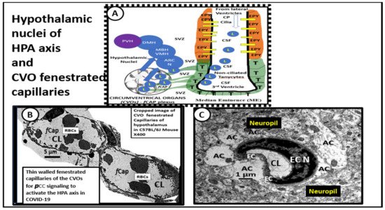
 2. COVID-19 Effects on the Hypothalamic-Pituitary-Adrenal Axis (HPA) and Adrenal Gland with Residual Effects in LC/PASC

In response to critical illnesses and marked psychogenic stress (including physical, emotional, or immunological stresses, such as excess PCC), the hypothalamic–pituitary–adrenal (HPA) axis is able to orchestrate the systemic release of cortisol in humans and corticosterone in rodents referred to as glucocorticoids (generally considered anti-inflammatory immunomodulators) [3]. The brain is known to respond to peripheral systemic cues, such as inflammation and the increased PCC that occurs due to the cytokine storm in acute COVID-19 and possibly LC/PASC. While cytokines and chemokines are known to have receptors on brain endothelial cells in the neurovascular unit blood-brain barrier and the blood-cerebrospinal fluid barrier, they are also known to signal the hypothalamus early on in COVID-19 via the leaky fenestrated circumventricular organ (CVO) capillaries (Figure 1).
Figure 1. Cropped images of the hypothalamic circumventricular organs (CVOs) fenestrated capillaries compared to neurovascular unit (NVU) blood–brain barrier (BBB) capillaries within the cortical hippocampal neuropil. Panel A illustrates the normal arrangement of the hypothalamic nuclei adjacent to the 3rd ventricle and how the tanycytes (T) and ependymal lining cells (EPY) of the ventricles may also be supplied by the CVO fenestrated capillaries in addition to BBB NVU capillaries in the subventricular zone of the third ventricle. Panel B depicts the phenotypic morphology of the CVOs thin-walled fenestrated brain endothelial cells (BECs) (fCap) that allow them to be leaky to the peripheral cytokines/chemokines (PCC). This arrangement allows the hypothalamus and its nuclei to be exposed to elevated systemic pCC and signals the hypothalamus to become activated. In turn, the activated hypothalamus will signal the adrenal gland to secrete increased glucocorticoids (cortisol in humans and corticosterone in rodent models), in addition to signaling the adrenal gland to produce increased epinephrine and norepinephrine. This excessive stimulation of the adrenal gland may result in adrenal fatigue (adrenal insufficiency) and/or even loss of corticosteroid production over time if the stimulation by pCC is excessive or prolonged, as in the cytokine storm in acute COVID-19 and possibly LC/PASC. Interestingly, autopsies of some COVID-19 patients have demonstrated necrosis, cortical lipid degeneration, hemorrhage, and ischemic necrosis, and focal inflammation of the adrenal glands. Panel C demonstrates the thicker wall of BECs with tight and adherens junctions (TJ/AJ) in the neurovascular unit with surrounding tightly adherent and abutting astrocytes foot processes (AC) from the cortical hippocampus region in a non-diabetic control model. Both Panels A and B are from normal control mice at 20 weeks of age. These vessels have TJ/AJs and also have pericytes (not shown in this image) to complete the formation of the neurovascular unit (NVU), i.e., the blood-brain barrier (BBB) capillaries of the neuropil.
Further, stress-induced glucocorticoids are an essential response to survive severe intensive care unit (ICU) illnesses, such as COVID-19 with SARS-CoV-2, as well as the excessive emotional and burdening stresses placed on the patients in these extremely stressful situations. Indeed, this stress-induced glucocorticoid secretion in severe disease may result in a relative adrenal insufficiency state [4].
In a series of autopsy studies, Zinserling et al. have been able to demonstrate in COVID-19 decedents that the adrenal gland may be infiltrated with CD3+ and CD8+ lymphocytes in perivascular regions due to SARS-CoV-2, resulting in impaired adrenal function [5]. The lymphocytic vasculitis they described would also increase the risk of the known endotheliitis that is associated with SARS-CoV-2 and, in turn, increase the inflammatory thromboembolic state within the adrenal that has also been reported by Santana et al in their autopsy studies [6]. Santana et al. reported frequent adrenal lesions consisting of an increase in ischemic necrosis, thrombus formation, hemorrhage, and cortical lipid degeneration with these abnormalities in 12 of 28 patients autopsied [6].
Hypoadrenalism and adrenal insufficiency may be plausible in LC/PASC [4]. Siejka and Barabutis [4] have proposed at least three mechanisms for the possible development of adrenal insufficiency due to SARS-CoV-2 as follows: (i) relative adrenal insufficiency is thought to be a common condition in critically ill patients [7] and increased PCC/CNSCC in acute COVID-19 may over time result in a negative feedback toward the hypothalamic-pituitary-adrenal axis [8]; (ii) SARS-CoV-2 generates certain amino acid sequences that may mimic the hosts' adrenocorticotropic hormone (ACTH) and, as a result of these corresponding antibodies, may contribute to the development of central adrenal insufficiency [4]; and (iii) the pituitary, hypothalamus, and adrenal glands each express angiotensin-converting enzyme 2 (ACE2), and SARS RNA has been identified in hypothalamic tissues of autopsied patients with COVID-19 that might cause hypophysitis and adrenalitis, and promote either secondary or primary adrenal insufficiency [4][9]. Therefore, LC/PASC patients should be carefully evaluated for adrenal insufficiency.

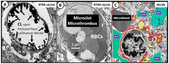
3. Central Nervous System Inflammatory Induced Thromboembolism: Microclots and Microbleeds in Acute COVID-19 and Possibly LC/PASC Due to Increased PCC and CNSCC

Systemic PCC and CNSCC in COVID-19 are known to induce endothelial cell activation and an inflammatory-related immunothrombotic or thromboembolic state within the vasculature. This immunothrombotic state ultimately causes macro-microthrombotic complications in multiple organ systems including the central nervous system, which may relate to either macrovascular (arteries and veins ≥ 200–300 μm) or microvascular (arterioles, venules or capillaries ≤ 20 μm) thrombi or clots [10].
Microvascular thrombi may also be associated with microbleeds especially in the white matter regions. These thrombi or microbleeds associated with cerebral small vessel disease (cSVD) will result in ischemic injury and ischemia/ischemia-reperfusion injury due to decreased cerebral blood flow and impaired metabolic delivery and metabolic byproduct uptake that may induce even further neuroinflammation due to increased CNSCC. This increase in CNSCC will develop in a response to injury wound healing mechanism due to activated neuroglia (activated microglia and reactive activated astrocytes) which secrete CNSCC and reactive oxygen-nitrogen species, resulting in ischemia-related regional injuries due to decreased cerebral blood flow to evoke even further neurodegeneration and or impaired cognition (Figure 2) [11].
Figure 2. Comparison of cerebral pial capillaries in the cortex and choroidal capillaries in choroid plexus villus with and without microclots, microthromus, and microbleeds—microhemorrhages. This image compares a normal healthy capillary to capillaries with a microclot and a microbleed. Panel A illustrates a morphological normal open capillary within a choroid plexus villus that is perfused fixed with lanthanum nitrate to stain the endothelial glycocalyx (ecGCx) (arrows) and note that the capillary lumen (CL) is open and depicts the electron-dense stained ecGCx (arrows) from a 20-week-old diabetic BTBR ob/ob model. Panel B resides within the same choroid plexus villus, as seen in Panel A; however, it does not have an ecGCx stain with lanthanum nitrate fixation and there appears to be a total occlusion of the capillary lumen with a microthrombus or microclot consisting of tightly packed adherent red blood cells, which are not usually seen when models have been perfusion-fixed prior to sacrifice, as seen in Panel A. This totally occluded capillary lumen will not allow for proper oxygenation or metabolic nutrition of surrounding tissues that will become ischemic and may result in adjacent basilar vascuolization/vesiculation in the basilar infoldings of the epithelial ependymal cell, as noted in previous Figure 10B. Panel C depicts a microbleed or hemorrhage adjacent to a dysmorphic highly remodeled capillary of the neurovascular unit of the BBB in cortical layer III of the 20-week-old db/db diabetic model with astrocyte activation detachment and retraction (drAC) (pseudo-colored cyan) and activated microglia (aMGC) (pseudo-colored red). Both the microthrombus microclot and microbleed will instigate the response to injury wound healing phase of increased inflammation and, thus, add to the pool of increased central nervous system cytokines/chemokines (CNSCC) and create a vicious cycle of micro-clot/bleeds and local inflammation. Scale bars = 2 μm in Panels A and B; 1 μm in Panel C. Figures adapted with permission by 4.0 [12][13]. EC N = endothelial cell nucleus; Pc N = pericyte nucleus.
Importantly, endothelial cell activation and dysfunction are tightly associated with cSVD and the endothelial activation in acute COVID-19 and LG/PASC with endotheliitis may certainly be playing a role with the development of microbleeds and/or clots [14][11].
Additionally, platelet activation due to endotheliitis with endothelial cell activation and dysfunction may also be a contributing factor since Pertorius et al [15] have demonstrated that, in LC/PASC, there is a persistent clotting protein pathology with increased thrombosis and an impaired fibrinolytic system resulting in impaired fibrinolysis that is associated with amyloid formation and increased antiplasmin [15][16]. Interestingly, serum amyloid A (SAA) is an amyloidogenic/fibrillogenic acute phase protein synthesized by the liver in inflammatory states (acute and chronic) that can affect coagulation by inducing amyloid formation in fibrin(ogen), as well as by inducing platelets to a more prothrombotic state [15]. Further, endothelial and platelet activation with a known endotheliitis [17] due to elevated PCC in acute COVD-19 may take a while to resolve, especially if there is an ongoing elevation of PCCs.
Recently, Chioh et al. were able to demonstrate that convalescent COVID-19 patients were susceptible to endothelial cell dysfunction due to persistent immune activation [18]. They were able to identify circulating endothelial cells in convalescent patients who had had COVID-19. Additionally, this group suggested that endothelial instability, damage, and activation/dysfunction may play a key mechanism that could be responsible for the development of post-COVID-19 vascular prothrombotic complications. Their findings, in combination with the previous discussion regarding increased inflammatory-induced thromboembolism, could also implicate an increased risk for the development of microclots and/or microbleeds, as discussed previously [10][17][18]. Further, they suggested that there may be a vicious cycle in regards to endothelial inflammation via PCC, vascular leakage, activation of the coagulation pathway, and inflammation. Certainly, the findings by Chioh et al. need to be carried out to longer time frames post-COVID-19 presentation.

3. Repurposing—Repositioning Approved Medications for LC/PASC

Our global societies have been facing the COVID-19 pandemic now for 17 months and the concerns regarding the disability of individuals with LC/PASC and their increasing numbers are of great concern. Therefore, researchers and clinicians are trying to better understand the multiple pathobiologies of LC/PASC in an attempt to be better informed as to how to treat these individuals. Indeed, repurposing or repositioning of existing medications is not a new concept [19] and one possible treatment is to suppress the chronic peripheral PCC and CNSCC neuroglia activation, neuroinflammation, and impaired cognition that may be central to the syndrome of LC/PASC. One example is to utilize anti-inflammatory modalities (such as methylprednisolone) along with the supplementation of micronutrients, such as vitamin B12 and B9–folic acid, to assist in one-carbon metabolism to treat individuals affected with this syndrome.
Importantly, Regland et al. have found that treatment with injectable vitamin B12 and oral folic acid (vitamin B9) have been successful in the treatment of patients with symptoms similar to LC/PASC who have suffered from myalgic encephalomyelitis and fibromyalgia, i.e., a chronic fatigue syndrome (ME/CFS) [20].

References

  1. Havervall, S.; Rosell, A.; Phillipson, M.; Mangsbo, S.M.; Nilsson, P.; Hober, S.; Thålin, C. Symptoms and functional impairment assessed 8 months after mild COVID-19 among health care workers. JAMA 2021, 325, 2015.
  2. Lerner, A.M.; Robinson, D.A.; Yang, L.; Williams, C.F.; Newman, L.M.; Breen, J.J.; Eisinger, R.W.; Schneider, J.S.; Adimora, A.A.; Erbelding, E.J. Toward Understanding COVID-19 Recovery: National Institutes of Health Workshop on Postacute COVID-19. Ann. Intern. Med. 2021, 174, 999–1003.
  3. Bellavance, M.-A.; Rivest, S. The HPA—Immune axis and the immunomodulatory actions of glucocorticoids in the brain. Front. Immunol. 2014, 5, 136.
  4. Siejka, A.; Barabutis, N. Adrenal insufficiency in the COVID-19 era. Am. J. Physiol. Metab. 2021, 320, E784–E785.
  5. Zinserling, V.A.; Semenova, N.; Markov, A.G.; Rybalchenko, O.V.; Wang, J.; Rodionov, R.N.; Bornstein, S.R. Inflammatory cell infiltration of adrenals in COVID-19. Horm. Metab. Res. 2020, 52, 639–641.
  6. Santana, M.F.; Borba, M.G.S.; Baía-Da-Silva, D.C.; Val, F.; Alexandre, M.A.A.; Brito-Sousa, J.D.; Melo, G.C.; Queiroga, M.V.O.; Farias, M.E.L.; Camilo, C.C.; et al. Case report: Adrenal pathology findings in severe COVID-19: An autopsy study. Am. J. Trop. Med. Hyg. 2020, 103, 1604–1607.
  7. Loriaux, D.L.; Fleseriu, M. Relative adrenal insufficiency. Curr. Opin. Endocrinol. Diabetes Obes. 2009, 16, 392–400.
  8. Annane, D.; Pastores, S.M.; Rochwerg, B.; Arlt, W.; Balk, R.A.; Beishuizen, A.; Briegel, J.; Carcillo, J.; Christ-Crain, M.; Cooper, M.S.; et al. Guidelines for the diagnosis and management of critical illness-related corticosteroid insufficiency (CIRCI) in critically ill patients (part I): Society of critical care medicine (SCCM) and european society of intensive care medicine (ESICM) 2017. Intensive Care Med. 2017, 43, 1751–1763.
  9. Pal, R. COVID-19, hypothalamo-pituitary-adrenal axis and clinical implications. Endocrine 2020, 68, 251–252.
  10. McFadyen, J.D.; Stevens, H.; Peter, K. The emerging threat of (Micro)thrombosis in COVID-19 and its therapeutic implications. Circ. Res. 2020, 127, 571–587.
  11. Poggesi, A.; Pasi, M.; Pescini, F.; Pantoni, L.; Inzitari, D. Circulating biologic markers of endothelial dysfunction in cerebral small vessel disease: A review. J. Cereb. Blood Flow Metab. 2015, 36, 72–94.
  12. Hayden, M.R. Type 2 diabetes mellitus increases the risk of late-onset Alzheimer’s disease: Ultrastructural remodeling of the neurovascular unit and diabetic gliopathy. Brain Sci. 2019, 9, 262.
  13. Hayden, M.; Banks, W. Deficient leptin cellular signaling plays a key role in brain ultrastructural remodeling in obesity and type 2 diabetes mellitus. Int. J. Mol. Sci. 2021, 22, 5427.
  14. Hayden, M.R. Endothelial activation and dysfunction in metabolic syndrome, type 2 diabetes and coronavirus disease 2019. J. Int. Med. Res. 2020, 48.
  15. Pretorius, E.; Vlok, M.; Venter, C.; Bezuidenhout, J.A.; Laubscher, G.J.; Steenkamp, J.; Kell, D.B. Persistent clotting protein pathology in long COVID/post-acute sequelae of COVID-19 (PASC) is accompanied by increased levels of antiplasmin. medRxiv 2021.
  16. Page, M.; Thomson, G.J.A.; Nunes, J.M.; Engelbrecht, A.-M.; Nell, T.A.; De Villiers, W.J.S.; De Beer, M.C.; Engelbrecht, L.; Kell, D.B.; Pretorius, E. Serum amyloid A binds to fibrin(ogen), promoting fibrin amyloid formation. Sci. Rep. 2019, 9, 3102.
  17. Varga, Z.; Flammer, A.J.; Steiger, P.; Haberecker, M.; Andermatt, R.; Zinkernagel, A.S.; Mehra, M.R.; Schuepbach, R.; Ruschitzka, F.; Moch, H. Endothelial cell infection and endotheliitis in COVID-19. Lancet 2020, 395, 1417–1418.
  18. Chioh, F.W.; Fong, S.-W.; E Young, B.; Wu, K.-X.; Siau, A.; Krishnan, S.; Chan, Y.-H.; Carissimo, G.; Teo, L.L.; Gao, F.; et al. Convalescent COVID-19 patients are susceptible to endothelial dysfunction due to persistent immune activation. eLife 2021, 10, e64909.
  19. Xue, H.; Li, J.; Xie, H.; Wang, Y. Review of drug repositioning approaches and resources. Int. J. Biol. Sci. 2018, 14, 1232–1244.
  20. Regland, B.; Forsmark, S.; Halaouate, L.; Matousek, M.; Peilot, B.; Zachrisson, O.; Gottfries, C.-G. Response to vitamin B12 and folic acid in myalgic encephalomyelitis and fibromyalgia. PLoS ONE 2015, 10, e0124648.
More
Information
Contributor MDPI registered users' name will be linked to their SciProfiles pages. To register with us, please refer to https://encyclopedia.pub/register :
View Times: 482
Revisions: 4 times (View History)
Update Date: 24 Sep 2021
1000/1000