Submitted Successfully!
To reward your contribution, here is a gift for you: A free trial for our video production service.
Thank you for your contribution! You can also upload a video entry or images related to this topic.
Version Summary Created by Modification Content Size Created at Operation
1 + 1916 word(s) 1916 2021-09-01 04:54:23 |
2 format correction Meta information modification 1916 2021-09-02 02:56:26 |

Video Upload Options

Do you have a full video?

Confirm

Are you sure to Delete?
Cite
If you have any further questions, please contact Encyclopedia Editorial Office.
Messa, P. Native Hypovitaminosis D and CKD. Encyclopedia. Available online: https://encyclopedia.pub/entry/13793 (accessed on 18 April 2024).
Messa P. Native Hypovitaminosis D and CKD. Encyclopedia. Available at: https://encyclopedia.pub/entry/13793. Accessed April 18, 2024.
Messa, Piergiorgio. "Native Hypovitaminosis D and CKD" Encyclopedia, https://encyclopedia.pub/entry/13793 (accessed April 18, 2024).
Messa, P. (2021, September 01). Native Hypovitaminosis D and CKD. In Encyclopedia. https://encyclopedia.pub/entry/13793
Messa, Piergiorgio. "Native Hypovitaminosis D and CKD." Encyclopedia. Web. 01 September, 2021.
Native Hypovitaminosis D and CKD
Edit

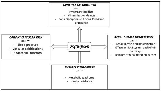
Native hypovitaminosis D (n-hVITD) is frequently found from the early stages of chronic kidney disease (CKD) and its prevalence increases with CKD progression. 

vitamin D mineral metabolism CKD cardiovascular risk vitamin D supplementation

1. Introduction

Vitamin D deficiency is considered to be an important public health problem. Despite the numerous warnings about the detrimental effects of native hypovitaminosis D (n-hVITD) in patients affected by chronic kidney disease (CKD), insufficient levels of 25(OH)VitD are still frequently found from the early stages of the disease [1]. Some evidence, mostly derived from experimental studies, have hypothesized a possible role for Vitamin D in the modulation of some physio-pathological processes beyond chronic kidney disease mineral bone disorder (CKD-MBD). Unfortunately, the lack of large and interventional studies focused on the so called “unconventional effects” (i.e., beyond CKDMBD) of 25(OH)VitD supplementation in CKD patients, does not conclusively demonstrate the beneficial effects of this supplementation in clinical practice. Conversely, the implications of n-hVITD in CKDMBD have been largely and well characterized in the literature, and for this reason in this review they will only be mentioned briefly [2].
Previously, our group addressed the impact of the supposed pleiotropic effects of 25(OH)VitD in renal transplanted patients [3].

2. Native Hypovitaminosis D during Chronic Kidney Disease (CKD): The Size of the Problem

2.1. Vitamin D Metabolism and Status Categorization in CKD Patients

Vitamin D is a fat soluble pre-hormone, obtained mainly through the skin photochemical conversion of 7-dehydrocholesterol after sunlight exposition (solar ultraviolet B radiation, 290–315 nm). The 7-dehydrocholesterol undergoes a temperature-dependent rearrangement to vitamin D3 (cholecalciferol) [4]. Vitamin D3 and vitamin D2 (ergocalciferol) may be supplied, in minor part, also by food intake (meat, eggs, fish, liver, cod liver oil, and dairy products).
Then, vitamin D undergoes 25-α-hydroxylation in the liver, which forms 25(OH)VitD and one subsequent additional 1-α-hydroxylation primarily in renal proximal tubular cells, responsible for the generation of the active form of vitamin D, the 1,25(OH)2VitD [5].
The generation of 1,25(OH)2VitD is also regulated by the activity of the vitamin D 24-hydroxylase (CYP24A1) in renal proximal tubules, which inactivates all the active vitamin D metabolites [6].
According to 25(OH)VitD levels, native vitamin D status is normally classified as: (1) deficiency: 25(OH)VitD < 20 ng/mL; (2) insufficiency: 21 < 25(OH)VitD < 29 ng/mL; (3) sufficiency: 25(OH)VitD > 30 ng/mL [7].
Several conditions may influence 25(OH)VitD levels in humans: aging, reduced skin synthesis, reduced dietary intake, malabsorption, obesity, glucocorticoid therapy, reduced liver function, and drugs interference with vitamin D metabolism. CKD patients present generally a higher prevalence of n-hVITD compared to the general population because of additional specific alterations of the mineral and bone metabolism present in those patients from the early stages of the disease [8][9]. In addition, the association between 25(OH)VitD urinary loss and the reduction of its renal hydroxylation further implicate the native and active hypovitaminosis D typically found in these patients. A proximal renal tubular impairment in the 25(OH)VitD megalin mediated reabsorption has been experimentally demonstrated in proteinuric and megalin knock out (KO) experimental models [10][11][12]. Recent published data, by Bohnert B et al. demonstrated clearly urinary loss of both 25(OH)VitD and 1,25(OH)2VitD and vitamin D binding protein in experimental nephrotic syndrome [13]. In addition, the low renal 1-α-hydroxylation capability, determined by the reduction of nephron mass, and the effects of phosphorus, FGF23 and αKlotho on the cytochromes CYP27B1 and CYP24A1 determine the additional decrease of 1,25(OH)2VitD [14].

2.2. Native Hypovitaminosis D Epidemiology in CKD Patients

The prevalence of n-hVitD in CKD has been evaluated in a monocentric American study, performed on a total of 43 CKD patients in which 86% of patients did not have sufficient 25(OH)VitD levels. Of note, 42% of them had 25(OH)VitD insufficiency, and 42% had mild 25(OH)VitD deficiency. Severe vitamin D deficiency (<5 ng/mL) was found in the remaining 2% of patients [1]. Recent data obtained in an Italian experience, including 450 prevalent CKD patients, report a mean level of 25(OH)VitD of 17.5 ± 11.2 ng/mL, and the presence of deficiency or insufficiency respectively in 66.4% and 16.5% of patients. In this cohort, the presence of diabetes mellitus was related to lower 25(OH)VitD levels. An inverse correlation between 25(OH)VitD and age, Parathormone (PTH), urinary protein excretion and Charlson index was also found, whereas a direct correlation with hemoglobin was demonstrated. In multivariate analysis only age and PTH levels impacted independently on 25(OH)VitD levels [15].
In our department, in Milan, we analyzed retrospectively 133 incident CKD stage 3–4 outpatients (males = 95; age 77 ± 11 years; eGFR-EPI 24 ± 10 mL/min). At the first evaluation, the mean levels of 25(OH)VitD were 17.5 ± 11.2 ng/mL. Not sufficient 25(OH)VitD levels, despite the native vitamin D supplementation prescribed in the 72% of patients, was found in 61% of patients, 42% had vitamin D insufficiency, and 19% had vitamin D deficiency. Interestingly, in our cohort, we did not find any difference in 25(OH)VitD according to the supplementary therapy. This result might support the evidence of the frequent inability to reach sufficient 25(OH)VitD levels in CKD patients with the simple supplementation. In this sense, a correction of life and alimentary habits can be important [16].

2.3. Native Vitamin D Supplementation: Dose, Interval,Toxicity

Despite the most important international guidelines suggesting 25(OH)VitD supplementation in presence of n-hVITD, at the moment the optimal strategy of supplementation and the optimal levels of 25(OH)VitD to reach are still debated worldwide. What KDIGO guidelines suggest, is that deficiency and insufficiency of 25(OH)VitD in CKD patients should be treated through strategies provided to the general population. No special considerations are present concerning which form of native vitamin D to use, and there is no strong evidence about a superiority in particular habits of dose and interval schedules [17]. In 2016, the Italian Society of Nephrology published, a report suggesting a supplementation in CKD patients with 25(OH)VitD < 30 ng/mL, using the scheme that nephrologists are most confident with. At the moment three different forms of native vitamin D are available: ergocalciferol, cholecalciferol and calcifediol. Among them, cholecalciferol seems to be superior in increasing 25(OH)VitD levels, with a safe dose of 10,000 UI/week [18][19]. A novel extended-release (ER) calcifediol formulation has been recently tested in 429 CKD patients. After a randomization 2 (daily pills containing oral ER calcifediol, 30 or 60 mcg):1 (placebo once daily), the cohort was followed for 26 weeks. In the sequent open label extension study, ER calcifediol was prescribed for an other 26 weeks in 298 patients. This study demonstrated a good correction of n-hVITD, a good effect on PTH, and a safe profile for oral ER calcifediol [20]. This formulation might in the future solve the problems of the adherence to the 25(OH)VitD therapy, burdened by the drop formulation and by the unusual assumption schemes prescribed [21]. Independently of the native form of vitamin D used, clinicians should pay close attention to vitamin D toxicity, avoiding too high levels of plasmatic 25(OH)VitD, and monitoring calcium and phosphorus levels. There should be particular attention during summer season, when the sunlight exposition is significantly higher [22]. According to our experience, levels of 25(OH)VitD of 30–60 ng/mL might represent a good general target in CKD population. Higher levels might be suggested to obtain effects on immunity, proteinuria etc., but unfortunately the little evidence and also the high risk of toxicity limit this suggestion. We agree with the Italian Nephrology of Society indications about the discontinuation of native vitamin D supplementation in presence of 25(OH)D levels >100 ng/mL and/or presence of hypercalcemia (Ca > 10.4 mg/dL) in the absence of active vitamin D therapy.

3. Native Hypovitaminosis D in CKD: Experimental and Clinical Evidence

The principal circulating form of vitamin D is 25(OH)VitD. In the early 2000s, the presence of 1-α-hydroxylase enzyme was noted in many tissues outside the kidneys, and a certain binding affinity of 25(OH)VitD for vitamin D receptor (VDR) was demonstrated [23][24]. Almost all tissues and cells, including brain, heart, skeletal muscle, smooth muscle cells, pancreas, activated T and B lymphocytes, and monocytes express the vitamin D receptor (VDR) [25]. Consequently, much experimental evidence has implicated the hypovitaminosis D (25(OH)VitD and/or 1,25(OH)2VitD) and the reduced activation of VDR in the modulation pathological processes also beyond CKDMBD [26]. The principal effects and their level of evidence (LOE) of n-hVitD in CKD patients are summarized in Figure 1.
Figure 1. Principal effects and their level of evidence of native hypovitaminosis D in chronic kidney disease (CKD) patients. LOE: level of evidence. ** slight level of evidence; *** moderate level of evidence; ***** strong level of evidence.

4. Native Vitamin D Supplementation in CKD Patients: Evidence from Randomized Interventional Studies

Despite the numerous data of the association previously reported, data derived by randomized prospective trials on the effect of native vitamin D supplementation on major clinical outcomes are still scanty and discordant with each other. The principal characteristics and results of the recent randomized interventional studies in patients with CKD stages 2–5 ND are summarized in Table 1. In 2002, by means of a randomized, double-blinded, placebo-controlled study, the ability of native vitamin D supplementation in delaying the onset of secondary hyperparathyroidism was tested in 72 children with CKD stages 2–4. No baseline differences were present between the two groups in 25(OH)VitD levels with a relatively native vitamin D insufficiency present in both groups (treated group 20.1 ± 7.1 ng/dL vs. placebo group 20.8 ± 7.2 ng/dL p = 0.69). Patients were followed up for 24 months. During the follow up time, in 9 of 20 children on placebo and 3 of 20 children on ergocalciferol treatment the development of hyperparathyroidism was found (odds ratio = 4.64, 95% confidence interval = 1.02−21.00), with a significantly longer time to the development of the anomaly in treated children (hazard ratio = 0.30, 95% confidence interval = 0.09–0.93, p = 0.05) [27].
Table 1. Principal characteristics and results of the recent randomized interventional studies in patients with chronic kidney disease (CKD) at stages 2–5 not dialysis (ND).

Type of Patients

Number of Patients

Type of Intervention

Results

Reference

CKD stages 2–4

87

GrA: Cholecalciferol 5000 IU/wk;

GrB: Cholecalciferol 20,000 IU/wk

In GrB vs. GrA: significant increase of 25OHVitD, 1,25OHD2 and reduction PTH

Oksa et al., 2008

CKD stages 1–5

25

GrA: Cholecalciferol 40,000 IU/wk;

GrB: placebo

In GrA vs. GrB: significant increase of 25OHVitD, 1,25(OH)D2, FGF23 and reduction PTH

Markmann et al., 2012

CKD 3–4

38

GrA: Ergocalciferol 50,000 IU/wk for one month followed by 50,000 IU/mth for 5 mths;

GrB: Placebo

In GrA vs. GrB: significant increase of 25(OH)VitD, endothelium dependent microcirculatory vasodilatation and reduction of pulse pressure. No effects on PWV and LVMI

Dreyer et al., 2014

CKD stages 3–4

429

Study 1 (n = 213): GrA: ER Calcifediol 12 wk at 30 g/daily + 14 wks 30 or 60 g/daily; GrB 26 wks placebo

Study 2 (n = 216): GrA: ER Calcifediol 12 wks 30 g/daily + 14 wks 30 or 60 g/daily; GrB: 26 wks placebo

In GrA vs. GrB in both studies: significant increase of 25OHVitD and reduction PTH

Sprague et al., 2016

CKD 3–5

44

GrA:Cholecalciferol 50.000 IU/wk;

GrB: Ergocalciferol 50.000 IU/wk

In GrA vs. GrB: significantly higher increase of 25(OH)VitD. No differences in 1,25(OH)D2 and PTH

Weltmore et al., 2016

CKD stages 3–4

128

GrA: Cholecalciferol 2000 IU/day;

GrB: Calcitriol 0.5 g/day

In GrA vs. GrB: significant increase of 25OHVitD in GrA vs. GrB. No effects on FMD

Kendrick et al., 2017

CKD stages 3–4

120

GrA: Cholecalciferol 300,000 UI mth;

GrB: Placebo

In GrA vs. GrB: significant increase of 25OHVitD, 1,25OHD2 and reduction PTH. Increase of FMD and PWV

Kumar et al., 2017

CKD stages 3–4

119

GrA: Placebo; GrB: Calcitriol 0.5 g 3×/wk;

GrC: Calcifediol 5.000 IU 3×/wk

In GrC vs. others: significant decrease of PWV. Patients in the highest 25(OH)VitD tertile at trial end had significant decreases in PWV

Levin et al., 2017

Footnotes: GrA: group A; GrB: Group B; GrC: Group C; wk: week; wks: weeks; mth: month; mths: months; PWV: pulse wave velocity; FMD: flow mediated dilation; LVMI: left ventricular mass index. PTH: Parathormone.

References

  1. González, E.A.; Sachdeva, A.; Oliver, D.A.; Martin, K.J. Vitamin D insufficiency and deficiency in chronic kidney disease. A single center observational study. Am. J. Nephrol. 2004, 24, 503–510.
  2. Cozzolino, M.; Ciceri, P.; Volpi, E.M.; Olivi, L.; Messa, P.G. Pathophysiology of calcium and phosphate metabolism impairment in chronic kidney disease. Blood Purif. 2009, 27, 338–344.
  3. Messa, P.; Regalia, A.; Alfieri, C. Nutritional vitamin D in renal transplant patients: Speculations and reality. Nutrients 2017, 9, 550.
  4. Holick, M.F.; MacLaughlin, J.A.; Clark, M.B.; Holick, S.A.; Potts, J.T.; Anderson, R.R.; Elias, P. Photosynthesis of previtamin D3 in human skin and the physiologic consequences. Science 1980, 210, 203–205.
  5. Nykjaer, A.; Dragun, D.; Walther, D.; Vorum, H.; Jacobsen, C.; Herz, J.; Willnow, T.E. An endocytic pathway essential for renal uptake and activation of the steroid 25-(OH) vitamin D3. Cell 1999, 96, 507–515.
  6. Jones, G.; Prosser, D.E.; Kaufmann, M. 25-Hydroxyvitamin D-24-hydroxylase (CYP24A1): Its important role in the degradation of vitamin D. Arch. Biochem. Biophys. 2012, 523, 9–18.
  7. Holick, M.F. Vitamin D deficiency. N. Engl. J. Med. 2007, 357, 266–281.
  8. Ginde, A.A.; Liu, M.C.; Camargo, C.A. Demographic differences and trends of vitamin D insufficiency in the US population, 1988–2004. Arch. Intern. Med. 2009, 169, 626.
  9. Kuro-o, M.; Moe, O.W. FGF23-αKlotho as a paradigm for a kidney-bone network. Bone 2017, 100, 4–18.
  10. Takemoto, F.; Shinki, T.; Yokoyama, K.; Inokami, T.; Hara, S.; Yamada, A.; Uchida, S. Gene expression of vitamin D hydroxylase and megalin in the remnant kidney of nephrectomized rats. Kidney Int. 2003, 64, 414–420.
  11. Matsui, I.; Hamano, T.; Tomida, K.; Inoue, K.; Takabatake, Y.; Nagasawa, Y.; Isaka, Y. Active vitamin D and its analogue, 22-oxacalcitriol, ameliorate puromycin aminonucleoside-induced nephrosis in rats. Nephrol. Dial. Transpl. 2009, 24, 2354–2361.
  12. Leheste, J.R.; Rolinski, B.; Vorum, H.; Hilpert, J.; Nykjaer, A.; Jacobsen, C.; Willnow, T.E. Megalin knockout mice as an animal model of low molecular weight proteinuria. Am. J. Pathol. 1999, 155, 1361–1370.
  13. Bohnert, B.N.; Daniel, C.; Amann, K.; Voelkl, J.; Alesutan, I.; Lang, F.; Artunc, F. Impact of phosphorus restriction and vitamin D-substitution on secondary hyperparathyroidism in a proteinuric mouse Model. Kidney Blood Press. Res. 2015, 40, 153–165.
  14. Messa, P.; Alfieri, C.; Rastaldi, M.P. Recent insights into vitamin D and its receptor. J. Nephrol. 2011, 24 (Suppl. 18), 30–37.
  15. Cupisti, A.; Vigo, V.; Baronti, M.E.; D’Alessandro, C.; Ghiadoni, L.; Egidi, M.F. Vitamin D status and cholecalciferol supplementation in chronic kidney disease patients: An Italian cohort report. Int. J. Nephrol. Renov. Dis. 2015, 8, 151–157.
  16. Jhee, J.H.; Kim, H.; Park, S.; Yun, H.R.; Jung, S.Y.; Kee, Y.K.; Yoo, T.H. Vitamin D deficiency is significantly associated with depression in patients with chronic kidney disease. PLoS ONE 2017, 12, e0171009.
  17. Kidney Disease: Improving global outcomes (KDIGO) CKD-MBD update work group. KDIGO 2017 clinical practice guideline update for the diagnosis, evaluation, prevention, and treatment of chronic kidney disease–mineral and bone disorder (CKD-MBD). Kidney Int. 2017, 7 (Suppl. 1), 1–59.
  18. Tripkovic, L.; Lambert, H.; Hart, K.; Smith, C.P.; Bucca, G.; Penson, S.; Lanham-New, S. Comparison of vitamin D2 and vitamin D3 supplementation in raising serum 25-hydroxyvitamin D status: A systematic review and meta-analysis. Am. J. Clin. Nutr. 2012, 95, 1357–1364.
  19. Vieth, R. Vitamin D toxicity, policy, and science. J. Bone Miner. Res. 2007, 22 (Suppl. S2), V64–V68.
  20. Sprague, S.M.; Crawford, P.W.; Melnick, J.Z.; Strugnell, S.A.; Ali, S.; Mangoo-Karim, R.; Bishop, C.W. Use of extended-release calcifediol to treat secondary hyperparathyroidism in stages 3 and 4 chronic kidney disease. Am. J. Nephrol. 2016, 44, 316–325.
  21. Alfieri, C.; Regalia, A.; Zanoni, F.; Vettoretti, S.; Cozzolino, M.; Messa, P. The importance of adherence in the treatment of secondary hyperparathyroidism. Blood Purif. 2019, 47, 37–44.
  22. Morrone, L.F.; Bolasco, P.; Camerini, C.; Cianciolo, G.; Cupisti, A.; Galassi, A.; Cozzolino, M. Vitamin D in patients with chronic kidney disease: A position statement of the Working Group; Trace elements and mineral metabolism. J. Nephrol. 2016, 29, 305–328.
  23. Townsend, K.; Evans, K.N.; Campbell, M.J.; Colston, K.W.; Adams, J.S.; Hewison, M. Biological actions of extra-renal 25-hydroxyvitamin D-1α-hydroxylase and implications for chemoprevention and treatment. J. Steroid. Biochem. Mol. Biol. 2005, 97, 103–109.
  24. Zehnder, D.; Bland, R.; Williams, M.C.; McNinch, R.W.; Howie, A.J.; Stewart, P.M.; Hewison, M. Extrarenal expression of 25-hydroxyvitamin D3 -1α-Hydroxylase 1. J. Clin. Endocrinol. Metab. 2001, 86, 888–894.
  25. Holick, M.F. Vitamin D in health and disease: Vitamin D for health and in chronic kidney disease. Semin. Dial. 2005, 18, 266–275.
  26. Jean, G.; Souberbielle, J.; Chazot, C. Vitamin D in chronic kidney disease and dialysis patients. Nutrients 2017, 9, 328.
  27. Shroff, R.; Wan, M.; Gullett, A.; Ledermann, S.; Shute, R.; Knott, C.; Rees, L. Ergocalciferol supplementation in children with CKD delays the onset of secondary hyperparathyroidism: A randomized trial. Clin. J. Am. Soc. Nephrol. 2012, 7, 216–223.
More
Information
Contributor MDPI registered users' name will be linked to their SciProfiles pages. To register with us, please refer to https://encyclopedia.pub/register :
View Times: 336
Revisions: 2 times (View History)
Update Date: 02 Sep 2021
1000/1000