Submitted Successfully!
To reward your contribution, here is a gift for you: A free trial for our video production service.
Thank you for your contribution! You can also upload a video entry or images related to this topic.
Version Summary Created by Modification Content Size Created at Operation
1 + 2878 word(s) 2878 2021-07-26 03:34:13 |
2 format correct -21 word(s) 2857 2021-08-05 03:06:38 |

Video Upload Options

Do you have a full video?

Confirm

Are you sure to Delete?
Cite
If you have any further questions, please contact Encyclopedia Editorial Office.
Quadros, E. FRAb in Autism Spectrum Disorders. Encyclopedia. Available online: https://encyclopedia.pub/entry/12774 (accessed on 20 April 2024).
Quadros E. FRAb in Autism Spectrum Disorders. Encyclopedia. Available at: https://encyclopedia.pub/entry/12774. Accessed April 20, 2024.
Quadros, Edward. "FRAb in Autism Spectrum Disorders" Encyclopedia, https://encyclopedia.pub/entry/12774 (accessed April 20, 2024).
Quadros, E. (2021, August 04). FRAb in Autism Spectrum Disorders. In Encyclopedia. https://encyclopedia.pub/entry/12774
Quadros, Edward. "FRAb in Autism Spectrum Disorders." Encyclopedia. Web. 04 August, 2021.
FRAb in Autism Spectrum Disorders
Edit

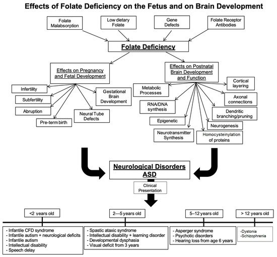
Folate deficiency and folate receptor autoimmune disorder are major contributors to infertility, pregnancy related complications and abnormal fetal development including structural and functional abnormalities of the brain. Food fortification and prenatal folic acid supplementation has reduced the incidence of neural tube defect (NTD) pregnancies but is unlikely to prevent pregnancy-related complications in the presence of folate receptor autoantibodies (FRAb). In pregnancy, these autoantibodies can block folate transport to the fetus and in young children, folate transport to the brain. These antibodies are prevalent in neural tube defect pregnancies and in developmental disorders such as cerebral folate deficiency (CFD) syndrome and autism spectrum disorder (ASD). In the latter conditions, folinic acid treatment has shown clinical improvement in some of the core ASD deficits. Early testing for folate receptor autoantibodies and intervention is likely to result in a positive outcome

autism spectrum disorders folate receptor alpha folates pregnancy brain development fetal development

1. Background

Folate, an umbrella term used for metabolically active forms of folic acid (B9), is an essential B-complex vitamin necessary for basic cellular metabolism including, but not limited to, essential cellular DNA synthesis, repair and methylation including regulation of synthesis and metabolism of monoamine neurotransmitters. As a nutrient found in green leafy vegetables, legumes and fruits, it is readily absorbed by the upper small intestine after breakdown from polyglutamates to monoglutamates. Folate in its active forms facilitates one-carbon transfer reactions and contributes to the synthesis of purines, pyrimidines and amino acids [1]. One of its most characterized roles is facilitating single carbon transfer to homocysteine to form methionine. This reaction is critical for maintaining intracellular S- adenosyl methionine, an essential compound for methylation reactions. Folate also has a co-dependent relationship with vitamin B12 in that both vitamins must be present in adequate amounts for conversion to the physiologic forms that participate in metabolic reactions. If folate and B12 are not adequate, cellular metabolism and replication is interrupted [2][3]. This is most critical during fetal and neonatal development because inadequate folate during this period can result in interruptions in brain development leading to structural abnormalities that produce functional deficits c of the CFD syndrome. Low cerebro-spinal fluid (CSF) folate is a characteristic feature of CFD syndrome, as first described by Ramaekers and Blau [4]. On rare occasions, CFD can also result from mutations in the FRα gene [5][6][7], but the most common cause of low CSF folate in CFD is the presence of anti-folate receptor antibodies (FRAb) that can block folate transport across the choroid plexus [8][9]. A recent report has identified mutations in the CIC transcription factor gene in children diagnosed with CFD syndrome. Mutations in the CIC gene decrease the expression of FRα to reduce folate transport across the choroid plexus [10]. No abnormalities of the FRα gene are found in ASD, but a majority of these children are positive for FRAb and have low CSF folate [11][12]. This is a priori proof that FRα is the primary transporter of folate into the brain under physiologic folate status.

2. Folate Requirements during Pregnancy

Since the discovery of its role in megaloblastic anemia and spina bifida, folate supplementation during pregnancy and fortification of food products have become two of the most globally accepted methods of treating and preventing folate deficiencyok. The basic folate requirement increases 75 to 100% (approximately 300–400 μg per day) in pregnancy because folate has a critical role in the growth and development of the embryo/fetus, especially during early stages of development [13]. It is, therefore, common practice to recommend that women supplement their diet with folate before conception and throughout pregnancy. The prevention of folate deficiency during pregnancy is achieved by consumption of at least 0.4 mg/day of folic acid during the first trimester of pregnancy [14][15]. In light of the recently discovered FRAb that can block folate transport, women positive for these antibodies may need additional supplementation with folinic acid to provide adequate folate to the developing fetus [16][17].

3. Folate and Fetal Brain Development

The importance of folate during embryonic and fetal brain development has been demonstrated in genetic animal models and dietary manipulations of folate deficiency [18][19]. If either folate transport or folate concentration in circulation is adversely manipulated, embryonic and fetal development is significantly altered. Mouse knockout models of genes such as FOLR1 that encode for folate receptor alpha (FRα) produce lethality in litters along with orbito-facial abnormalities, congenital heart defects and/or neural tube defects [20]. In FOLR1 knockout mouse, these lethalities can be prevented with adequate folinic acid (N5-formyltetrahydrofolate, a reduced form of folate) supplementation. These dramatic results occur because folate transport is lacking in the KO mouse during the early stages of neurulation and in regions where abnormalities arise [21]. In rodent models, folate deficiency causes a decrease in progenitor cells and an increase in apoptosis, and this could lead to infertility or resorption of embryos or fetal malformations [22]. Behavioral deficits are seen in rat pups born to folate-deficient mothers [23] and on methyl donor deficient diet during pregnancy [24]. In a rat model of exposure to rat folate receptor antibodies during pregnancy, resorption of embryos and malformations of the cranio-facial region and the brain were reported [25]. When the antibodies were administered at lower doses, embryos were carried to term with normal appearing pups born. However, these pups showed severe behavioral deficits [23][26]. The behavioral phenotype can be rescued by treatment with folinic acid and dexamethasone prior to antibody exposure [27]. These studies provide strong evidence in support of the pathologic consequences of exposure to FRα antibodies and the protective role of folinic acid.

4. Folate and Neonatal Brain Development

After birth, it is crucial for the offspring to have an adequate amount of folate in their diet. Instead of rapid cell division as embryogenesis calls for, postnatal development requires folate for neural progenitor differentiation as well as proliferation [28]. It has yet to be fully elucidated what the detailed mechanisms of folate action are, but the folate deficiency produced in animal models during early postnatal development illustrates the importance of folate in preventing developmental and cognitive deficits [23][27]. Researchers have also reported changes in neuronal excitability and maintenance that arise with a decrease in brain folate in a rat model [29]. Others have reported an increase in p53 and signs of homocysteine accumulation in the neurons and astrocytes [30]. There was a long-term effect on locomotor function and cognition in these animals. Therefore, folate is necessary for maintenance of neuronal function, as well. Based on this, further investigations into the mechanisms of folate metabolism in neurons and support cells of the brain are necessary. Thus far, folate has been linked to neuronal repair and differentiation after injury, myelin formation and maintenance and neuronal plasticity [30][31][32]. Figure 1 provides a summary of the effects of folate deficiency on fetal and post-natal brain development and the consequent sequelae that contribute to neurologic deficits.
Figure 1. Effects of folate deficiency on the fetus and on brain development. Multiple causes lead to systemic as well as fetal folate deficiency. Folate receptor autoantibodies can block folate transport to the fetus and to the fetal as well as neonatal brain. In addition to folate deficiency, immune-mediated inflammation can contribute to the pathology. This has multipronged effects on brain development and function.

5. Folate Receptors: Expression and Function

In humans, there are four genes that code for folate receptors (see Table 1). The most characterized of these receptors is folate receptor alpha (FRα). As extracellular receptors, FRα, FRβ, FRγ and FRδ function as transporters of folate across different target tissues [33][34]. FRα can also act as a transcription factor [33]. Other transporters of folate include the reduced folate carrier (RFC), which requires high local concentrations (micromolar) of biologically active reduced forms of folates, and the proton-coupled folate transporter (PCFT), which can only transport folates and folic acid under acidic conditions and is the primary transporter involved in folate absorption in the gut [35].
Table 1. Summary of folate transporters.
Protein Gene Chromosome GPI Anchor? Localization Cofactors? Refs.
FRα FOLR1 11q13.3 Yes Liver, kidney, uterus, placenta, choroid plexus, retinal pigment epithelium LRP2 [33][35][36][37][38]
FRβ FOLR2 11q13.4 Yes Placenta, spleen, bone marrow, thymus, macrophages NA [33][35][36][37][38][39]
FRγ FOLR3 11q13.4 NA Secretory granules of neutrophil granulocytes NA [33][35][36][37][38][39]
FRδ FOLR4 11q14 Yes Oocytes NA [33][35][36][37][38][39]
RFC SLC19A1 21q22.3 No Liver, kidney, placenta, choroid plexus, intestinal tract Vitamin D, thiamine pyrophosphate [34][40]
PCFT SLC46A1 17q11.2 No Liver, kidney, choroid plexus, placenta, intestinal epithelium, human tumors Proton gradient [34][40]

6. FRα Role in Maternofetal Transport of Folate

The high demand for folate during pregnancy requires homeostatic mechanisms to ensure that sufficient folate is provided to the fetus throughout development. As the most characterized receptor in the folate transporter family of proteins, the accepted mechanism of FRα-mediated transport is translocation/endocytosis of the holo receptor subsequent to folate binding [35]. FRα is expressed on all epithelial cells including the choroid plexus. It is highly expressed in the reproductive tissues including the placenta and the fetus. To determine the mechanism of folate transport in the placenta during pregnancy, Yasuda et al. [41] manipulated osmolarity, concentrations of phosphatidylinositol-specific phospholipase C inhibition and concentrations of 3H-folic acid in vitro culture of human placental brush border membrane vesicles and determined that FRα, RFC and PCFT could transport various forms of folate, but that approximately 60% of folate was binding to FRα. They also noted that the folate requirements of Wistar rats increased across gestation, and expression of the mRNA of the transporters increased as well.

7. FRα Role in Folate Transport to the Brain

FRα is accepted as the main transporter of folate into the brain. However, there have been limitations to studying how FRα transports folate across the blood–brain barrier. A potential mechanism of folate transport across the choroid plexus and into the brain has been described by Grapp et al. [42]. In their experiments using immortalized Z310n rat choroid plexus cells in culture and a mouse model, they determined that transport of folate required shuttling of folates via exosomes from the basolateral side of the choroid plexus to the brain parenchyma of the apical side. Alternative transporters such as RFC and PCFT may only play a role when there is a disruption of FRα expression and transport, and adequate folate concentration is made available locally at the receptor [43]. The shuttling across the epithelial lining of the choroid plexus is a mechanism presumed to be conserved in all tissues that express FRα [44].

8. Folate Receptor Autoantibodies: Their Role in Disrupting Folate Transport

In some conditions, there is disruption in folate utilization that is not related to a dietary deficiency but is most likely due to a disturbance in the folate’s transport due to genetic or metabolic abnormalities. An emerging culprit of folate transport disruption is folate receptor autoimmune disorder, where autoantibodies against the FRα can interfere with folate transport to the fetus; it has been associated with subfertility, difficulty in conceiving, miscarriage and neural tube defects in the fetus [16][17][45][46].
In infants and young children, these antibodies can block folate transport to the brain. Approximately 70% of the children diagnosed with cerebral folate deficiency syndrome or autism spectrum disorder have low CSF folate and respond to folinic acid treatment [47][48]. The majority of the autoantibodies are of the IgG class and, therefore, can readily cross the placenta and affect the fetus. Two distinct types of antibodies have been identified. One binds to FRα at the active site where folate binds and, as a consequence, blocks folate binding (blocking Ab). Another type of antibody binds to an antigenic site not involved in folate binding (binding Ab) but can trigger an immune reaction and inflammation and render the receptor nonfunctional. In most cases, one or both types of antibodies are present [49][50]. Thus, functional blocking of folate transport and inflammation are an integral part of the pathology [44].

9. Pathologic Consequences of Folate Receptor Antibodies

The presence of folate receptor autoantibodies can disrupt the transport of folate, and the consequences of decreased folate uptake by cells can impact development of the fetus, especially the central nervous system. There is also a correlation of folate receptor antibodies with neural tube defect pregnancy [16]. In less severe cases, a subset of children born with exposure to maternal FRα autoantibodies in utero develop low-functioning autism with or without neurological deficits after birth. Recent studies show significant association of folate receptor autoantibodies with autism spectrum disorder in children [11][51][52].

10. Diagnosis of Folate Receptor Autoimmune Disorder

Early indications of cerebral folate deficiency that are potentially due to maternal folate deficiency or folate receptor autoantibodies can be deduced by measuring serum folate and homocysteine and folate receptor autoantibodies in the mother during pregnancy. Other than dietary folate deficiency, folate receptor autoantibodies in the pregnant mother can contribute to fetal folate deficiency. In the latter case, blocking of folate transport across the placenta and antibody-mediated inflammation could contribute to the pathology, as shown in the rat model of exposure to rat folate receptor antibodies during pregnancy [26][27]. In infants, the presence of folate receptor autoantibodies in the blood could provide a mechanism by which folate transport to the brain via the choroid plexus could be blocked, thus leading to cerebral folate deficiency [51][52]. Therefore, determining the presence of folate receptor autoantibodies in the blood of pregnant mothers and children becomes a necessary test to prove or rule out folate receptor autoimmune disorder.
Methodology for determination of the antibody titer in serum is well-established. Two distinct types of IgG and/or IgM antibodies have been described [50]. These antibodies can be blocking and/or binding antibodies. Both types of antibodies are capable of triggering an immune reaction due to antigen/antibody interaction, leading to local inflammation, and this could interfere with folate transport via the FR protein. Both types of assays can be performed in a laboratory setting as described below.

11. Assay for Blocking Antibodies

Blocking autoantibodies to FRα are determined using a functional binding radio assay. Patient’s serum (200 μL) is acidified with 300 μL 0.1 M glycine/HCl pH 2.5/0.5% Triton X-100/10 mM EDTA and added to 12.5 mg charcoal pellets in a separate tube (250 μL of 5% charcoal/1% dextran in 0.1 M Na PO4 pH 7.4/0.5% Triton X-100/10 mM EDTA, spun down and supernatant-aspirated) to remove any endogenous folate, and the pH of the supernatant fluid is neutralized with 40 μL of 1 M dibasic NaPO4 prior to using it in the assay. This assay is performed by adding purified apo human FRα protein (40 ng) to the processed serum and incubating overnight at 4 °C. The next day, 3H-folic acid (700 pg) (Moravek) is added and incubated for 20 min at room temperature. Unbound 3H folic acid is removed with dextran-coated charcoal (200 μL) and the 3H folate bound to FRα determined by counting the sample in a liquid scintillation counter. The reduction in binding of 3H-folic acid to the apo human FRα when compared to the negative control serum sample provides a measure of the blocking autoantibody present in the sample [50]. Blocking antibody can be IgG or IgM; the values are expressed as pico moles of 3HPGA blocked per ml serum, and the titer can range from >0.2 to 0.5 (low titer), >0.5 to 1.0 (medium titer) or >1.0 (high titer).

12. Assay for Binding Antibody

Binding of the IgG autoantibody to folate receptor alpha (FRα) is determined by an ELISA-based method. FRα (1 ug in 100 μL) purified from human milk is added to each well of an ELISA plate to covalently bind the protein to maleic anhydride-coated wells (Thermo Fisher, Waltham, MA, USA). Following blocking of additional sites by treatment with normal goat serum (200 μL) overnight to prevent non-specific binding to the wells, serum samples (4 and 8 μL) (negative control, positive control and patient samples) are added to wells along with 100 μL fresh goat serum and incubated at 4 °C overnight to facilitate binding of autoantibodies to the FRα in the wells. Following washing of the wells to remove unbound proteins, the specific IgG autoantibody bound in each well is detected by incubating with a peroxidase-conjugated, anti-human IgG secondary antibody (1:6000 dilution) (Vector Labs) for 1 h at room temperature. After washing to remove the unbound secondary antibody, the bound peroxidase-conjugated secondary antibody is determined by incubation with ultra TMB (Thermo Fisher) for 10 min. The resultant blue colored reaction is converted to yellow with 100 μL of 1.0 M HCl, and then absorbance is read at 450 nm in an ELISA plate reader. In a second set of wells, known amounts of human IgG captured in protein A-coated plates are used to construct a standard curve [50]. Values are expressed as pico moles of IgG antibody per ml serum and can range from >0.1 to 0.5 (low titer); >0.5 to 2.0 (medium titer) and >2.0 (high titer).
Among other criteria, specific diagnosis of folate receptor autoimmune disorder is confirmed using the above tests. After correcting for background, for the blocking antibody, values of 0.2 pmoles or greater are considered positive and for the binding antibody, 0.1 pmoles or greater are considered positive. Because folate receptor alpha is a peripheral membrane protein, the antibody titer measured in the serum should be considered as excess antibody appearing in the circulation after saturating the membrane-bound antigen. Fluctuations in antibody titer have been reported in the same individual over time and can range from low to medium titer or to undetectable levels. While the reason for these changes in antibody titer are not identified, it is likely that changes in FR antigen on cells, exposure to milk FR antigen in the gut and the specific B-cell population may be contributory factors.

References

  1. Rucker, R.B.; Zempleni, J.; Suttie, J.W.; McCormick, D.B. Handbook of Vitamins, 4th ed.; Taylor & Francis: Boca Raton, FL, USA, 2007; Available online: https://books.google.com/books?id=AasGngEACAAJ (accessed on 4 November 2019).
  2. Mikkelsen, K.; Apostolopoulos, V. Vitamin B12, Folic Acid, and the Immune System. In Nutrition and Immunity; Mahmoudi, M., Rezaei, N., Eds.; Springer: Cham, Switzerland, 2019.
  3. Scott, J.M. Folate and vitamin B12. Proc. Nutr. Soc. 1999, 58, 441–448.
  4. Ramaekers, V.T.; Blau, N. Cerebral folate deficiency. Dev. Med. Child. Neurol. 2004, 46, 843–851.
  5. Cario, H.; Bode, H.; Debatin, K.M.; Opladen, T.; Schwarz, K. Congenital null mutations of the FOLR1 gene: A progressive neurologic disease and its treatment. Neurology 2009, 73, 2127–2129.
  6. Pérez-Dueñas, B.; Toma, C.; Ormazábal, A.; Muchart, J.; Sanmartí, F.; Bombau, G.; Serrano, M.; García-Cazorla, A.; Cormand, B.; Artuch, R. Progressive ataxia and myoclonic epilepsy in a patient with a homozygous mutation in the FOLR1 gene. J. Inherit. Metab. Dis. 2010, 33, 795–802.
  7. Delmelle, F.; Thöny, B.; Clapuyt, P.; Blau, N.; Nassogne, M.C. Neurological improvement following intravenous high-dose folinic acid for cerebral folate transporter deficiency caused by FOLR-1 mutation. Eur. J. Paediatr. Neurol. 2016, 20, 709–713.
  8. Ramaekers, V.T.; Rothenberg, S.P.; Sequeira, J.M.; Opladen, T.; Blau, N.; Quadros, E.V.; Selhub, J. Autoantibodies to folate receptors in the cerebral folate deficiency syndrome. N. Engl. J. Med. 2005, 352, 1985–1991.
  9. Ramaekers, V.T.; Segers, K.; Sequeira, J.M.; Koenig, M.; Van Maldergem, L.; Bours, V.; Kornak, U.; Quadros, E.V. Genetic assessment and folate receptor autoantibodies in infantile-onset cerebral folate deficiency (CFD) syndrome. Mol. Genet. Metab. 2018, 124, 87–93.
  10. Cao, X.; Wolf, A.; Kim, S.E.; Cabrera, R.M.; Wlodarczyk, B.J.; Zhu, H.; Parker, M.; Lin, Y.; Steele, J.W.; Han, X.; et al. CIC de novo loss of function variants contribute to cerebral folate deficiency by downregulating FOLR1 expression. J. Med. Genet. 2020, 1–11.
  11. Ramaekers, V.T.; Blau, N.; Sequeira, J.M.; Nassogne, M.C.; Quadros, E.V. Folate receptor autoimmunity and cerebral folate deficiency in low-functioning autism with neurological deficits. Neuropediatrics 2007, 38, 276–281.
  12. Ramaekers, V.T.; Sequeira, J.M.; Thöny, B.; Quadros, E.V. Oxidative Stress, Folate Receptor Autoimmunity, and CSF Findings in Severe Infantile Autism. Autism Res. Treat. 2020, 2020, 9095284.
  13. Fekete, K.; Berti, C.; Trovato, M.; Lohner, S.; Dullmeijer, C.; Souverein, O.W.; Cetin, I.; Decsi, T. Effect of folate intake on health outcomes in pregnancy: A systematic review and meta-analysis on birth weight, placental weight and length of gestation. Nutr. J. 2012, 11, 1–8.
  14. Greenberg, J.A.; Bell, S.J.; Guan, Y.; Yu, Y. Folic acid supplementation and pregnancy—More than just neural tube defect prevention. Rev. Obs. Gynecol. 2011, 4, 52–59.
  15. Bailey, L.B.; Stover, P.J.; McNulty, H.; Fenech, M.F.; Gregory, J.F., 3rd; Mills, J.L.; Pfeiffer, C.M.; Fazili, Z.; Zhang, M.; Ueland, P.M.; et al. Biomarkers of nutrition for development-Folate Review. J. Nutr. 2015, 145, 1636S–1680S.
  16. Rothenberg, S.P.; da Costa, M.P.; Sequeira, J.M.; Cracco, J.; Roberts, J.L.; Weedon, J.; Quadros, E.V. Autoantibodies against folate receptors in women with a pregnancy complicated by a neural-tube defect. N. Engl. J. Med. 2004, 350, 134–142.
  17. Shapira, I.; Sequeira, J.M.; Quadros, E.V. Folate receptor autoantibodies in pregnancy related complications. Birth Defects Res. Part A Clin. Mol. Teratol. 2015, 103, 1028–1030.
  18. Peng, L.; Dreumont, N.; Coelho, D.; Guéant, J.L.; Arnold, C. Genetic animal models to decipher the pathogenic effects of vitamin B12 and folate deficiency. Biochimie 2016, 126, 43–51.
  19. Kappen, C. Folate supplementation in three genetic models: Implications for understanding folate-dependent developmental pathways. Am. J. Med. Genet. Part C Semin Med. Genet. 2005, 135C, 24–30.
  20. Piedrahita, J.A.; Oetama, B.; Bennett, G.D.; van Waes, J.; Kamen, B.A.; Richardson, J.; Lacey, S.W.; Anderson, R.G.; Finnell, R.H. Mice lacking the folic acid-binding protein Folbp1 are defective in early embryonic development. Nat. Genet. 1999, 23, 228–232.
  21. Tang, L.S.; Santillano, D.R.; Wlodarczyk, B.J.; Miranda, R.C.; Finnell, R.H. Role of Folbp1 in the regional regulation of apoptosis and cell proliferation in the developing neural tube and craniofacies. Am. J. Med. Genet. Part C Semin. Med. Genet. 2005, 135C, 48–58.
  22. Craciunescu, C.N.; Brown, E.C.; Mar, M.H.; Albright, C.D.; Nadeau, M.R.; Zeisel, S.H. Folic acid deficiency during late gestation decreases progenitor cell proliferation and increases apoptosis in fetal mouse brain. J Nutr. 2004, 134, 162–166.
  23. Berrocal-Zaragoza, M.I.; Sequeira, J.M.; Murphy, M.M.; Fernandez-Ballart, J.D.; Abdel Baki, S.G.; Bergold, P.J.; Quadros, E.V. Folate deficiency in rat pups during weaning causes learning and memory deficits. Br. J. Nutr. 2014, 112, 1323–1332.
  24. Blaise, S.A.; Nédélec, E.; Schroeder, H.; Alberto, J.M.; Bossenmeyer-Pourié, C.; Guéant, J.L.; Daval, J.L. Gestational vitamin B deficiency leads to homocysteine-associated brain apoptosis and alters neurobehavioral development in rats. Am. J. Pathol. 2007, 170, 667–679.
  25. da Costa, M.; Sequeira, J.M.; Rothenberg, S.P.; Weedon, J. Antibodies to Folate Receptors Impair Embryogenesis and Fetal Development in the Rat. Birth Defects Res. Part A Clin. Mol. Teratol. 2003, 67, 837–847.
  26. Sequeira, J.M.; Desai, A.; Berrocal-Zaragoza, M.I.; Murphy, M.M.; Fernandez-Ballart, J.D.; Quadros, E.V. Exposure to folate receptor alpha antibodies during gestation and weaning leads to severe behavioral deficits in rats: A pilot study. PLoS ONE 2016, 11, e0152249.
  27. Desai, A.; Sequeira, J.M.; Quadros, E.V. Prevention of behavioral deficits in rats exposed to folate receptor antibodies: Implication in autism. Mol. Psychiatry 2017, 22, 1291–1297.
  28. Balashova, O.A.; Visina, O.; Borodinsky, L.N. Folate action in nervous system development and disease. Dev. Neurobiol. 2018, 78, 391–402.
  29. Mann, A.; Portnoy, E.; Han, H.; Inbar, D.; Blatch, D.; Shmuel, M.; Ben-Hur, T.; Eyal, S.; Ekstein, D. Folate homeostasis in epileptic rats. Epilepsy Res. 2018, 142, 64–72.
  30. Kruman, I.I.; Mouton, P.R.; Emokpae, R., Jr.; Cutler, R.G.; Mattson, M.P. Folate deficiency inhibits proliferation of adult hippocampal progenitors. NeuroReport. 2005, 16, 1055–1059.
  31. Weng, Q.; Wang, J.; Wang, J.; Tan, B.; Wang, J.; Wang, H.; Zheng, T.; Lu, Q.R.; Yang, B.; He, Q. Folate Metabolism Regulates Oligodendrocyte Survival and Differentiation by Modulating AMPKα Activity. Sci. Rep. 2017, 7, 1705.
  32. Kim, G.B.; Chen, Y.; Kang, W.; Guo, J.; Payne, R.; Li, H.; Wei, Q.; Baker, J.; Dong, C.; Zhang, S.; et al. The critical chemical and mechanical regulation of folic acid on neural engineering. Biomaterials. 2018, 178, 504–516.
  33. Mayanil, C.S.; Siddiqui, M.R.; Tomita, T. Novel functions of folate receptor alpha in CNS development and diseases. Neurosci. Discov. 2014, 2, 5.
  34. Hou, Z.; Matherly, L.H. Biology of the major facilitative folate transporters SLC19A1 and SLC46A1. Curr. Top. Membr. 2014, 73, 175–204.
  35. Antony, A.C. Folate receptors. Annu. Rev. Nutr. 1996, 16, 501–521.
  36. Machacek, C.; Supper, V.; Leksa, V.; Mitulovic, G.; Spittler, A.; Drbal, K.; Suchanek, M.; Ohradanova-Repic, A.; Stockinger, H. Folate Receptor β Regulates Integrin CD11b/CD18 Adhesion of a Macrophage Subset to Collagen. J. Immunol. 2016, 197, 2229–2238.
  37. Kelemen, L.E. The role of folate receptor alpha in cancer development, progression and treatment: Cause, consequence or innocent bystander? Int. J. Cancer 2006, 119, 243–250.
  38. Spiegelstein, O.; Eudy, J.D.; Finnell, R.H. Identification of two putative novel folate receptor genes in humans and mouse. Gene 2000, 258, 117–125.
  39. Holm, J.; Hansen, S.I. Characterization of soluble folate receptors (folate binding proteins) in humans. Biological roles and clinical potentials in infection and malignancy. Biochim. Biophys. Acta Proteins Proteom. 2020, 1868, 140466.
  40. Alam, C.; Hoque, M.T.; Finnell, R.H.; Goldman, I.D.; Bendayan, R. Regulation of Reduced Folate Carrier (RFC) by Vitamin D Receptor at the Blood-Brain Barrier. Mol. Pharm. 2017, 14, 3848–3858.
  41. Yasuda, S.; Hasui, S.; Yamamoto, C.; Yoshioka, C.; Kobayashi, M.; Itagaki, S.; Hirano, T.; Iseki, K. Placental folate transport during pregnancy. Biosci. Biotechnol. Biochem. 2008, 72, 2277–2284.
  42. Grapp, M.; Wrede, A.; Schweizer, M.; Hüwel, S.; Galla, H.J.; Snaidero, N.; Simons, M.; Bückers, J.; Low, P.S.; Urlaub, H.; et al. Choroid plexus transcytosis and exosome shuttling deliver folate into brain parenchyma. Nat. Commun. 2013, 4, 2123.
  43. Alam, C.; Kondo, M.; O’Connor, D.L.; Bendayan, R. Clinical Implications of Folate Transport in the Central Nervous System. Trends Pharmacol. Sci. 2020, 41, 349–361.
  44. Desai, A.; Sequeira, J.M.; and Quadros, E.V. The metabolic basis for developmental disorders due to defective folate transport. Biochimie 2016, 126, 31–42.
  45. Berrocal-Zaragoza, M.I.; Fernandez-Ballart, J.D.; Murphy, M.M.; Cavallé-Busquets, P.; Sequeira, J.M.; Quadros, E.V. Association between blocking folate receptor autoantibodies and subfertility. Fertil. Steril. 2009, 91 (Suppl. 4), 1518–1521.
  46. Vo, H.D.; Sequeira, J.M.; Quadros, E.V.; Schwarz, S.M.; Perenyi, A.R. The role of folate receptor autoantibodies in preterm birth. Nutrition 2015, 31, 1224–1227.
  47. Ramaekers, V.; Sequeira, J.M.; Quadros, E.V. Clinical recognition and aspects of the cerebral folate deficiency syndromes. Clin. Chem. Lab. Med. 2013, 51, 497–511.
  48. Frye, R.E.; Slattery, J.; Delhey, L.; Furgerson, B.; Strickland, T.; Tippett, M.; Sailey, A.; Wynne, R.; Rose, S.; Melnyk, S.; et al. Folinic acid improves verbal communication in children with autism and language impairment: A randomized double-blind placebo-controlled trial. Mol. Psychiatry 2018, 23, 247–256.
  49. Frye, R.E.; Delhey, L.; Slattery, J.; Tippett, M.; Wynne, R.; Rose, S.; Kahler, S.G.; Bennuri, S.C.; Stepan, M.; Sequeira, J.M.; et al. Blocking and binding folate receptor alpha autoantibodies identify novel autism spectrum disorder subgroups. Front. Neurosci. 2016, 10, 80.
  50. Sequeira, J.M.; Ramaekers, V.T.; Quadros, E.V. The diagnostic utility of folate receptor autoantibodies in blood. Clin. Chem. Lab. Med. 2013, 51, 545–554.
  51. Ramaekers, V.T.; Sequeira, J.M.; DiDuca, M.; Vrancken, G.; Thomas, A.; Philippe, C.; Peters, M.; Jadot, A.; Quadros, E.V. Improving Outcome in Infantile Autism with Folate Receptor Autoimmunity and Nutritional Derangements: A Self-Controlled Trial. Autism Res. Treat. 2019, 2019, 7486431.
  52. Zhou, J.; Liu, A.; He, F.; Jin, Y.; Zhou, S.; Xu, R.; Guo, H.; Zhou, W.; Wei, Q.; Wang, M. High prevalence of serum folate receptor autoantibodies in children with autism spectrum disorders. Biomarkers 2018, 23, 622–624.
More
Information
Subjects: Pathology
Contributor MDPI registered users' name will be linked to their SciProfiles pages. To register with us, please refer to https://encyclopedia.pub/register :
View Times: 394
Revisions: 2 times (View History)
Update Date: 05 Aug 2021
1000/1000