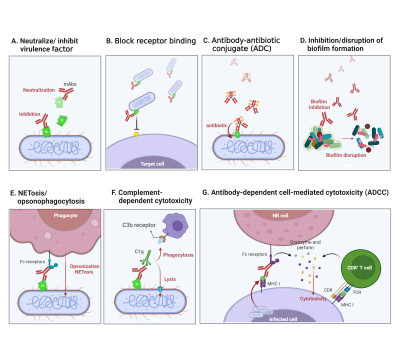
Multifaceted mechanisms of mAbs against bacterial figure illustrates the complex mechanisms through which mAbs counteract bacterial infections. (A) Highlights the neutralization or inhibition of bacterial virulence factors by mAbs, mitigating their pathogenic effects. (B) The process of mAbs blocking receptor-mediated adhesion is depicted, preventing bacterial adherence to host cells and hindering the progression of infection. (C) Portrays the Antibody–Antibiotic Conjugate (ADC) strategy, where mAbs conjugated with antibiotics enhance the precision and effectiveness of bacterial targeting and elimination. (D) Focuses on the role of mAbs in inhibiting or disrupting biofilm formation, a primary bacterial defense mechanism, facilitating bacterial clearance. (E) Illustrates the mAb-mediated NETosis/opsonophagocytosis pathway, promoting bacterial clearance through the facilitation of neutrophil extracellular traps (NETs) and enhanced phagocytosis. (F) Delineates the activation of complement-dependent cytotoxicity by mAbs, leading to bacterial cell lysis. (G) Illustrates the synergy between innate and adaptive immune responses facilitated by mAbs in Antibody-Dependent Cell-Mediated Cytotoxicity (ADCC), enhancing the clearance of bacterial infections. [1]
19 Feb 2024
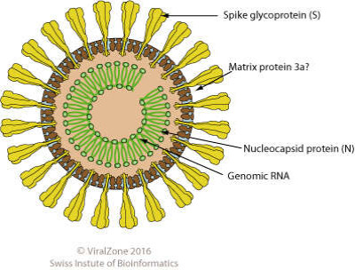
Diagram of mesonivirus virion. Enveloped, spherical, about 60-80 nm in diameter.
ViralZone, SIB Swiss Institute of Bioinformatics, Wikimedia Commons
22 Feb 2024
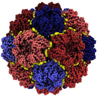
The yellow boundaries separate the capsid's mechanical units, which were identified with the algorithm of Polles et al. [1] The partitioning involves 32 units, which are further grouped in two distinct types: pentamers (blue) and hexamers (red).
Guido Polles, Wikimedia Commons
08 Mar 2024
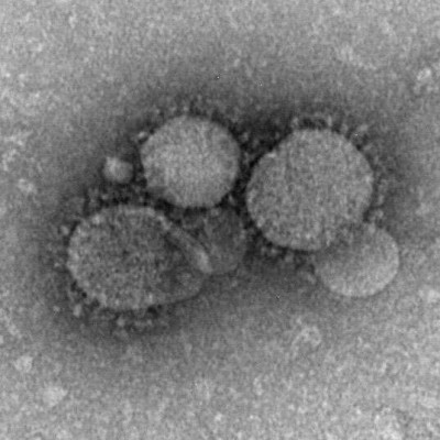
MERS-CoV particles as seen by negative stain electron microscopy. Virions contain characteristic club-like projections emanating from the viral membrane.
Maureen Metcalfe/Cynthia Goldsmith/Azaibi Tamin, Wikimedia Commons
11 Mar 2024
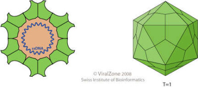
Schematic drawing of a virion of the genus Circovirus, cross section and side view. Non-enveloped, round, T=1 icosahedral symmetry, about 17 nm in diameter. The capsid consists of 12 pentagonal trumpet-shaped pentamers.
ViralZone, SIB Swiss Institute of Bioinformatics, Wikimedia Commons
11 Mar 2024
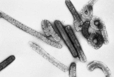
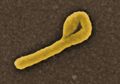
This negative stained transmission electron micrograph (TEM) depicts a number of filamentous Marburg virions, which had been cultured on Vero cell cultures, and purified on sucrose, rate-zonal gradients. Note the virus’s morphologic appearance with its characteristic “Shepherd’s Crook” shape; Magnified approximately 100,000x. Marburg hemorrhagic fever is a rare, severe type of hemorrhagic fever which affects both humans and non-human primates. Caused by a genetically unique zoonotic (that is, animal-borne) RNA virus of the filovirus family, its recognition led to the creation of this virus family. The four species of Ebola virus are the only other known members of the filovirus family. Marburg virus was first recognized in 1967, when outbreaks of hemorrhagic fever occurred simultaneously in laboratories in Marburg and Frankfurt, Germany and in Belgrade, Yugoslavia (now Serbia).
CDC/ Dr. Erskine Palmer, Russell Regnery, Wikimedia Commons
11 Mar 2024
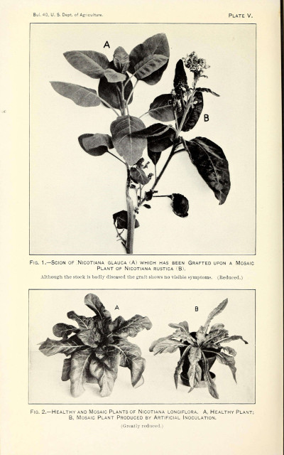
The mosaic disease of tobacco. [1]
Wikimedia Commons, Allard, H. A
14 Mar 2024
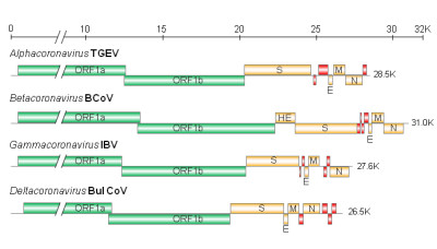
Genome organizations of various Coronavirus genera. The first two-thirds of the genome contains open reading frame (ORF)1a and ORF1b, which encode the replicase/transcriptase proteins (Green). The remaining one-third of the genome encodes four structural proteins: spike (S), envelope (E), membrane (M), and nucleocapsid (N) proteins (Orange). Some betaoronaviruses have an additional hemagglutinin-esterase (HE) gene (Orange). The genome of each genus or species has a set of unique accessory proteins (red).
Wikimedia Commons, Makoto Ujike and Fumihiro Taguchi
02 Apr 2024
Scanning electron micrograph of Ebola virus budding from the surface of a Vero cell (African green monkey kidney epithelial cell line.
Wikimedia Commons, NAID
03 Apr 2024
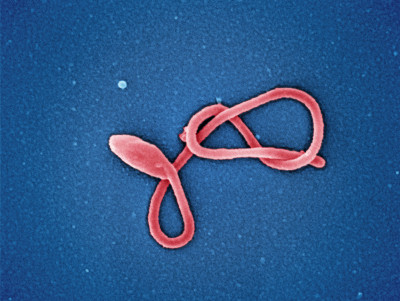
Colorized scanning electron micrograph of a single filamentous Ebola virus particle (colorized red). Image captured and color-enhanced at the NIAID Integrated Research Facility in Ft. Detrick, Maryland.
Wikimedia Commons, NIAID
03 Apr 2024
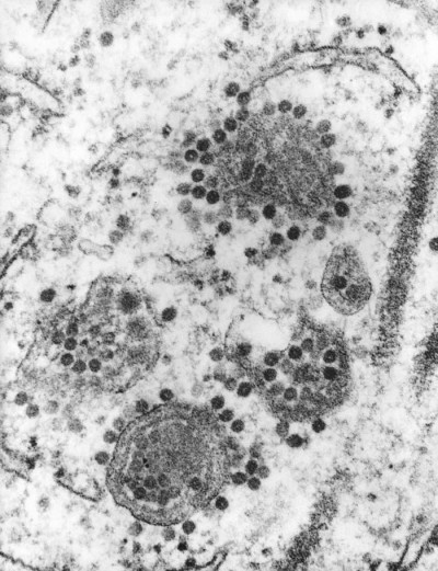
This 1975 transmission electron micrograph (TEM) revealed the presence of a number of Eastern Equine Encephalitis (EEE) virus virions that happened to be in a specimen of central nervous system tissue. EEE is an zoonotic arbovirus, which means that it is spread to human beings through the bite of an infected mosquito. EEE virus (EEEV) occurs in the eastern half of the United States where it causes disease in humans, horses, and some bird species. Because of the high mortality rate, EEE is regarded as one of the most serious mosquito-borne diseases in the United States. EEE is a Togaviridae virus family member, and the genus Alphavirus.
Public Health Image Library
11 Apr 2024
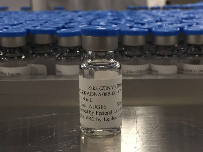
A vial of the NIAID Zika Virus Investigational DNA Vaccine, taken at the NIAID Vaccine Research Center’s Pilot Plant in Frederick, Maryland.
Wikimedia Commons, NAID
24 Apr 2024
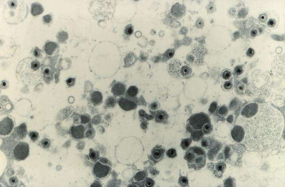
Magnified 49,200X, this transmission electron microscopic (TEM) image depicts numbers of cytomegalovirus (CMV) virions that were present in an unknown tissue sample.
Wikimedia Commons, CDC
24 Apr 2024
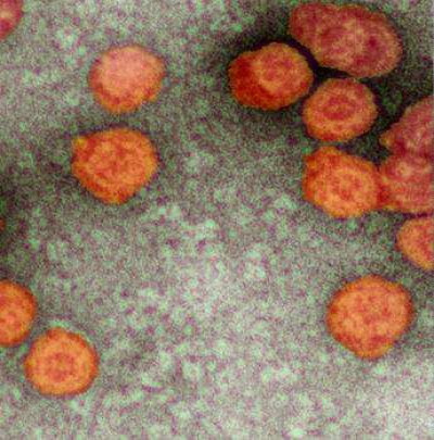
Virions of Pseudomonas virus phi6, colored.
Wikimedia Commons, Shankar Chellam
28 May 2024
The need for a color electron microscopy
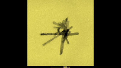
A black & white SEM/TEM micrograph is not simply the output of a scientific equipment but, similarly to any other type of photography, it is a form of art. Color is one of the fundamental elements of all artistic images and therefore it should be artificially put also in electron microscopy. As an example, in the above picture the TEM micrograph background has been dyed of yellow for better evidencing the outline of the nanostructure (i.e., a small bundle of graphite nanoscrolls). To add color to an electronic micrograph allows to associate one further information to the image. For example, to allow distinguishing an organic nanostructure from the inorganic one, a conventional coloration could be established for the micrograph background. All colors could have a different meaning to be transferred SEM/TEM micrographs by artificially coloring the image background.
27 Aug 2024
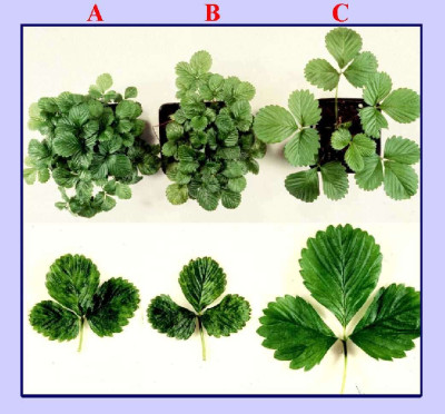
Symptoms of strawberry vein banding virus (SVBV) on leaves from indicator UC-5 strawberries. A plant vegetatively propagated from an SVBV infected plant. B, plant inoculated by particle gun bombardment of pSVBV-E3 insert DNA. C, non-infected healthy control.
Wikimedia Commons
25 Jan 2024
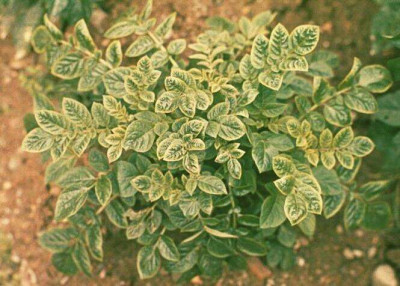
Potato plant displaying symptoms of Potato Leaf Roll Virus.
Wikimedia Commons, William M. Brown Jr., Bugwood.org
29 Jan 2024
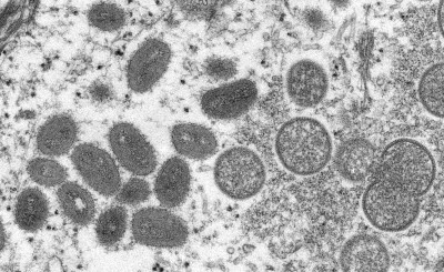
This electron microscopic (EM) image depicted a monkeypox virion, obtained from a clinical sample associated with the 2003 prairie dog outbreak. It was a thin section image from of a human skin sample. On the left were mature, oval-shaped virus particles, and on the right were the crescents, and spherical particles of immature virions.
Cynthia S. Goldsmith, Russell Regnery, CDC
31 Jan 2024
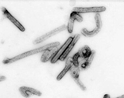
Negative stain image of an isolate of Marburg virus, showing filamentous particles as well as the characteristic "Shepherd's Crook." Magnification approximately 100,000 times.
Russell Regnery, CDC, Wikimedia Commons
31 Jan 2024
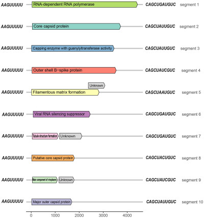
Schematic representation of the organization of rice black‐streaked dwarf virus (RBSDV) genomic RNA segments (linear lines); open reading frames (ORFs) (boxes), and products of each segment. [1]
Nan Wu, Lu Zhang, Yingdang Ren, and Xifeng Wang
01 Feb 2024
 Encyclopedia
