
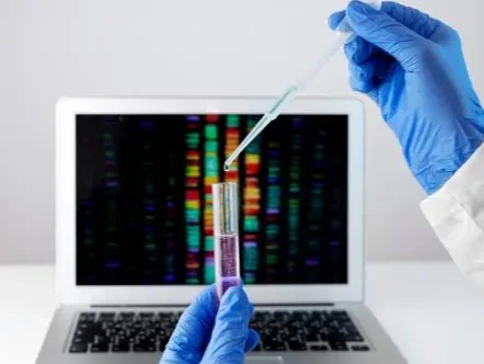
Summary

Biosynthesis and synthetic biology explore the enzymatic formation of biological molecules and the design, engineering, and reconstruction of biological systems. This collection covers biosynthetic pathways, enzyme-mediated reactions, metabolic engineering, genetic circuits, engineered microorganisms, cellular factories, genome editing, and the production of valuable compounds through programmable biological processes.

Expand All
Entries
Topic Review
Secosteroid Biosynthesis
Secosteroid biosynthesis denotes the formation of secosteroids—steroidal compounds in which one of the parent rings of the tetracyclic nucleus, most commonly the B-ring, has undergone scission—through photochemical or enzymatic mechanisms [1]. The prototypical pathway is the UV-B–driven photolysis of a 5,7-diene sterol precursor: in vertebrates, 7-dehydrocholesterol in the skin absorbs UV-B radiation (290–315 nm), causing cleavage of the B-ring between C9 and C10 to yield previtamin D3, which then undergoes a temperature-dependent rearrangement to cholecalciferol (vitamin D3); an analogous photoconversion of ergosterol yields vitamin D2 in fungi [2]. Secosteroid biosynthesis also encompasses enzyme-catalyzed routes, notably cytochrome P450 CYP11A1–mediated hydroxylation of 7-dehydrocholesterol or vitamin D3, which generates hydroxylated secosteroid metabolites with modified A-ring or side-chain structures that can be further transformed by UV-B [3]. The term additionally covers the formation of diverse 9,11-, 5,6-, and other secosteroid classes in marine invertebrates and microorganisms, where oxidative cleavage of sterol rings produces structurally distinct secosteroid scaffolds [1][4]. Secosteroid biosynthesis thus spans non-enzymatic photochemical ring opening and enzyme-dependent oxidative routes that together generate the secosteroid class from sterol precursors [1][2][3].
  • 7
  • 25 Oct 2026
Topic Review
Peptide Biosynthesis
Peptide biosynthesis is the biological formation and maturation of molecules containing amino-acid residues connected by peptide bonds. Ribosomal pathways translate an encoded precursor sequence, after which biosynthetic enzymes can modify the peptide to establish its mature chemical structure [1]. Leader regions within precursor peptides provide recognition elements that direct modifying enzymes toward the product-forming region [2]. Processing reactions can generate cyclic structures, altered side chains, and other chemical features within ribosomally produced natural peptides [3]. Multiple modifications may occur on a single precursor, including changes in amino-acid stereochemistry and the addition of methyl groups [4]. The mature product thus arises through an ordered relationship between precursor synthesis, enzyme recognition, chemical modification, and release of the processed peptide from any associated precursor regions [1].
  • 12
  • 25 Oct 2026
Topic Review
Hemoglobin Biosynthesis
Hemoglobin biosynthesis denotes the coordinated cellular process by which erythroid precursor cells assemble the tetrameric oxygen-transport protein hemoglobin from its two constituent parts: the prosthetic group heme and the globin polypeptide chains. The process is defined by the convergence of two biosynthetic streams that must be stoichiometrically balanced to avoid the accumulation of unliganded, toxic intermediates [1]. Heme is synthesized de novo through an eight-enzyme pathway that, in developing erythroid cells, must support an enormous demand for the prosthetic group; this pathway spans the mitochondrial matrix and the cytoplasm, beginning with the condensation of glycine and succinyl-CoA by 5-aminolevulinate synthase (ALAS) and concluding with the insertion of ferrous iron into protoporphyrin IX by ferrochelatase [2]. In parallel, the globin chains are synthesized on cytoplasmic ribosomes under the control of the erythroid transcriptional program, with the specific globin genes expressed—embryonic, fetal, or adult α- and β-type—depending on the developmental stage of the erythroid cell [3]. The defining feature of hemoglobin biosynthesis is the assembly phase, in which heme becomes associated with globin chains; this association has been proposed to occur co-translationally, with heme binding to the nascent α-globin chain as it emerges from the ribosome [4], and is facilitated by chaperone proteins such as alpha-hemoglobin stabilizing protein (AHSP) that maintain the globin chain in a soluble, assembly-competent state. The term encompasses the entire trajectory—from the enzymatic synthesis of heme and the regulated translation of globin mRNAs through the chaperone-assisted folding and tetramer assembly of the α₂β₂ hemoglobin molecule—as an integrated biosynthetic pathway unique to the erythroid lineage [5][6].
  • 15
  • 25 Oct 2026
Topic Review
Recombinant Antigens
Recombinant antigens are antigenic proteins or protein-derived constructs produced through expression of engineered genetic sequences. Their molecular identity includes the encoded amino-acid sequence and any structural or chemical features introduced during biosynthesis. Glycosylation can contribute directly to recognized epitopes or influence the folding and accessibility of antigenic surfaces [1]. Recombinant expression can generate a selected antigenic protein as part of a larger fusion polypeptide [2]. The spatial arrangement of exposed residues establishes conformational epitopes recognized by antibodies, making folding and structural integrity integral properties of the expressed antigen [3]. Genetic fusion can also join an antigenic sequence to another protein domain within a single translation product [4]. 
  • 8
  • 25 Oct 2026
Topic Review
Androgen Biosynthesis
Androgen biosynthesis is the enzymatic formation of androgenic steroids from cholesterol-derived precursors. Steroidogenic pathways use cytochrome P450 enzymes and hydroxysteroid dehydrogenases to modify the carbon skeleton and oxygen-containing functional groups of steroid intermediates [1]. Cholesterol delivery to the mitochondrial steroidogenic machinery initiates precursor formation, with steroidogenic acute regulatory protein participating in the transfer process [2]. The activity of this protein at the outer mitochondrial membrane links the supply of substrate with acute regulation of steroid production [3]. Subsequent reactions in steroidogenic tissues generate androgen precursors and active androgens through tissue-specific combinations of enzymes [4]. The biosynthetic sequence integrates precursor availability, intracellular transport, enzyme expression, and catalytic conversion, establishing the identities and relative amounts of androgenic products formed by a cell [5].
  • 5
  • 25 Oct 2026
Topic Review
Microsomal Enzyme
Microsomal enzymes are the drug-metabolizing enzymes that remain associated with fragments of the smooth endoplasmic reticulum after liver tissue is homogenized and subjected to differential centrifugation, yielding a membrane-bound vesicle fraction termed microsomes [1]. The microsomal drug-metabolizing system of the liver consists of two protein components—cytochrome P450 and NADPH-cytochrome c reductase—together with the lipid phosphatidylcholine; cytochrome P450 serves as the binding site for oxygen and substrate, the reductase shuttles electrons from NADPH to cytochrome P450, and the phospholipid facilitates electron transfer between them [2]. This system is classified as a mixed-function oxidase or monooxygenase: it requires both NADPH and molecular oxygen, and it incorporates one atom of oxygen into the substrate while reducing the second oxygen atom to water [3]. Microsomal enzymes occupy the principal position in phase I xenobiotic biotransformation, catalyzing aromatic and aliphatic hydroxylation, N-, O-, and S-dealkylation, deamination, dehalogenation, sulfoxidation, N-oxidation, and azo- and nitro-reduction [2][3]. Within the taxonomy of pharmacogenetics and drug metabolism, the term designates this membrane-associated, cytochrome P450–centered enzyme system as the object of studies on substrate specificity, enzyme induction, competitive and mechanism-based inhibition, and interindividual variability in hepatic drug clearance [3]. The microsomal fraction also retains phase II enzymes situated in the endoplasmic reticulum, notably UDP-glucuronosyltransferases, whose activity is influenced by the membrane environment in which the microsomal preparation embeds them [4].
  • 7
  • 25 Oct 2026
Topic Review
Enzyme Formulation
Enzyme formulation is the composition and physical presentation of a preparation containing an enzyme and the materials surrounding it. The formulation establishes chemical conditions such as pH, ionic composition, and hydration that influence the persistence of functional protein structure [1]. Protein concentration and interactions with the surrounding medium can also alter the formation of aggregates from unfolded or partially unfolded molecules [2]. Thermal unfolding measurements can characterize changes in protein stability associated with particular solution components or bound ligands [3]. Formulations may present the protein in solution or incorporate it within a structured material that controls its physical environment and release [4]. Composition, physical state, catalytic activity, and stability are therefore complementary properties of an enzyme formulation, linking the molecular condition of the catalyst with the characteristics of the complete preparation [5].
  • 7
  • 25 Oct 2026
Topic Review
Peptide Hormone Biosynthesis
[1]Peptide hormone biosynthesis is the cellular production and molecular maturation of peptides that act as hormonal signals. Translation generates precursor proteins that enter the secretory pathway and undergo processing to produce mature peptide products. Proprotein convertases catalyze specific cleavage reactions within these precursors, frequently at sites containing basic amino-acid residues [2]. The intracellular localization of a processing enzyme positions its catalytic activity within the appropriate secretory compartment [3]. Recognition of paired basic residues is a molecular feature of several precursor-processing reactions and contributes to the selection of cleavage sites . Compartmental pH and calcium conditions can further regulate the activity of processing enzymes, as in proinsulin conversion [4]. Precursor sequence, protease specificity, and secretory-compartment chemistry together organize the formation and maturation of the hormonal peptide [5].
  • 7
  • 25 Oct 2026
Topic Review
Metabolic Flux Profiling
Metabolic flux profiling designates the quantitative determination of the rates at which metabolites are converted through intracellular biochemical reactions—the metabolic fluxes—in a living biological system, yielding a flux map that describes the functional state of the metabolic network under defined genetic or environmental conditions [1]. Its conceptual core rests on a distinction between two classes of flux: extracellular fluxes, such as substrate uptake and product secretion rates, which cross the cell membrane and are directly measurable by tracking extracellular metabolite concentrations or biomass over time; and intracellular fluxes, which do not cross the membrane and cannot be measured directly [2]. Intracellular flux profiling therefore proceeds inferentially: an isotopically labeled substrate (for example, ¹³C-glucose) is supplied, the resulting labeling patterns of downstream metabolites—expressed as mass distribution vectors describing the fractional enrichment of isotopologues and isotopomers—are measured by mass spectrometry or nuclear magnetic resonance, and a metabolic network model is used to compute the flux distribution that best accounts for those patterns [1][2]. The computational procedure integrating isotope labeling data with a stoichiometric network model to generate a quantitative flux map is termed isotope-assisted metabolic flux analysis (iMFA) [3]. Profiling may be conducted under metabolic and isotopic steady state, in which labeling patterns are stable and fluxes are assumed constant, or under isotopically nonstationary conditions, in which transient labeling kinetics are modeled to resolve fluxes in systems that do not reach steady state [4]. A defining feature of metabolic flux profiling is that its output—the flux distribution—constitutes a phenotype-level description that integrates the effects of gene expression, enzyme abundance, and allosteric regulation, capturing the net functional behavior of the metabolic network rather than the abundance of any individual component [2][5].
  • 16
  • 25 Oct 2026
Topic Review
Genome Assembly Techniques
Genome assembly techniques comprise the algorithmic and computational protocols by which a genome sequence is reconstructed de novo from a set of sequencing reads, without reliance on a pre-existing reference sequence [1]. The defining operational feature is the inference of the original linear or haplotype-resolved chromosome sequence from short, overlapping or non-overlapping fragments, a problem whose solution requires resolving repeats, distinguishing allelic variants from paralogous sequences, and ordering and orienting contigs into chromosome-scale scaffolds [2]. The methodological space is conventionally organized by the graph paradigm employed for reconstruction: short-read assembly predominantly uses the de Bruijn graph, in which reads are decomposed into fixed-length k-mers and the genome is traversed as a path through a compact graph whose edges represent (k−1)-overlaps, whereas long-read assembly employs overlap–layout–consensus (OLC) approaches that exploit the greater span of individual reads to bridge repetitive regions that are unresolvable with short fragments [2][3]. Contemporary chromosome-scale assembly integrates multiple data types: long accurate reads (for example, PacBio HiFi) provide high per-base accuracy across multi-kilobase spans, ultralong reads (for example, Oxford Nanopore Technologies) traverse large repetitive blocks, and proximity-ligation or Hi-C data supply long-range linkage information for phasing haplotypes and scaffolding contigs into telomere-to-telomere (T2T) chromosome arms [4]. The term further encompasses assembly paradigms for non-clonal or mixed samples, including single-cell amplified genome assembly, in which the genome of an individual uncultivated cell is reconstructed from whole-genome amplification products, and metagenome assembly, in which reads from a microbial community are partitioned and assembled into metagenome-assembled genomes (MAGs) [5]. Across these settings, the output of a genome assembly technique is a set of contigs or scaffolds accompanied by quantitative metrics of continuity and completeness that describe the extent to which the reconstructed sequence recovers the underlying chromosome architecture [1].
  • 18
  • 25 Oct 2026
  • Page
  • of
  • 9
>>