Stents are tubular ducts made of non-invasive materials designed to maintain the continuous flow of air through the airway lumen, or various types of fluid in the case of the urinary and circulatory systems. Stenting in veterinary medicine has been a rapidly growing method of interventional surgery. This procedure is usually performed in the respiratory and urinary tracts, but there are cases of stenting of blood vessels or gastrointestinal structures. It is based on maintaining the permeability of a given tubular structure, thus allowing the passage of gas or liquid. This procedure is often performed as a first-line treatment in situations where pharmacological agents do not work and as an alternative method, often cheaper than the classically performed ones. There are also cases where stenting is used as a palliative treatment, e.g., to enable defecation in colonic obstruction due to tumour infiltration of the colon wall. Stenting is often a life-saving or comfort-improving procedure for animals, but one should also be aware of possible postoperative complications and be prepared for any adversity.
1. Introduction
Their use in medicine has become increasingly important since 1986, when one of the first self-expandable stents was implanted in coronary arteries with long-term positive results [
1]. At the same time, stenting was being attempted in dogs but with unsatisfactory results [
2,
3]. This encouraged researchers to explore new possibilities for the use of stents in human medicine and later in veterinary medicine. They have become an attractive alternative for several procedures, including tracheal collapse or urinary tract obstruction [
4,
5,
6], due to the reduction in adverse effects compared to previous procedures. Stents have also found application in procedures where traditional methods have become ineffective due to the atypical morphology of the structure in question, e.g., during the occurrence of a persistent arterial duct with two different diameters at the ends of the vessel [
7]. Stent placement has also found its way into palliative treatment, which has significantly alleviated the clinical symptoms of concomitant disease, i.e., in the case of stenting of a part of the colon affected by mucosal cancer, making defecation difficult [
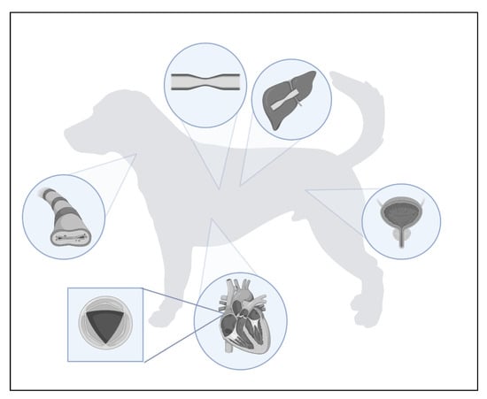
8]. The locations of the described stent sites are shown in
Figure 1.
Figure 1. Use of stents in the treatment of canine and feline diseases.
Currently, stents made of different types of materials, including various metallic, polymeric, and silicone materials, as well as biodegradable fibres, are used in veterinary medicine [
9,
10]. In new generations of stents, these materials are coated with various types of layers that perform additional functions. These include stents that release immunomodulatory drugs (paclitaxel, biolimus, sirolimus, arsenic trioxidate, or zotarolimus) or antibiotics (cefotaxime, triclosan), which are particularly useful in highly invasive procedures that often induce inflammation [
10,
11,
12,
13,
14,
15,
16,
17,
18,
19]. The most commonly used seems to be the self-expandable nitinol stent (nickel-titanium alloy with a ratio of 55:45%) with different types of plexus, as shown in
Figure 2 [
20]. In favour of its use is spontaneous passivation, which prevents corrosion of the material and significantly increases its biocompatibility with surrounding tissues [
21]. Passivation is a chemical process that generally only affects metals and involves the production of a film by reaction with the surrounding environment. This thin layer insulates the stent and makes it neutral to the organism. In addition to choosing the right type of stent, the size of the stent is also very important. In determining the diameter of stents, a scale defined by the French surgical instrument maker Joseph-Frédéric-Benoît Charrière is commonly used [
22]. The abbreviation CH, or Ch, from his last name, was used to refer to the unit of measurement, but nowadays it is more common to use the abbreviation from the French word ‘Fr’ or ‘FR’. The abbreviations ‘Fg’ or ‘F’ may also be encountered, although these are rarely used. This unit defines the outer diameter of the catheter. To convert the diameter of the catheter into millimetres, the French size is divided by 3 (1 mm = 3 Fr).
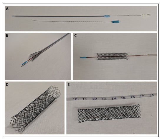
Figure 2. (A) Tracheal stenting kit containing a nitinol stent with a cross-braid and a measure to assess the tracheal length. (B) View of the initial stent deployment process. (C–E) Different views of the fully expanded stent. The photographs come from the private archives of Dr Dariusz Jagielski.
2. Types of Stents
The selection of proper material in relation to the stent placement site has enormous significance. The creation of the ideal stent has been a priority in interventional surgery for more than 30 years, but it is still difficult to combine all the characteristics that meet this criterion: elasticity, biocompatibility, thermostability, resistance to pressure forces, and a lack of reaction to surrounding tissue. Its properties determine the subsequent possibility of postoperative complications, which sometimes necessitate a second procedure. Currently, the most commonly used metal material in veterinary medicine is nitinol. It has gained its popularity due to two important characteristics, super-elasticity and shape memory. The super-elasticity of nitinol stents is defined by their incredible ability to adapt their shape in relation to the high mobility of a given structure, such as a vessel or trachea [
24]. However, in some cases, defects related to stent flexibility are still reported, as in the case of peripheral artery disease in humans during stenting of the femoro-popliteal arteries [
25]. Although in animals there is no predilection for atherosclerotic plaque accumulation in vessels, the site associated with high mobility is the trachea during rapid head movements, especially in impulsive dogs [
26]. Shape memory, on the other hand, prevents any deformation of the stent due to the high temperature > 20 °C, thus disallowing any shape change after insertion due to the constant body temperature of the animals [
24]. Another equally important property is its biocompatibility with surrounding tissues, which prevents inflammatory reactions and responses [
24]. Other metals are not as common as nitinol, however, the use of materials such as stainless steel or elgiloy, which has very similar properties, has been reported [
27,
28].
Polymeric fibres are a forward-looking material used in human medicine and, more importantly, in veterinary medicine. In animals, they are used for urinary tract stenting with double-pigtail stents or for stenting the blood vessel wall with drug-eluting stents, which can be temporary—biodegradable—or permanent. Polymer stent materials include polyurethane, silicone, poly-lactide-co-glycolide, or l-lactide-glycolic acid copolymer [
29]. Maintaining vessel patency in animals is rarely performed because vessel wall obstruction in animals generally occurs when other structures such as cancerous tumours are compressed [
30]. In humans, the problem is much more exacerbated due to a predilection for the accumulation of atherosclerotic plaques concentrating on the inner vessel wall, leading to stenosis of the vessel lumen [
31]. Stenting of the urinary tract is observed much more frequently in everyday veterinary practice and polymeric materials are the main raw material for ureteral stents. Postoperative complications related to restenosis, stent migration, or tissue encrustation are still a major problem. For this reason, developers are looking for newer and better solutions, with an emphasis on greater biocompatibility, durability, and flexibility. Future solutions using biodegradable polymers or metal drug-eluting stents for specific conditions are being considered [
32]. However, the use of drug-eluting stents in veterinary medicine is poorly understood and requires much research into their efficacy and performance in animals, as evidenced by the small amount of documented work in this area. Materials for the production of stents in veterinary medicine are listed in
Table 1. The data presented suggest that nitinol is the most commonly used material, mainly used for stenting of the respiratory tract, urethra, and some vessels, but in the latter case, it is much less common. This choice is undoubtedly due to the properties of nitinol described above, as well as the many papers available in the literature on efficacy and post-operative complications, which only confirms that nitinol may be the principal material for stents. The choice of this material in the context of ureteral stenting is not so obvious anymore, as the most commonly used materials in this type of surgery are polymeric fibres, mainly polyethylene. However, there is still too little information available on the efficacy of biodegradable and drug-eluting stents in the context of successful therapy and the impact on the animal body, which should certainly become a topic for future research.
This entry is adapted from the peer-reviewed paper 10.3390/ma16041480