S-Glutathionylation and S-Nitrosylation in Mitochondria: Homeostasis and Neurodegeneration: History
Please note this is an old version of this entry, which may differ significantly from the current revision.

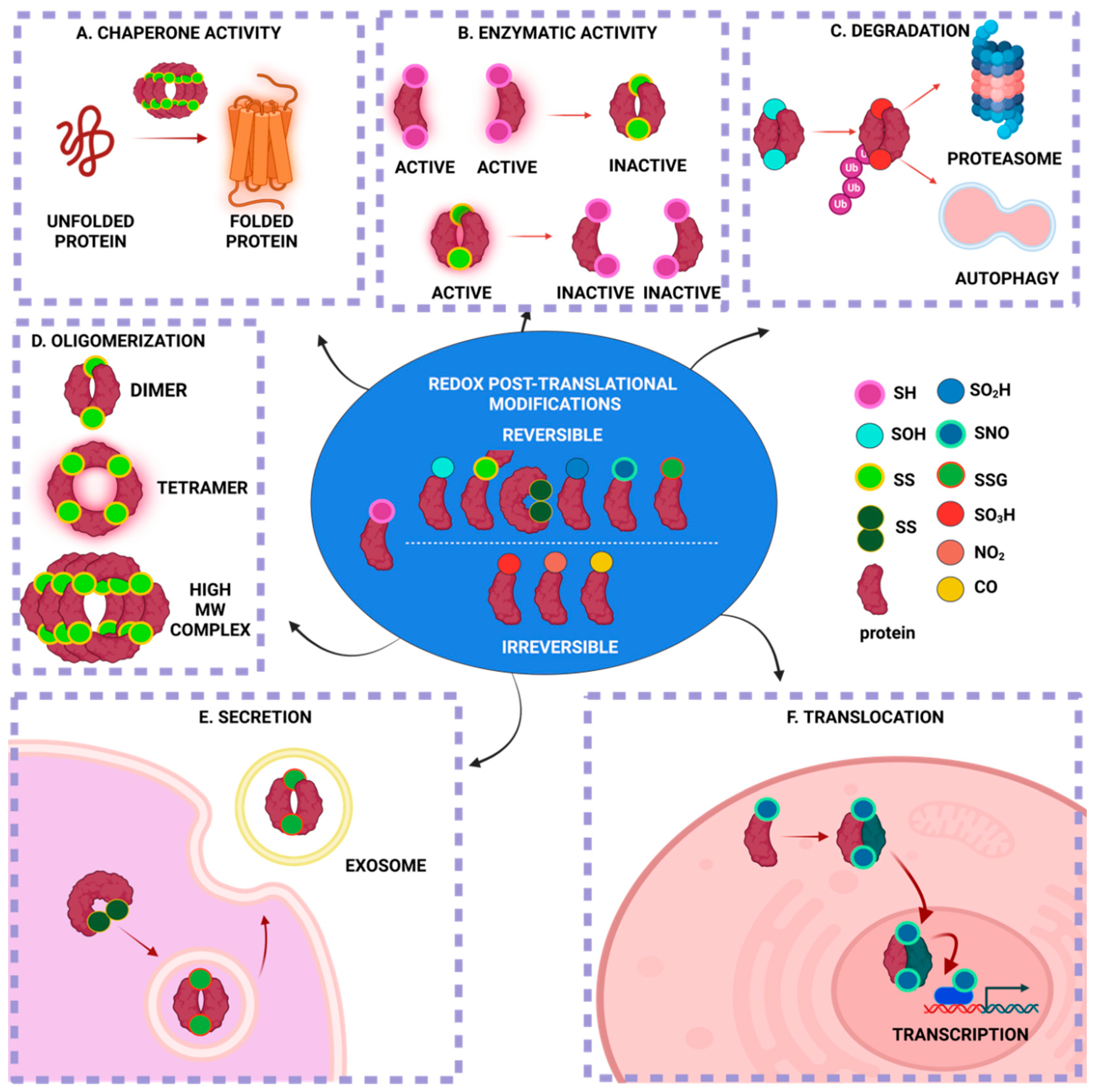
Redox post-translational modifications are derived from fluctuations in the redox potential and modulate protein function, localization, activity and structure. Amongst the oxidative reversible modifications, the S-glutathionylation of proteins was the first to be characterized as a post-translational modification, which primarily protects proteins from irreversible oxidation. S-nitrosylation, another post-translational modification, was identified in the past, but it was re-introduced as a prototype cell-signaling mechanism, one that tightly regulates core processes within the cell’s sub-compartments, especially in mitochondria. S-glutathionylation and S-nitrosylation are modulated by fluctuations in reactive oxygen and nitrogen species and, in turn, orchestrate mitochondrial bioenergetics machinery, morphology, nutrients metabolism and apoptosis.

  • glutathionylation
  • nitrosylation
  • redox
  • mitochondria
  • neurodegeneration
  • Alzheimer's
  • Parkinson's
  • ALS
  • Friedreich's

1. Introduction

Reactive oxygen and nitrogen species (RONS) are byproducts of aerobic metabolism and constitute essential signaling molecules [1]. The RONS signaling cascade is mediated by the oxidation–reduction (redox)-based post-translational modifications (PTMs) of proteins [2].
The mammalian brain is a major source of RONS due to its high metabolic activity [1]. Consequently, redox PTM-mediated signaling is pivotal for the brain under normal physiology [3]. In contrast, under pathological conditions, aberrantly produced RONS cause oxidative and/or nitrosative stress, which, in turn, damages DNA, proteins and lipids and leads to impaired cellular function [4][5]. The mitochondrial oxidative phosphorylation (OXPHOS) system and enzymes such as NADPH oxidases (NOXs) and nitric oxide synthases (NOSs) are the main sources of RONS in aerobic organisms [6][7][8]. Some major RONS are hydrogen peroxide (H2O2), hydroxyl radical (OH), superoxide anion (O2), nitric oxide (NO) and peroxynitrite (ONOO) [9]. Concomitantly, there is a tightly modulated machinery that reduces the levels of RONS in the cell, which is constituted by small molecules (ascorbate, cysteine, glutathione (GSH) and proteins glutathione peroxidases (GPxs), catalases (CATs), superoxide dismutases (SODs) and peroxiredoxins (PRDXs)) [10][11][12][13]. Under eustress (normal physiological conditions), there is a balance between RONS production and reduction, whereas any imbalance in this system causes oxidative/nitrosative stresses [4][11]. The complex crosstalk between reactive oxygen species (ROS) and reactive nitrogen species (RNS) triggers a downstream cascade of events that collectively are termed redox signaling [4][5][11].
The main targets of redox PTMs are proteins due to their high abundance and increased rate constants for oxidation reactions [11][14][15][16]. While redox PTMs can happen on almost every amino acid, redox modifications of thiol happen only on cysteine (Cys) residues. Cys residues constitute the primary drivers of redox signaling due to their high rates of oxidation, which are mostly reversible, and reduction [17]. Cys residues can be targets of multiple redox PTMs due to the electronic structure of the thiol (-SH) group, which allows for multiple oxidation states (ranging from −2 to +6) [17]; for example, protein disulfide isomerase (PDI) can be both S-nitrosylated and S-glutathionylated at its reactive thiols [18]. The redox PTMs of Cys in proteins have multiple effects on the particular protein (Figure 1). Under eustress, only a subset of redox-sensitive Cys residues within proteins is susceptible to multiple redox PTMs, whereas, under oxidative and/or nitrosative stress, even lowly reactive Cys residues undergo multiple redox PTMs, which can be excessive and/or irreversible [19]. Redox PTMs in proteins might act as a “redox-switch” that modifies the protein’s function: for instance, rendering an enzyme active or inactive [20]. Redox PTMs can alter the protein structure via disulfide bond formations, Cys-dependent metal cofactors interactions and the modulation of the topography of the protein [17][21]. Moreover, redox PTMs can modify the stability and degradation of the target protein, which depends on the oxidation status, which is reversible or irreversible, via ATP- or ubiquitin-dependent or independent mechanisms [22][23]. Finally, the redox status of a protein can affect its localization and translocalization: for instance, by changing the conformation of a protein and rendering it available for interactions with other proteins that translocate into other subcompartments [17][24]. Collectively, this continuum of the reactive Cys residues of proteins constitutes a redox-sensing mechanism with which the cell reacts to the differential level of RONS and adjusts to its redox status via multiple downstream effects (Figure 1).
Figure 1. Redox post-translational modifications effects on proteins structure, function, location and turnover. ROS/RNS can modify amino acid residues of a protein by S-sulfenylation (–SOH), intermolecular and intramolecular disulfide bonds (–SS–), S-sulfinylation (–SO2H), S-nitrosylation (–SNO), S-glutathionylation (–SSG), S-sulfonylation (–SO3H), nitration (–NO2) and carbonylation (–CO: carbonyl groups). Based on its redox PTMs, a protein can act as a chaperone (A), change the conformation that activates or deactivates its enzymatic function (B), irreversibly oxidize it and trigger ubiquitin-mediated proteasomal degradation (C), modulate its conformation and oligomerization (D), regulate its secretion in extracellular space (E) and trigger its translocation into sub-compartments, thus acting as a signaling molecule (F). Created with BioRender.com.
Aberrant redox PTMs, including S-glutathionylation and S-nitrosylation, can alter multiple processes within the cell and have been extensively studied, mainly separately, in mitochondria homeostasis and neurodegenerative diseases [25][26][27][28][29]. Of particular importance and extensively reviewed in the following sections is the trigonal interaction of oxidative/nitrosative stress, mitochondrial dysfunction and neurodegeneration. Both S-glutathionylated and S-nitrosylated proteins were incorporated here, with a particular focus on mitochondrial proteins or proteins that directly affect mitochondria and that have been extensively investigated (or their role is still elusive) in neurodegenerative disorders, including Alzheimer’s disease (AD), Parkinson’s disease (PD), amyotrophic lateral sclerosis (ALS) and Friedreich’s ataxia [30][31][32][33].
Here, it is to bring together and compare S-glutathionylation and S-nitrosylation processes that regulate mitochondria and interfere with or exacerbate neurodegenerative disorders. By doing so, the converging redox-induced mechanisms between neurodegenerative disorders might unravel commonalities in neurodegeneration or aging processes and, thus, contribute to novel therapeutic approaches beyond antioxidant supplementation, which has failed in clinical trials [34]. While the focus was mainly on AD, PD, ALS and Huntington’s disease in the majority of other reviews that had the same purposes, the focus is on neurodegenerative disorders where mitochondria dysfunction and S-glutathionylation/S-nitrosylation have been proposed to interfere with the disease’s pathogenesis. In addition, the focus is brought to Friedrich’s ataxia, which is a mitochondrial neurodegenerative disorder in which nitric oxide-mediated S-nitrosylation signaling has not yet been investigated, and the research field is urged to shed light on that aspect of the disorder by discussing the data that indirectly indicate its possible importance. The major objectives of here are to re-introduce redox PTMs as ROS/RNS-induced cell signaling processes with pivotal roles in mitochondria homeostasis and neurodegeneration and as converging mechanisms between homeostasis, aging and neurodegenerative disorders. Given the significant leap in authors' scientific knowledge about their multiverse roles in the last two decades, particularly in neurodegenerative disorders, herein, it is to combine previous and new data demonstrating not only their independent roles but also their interdependent function that orchestrates mitochondria regulation and triggers or exacerbates neuronal degeneration.
Herein, it has some limitations concerning the extent of biochemical principles governing the multiple ways that can lead to S-glutathionylation and S-nitrosylation of proteins. For this reason, appropriate literature was cited for the particular cases where biochemical processes are not extensively described. Despite that, the topics covered herein focus on signaling pathways for which the information stated is adequate for the reader to comprehend thoroughly the cellular processes implicated in mitochondria homeostasis and disease.

2. Protein S-Glutathionylation

Protein S-glutathionylation (PSSG) is a reversible oxidative post-translational modification that is mediated by the conjugation of glutathione (GSH) to an exposed Cys residue (-SH). The S-glutathionylation of mitochondria proteins is highly sensitive to local fluctuations in redox status, particularly via the alterations of the ratio between reduced and oxidized glutathione (GSH/GSSG) [35]. Proteins can be S-glutathionylated either non-enzymatically or enzymatically (Figure 2). Non-enzymatic protein S-glutathionylation takes place via different mechanisms that have been extensively reviewed by many authors [35][36][37]. Non-enzymatic protein S-glutathionylation is very non-specific and primarily occurs under oxidative stress. In mitochondria under oxidative stress, proteins can be hyper-glutathionylated, and this effect can be utilized as an oxidative stress marker [38]. Under normoxia, a number of mitochondrial proteins are S-glutathionylated at specific Cys residues in a reversible, sensitive-to-redox-fluctuations and enzymatically driven manner. S-glutathionylation reactions have been particularly investigated in cytosol where glutaredoxin 1 (GRX1) resides while being the chief enzyme responsible for PSSG de-glutathionylation [39]. The mitochondrial GRX1 isozyme, GRX2, is only ~34% identical to the sequence of GRX1 but shares the exact same catalytic mechanism [40]. The sequence differences between GRX1 and GRX2 are important for their regulation. GRX2, unlike GRX1, has no exposed thiol, making it less susceptible to ROS-mediated deactivation. Moreover, GRX2 forms a homodimer that is stabilized by a 2Fe-2S cluster, for which its disassembly by ROS superoxide, O2, leads to the release of active monomeric GRX2. This regulation potentiates the link between S-glutathionylation and mitochondrial ROS [41]. Both GRX2 and GRX1 can mediate S-glutathionylation, but the exact mechanisms are still elusive. GRX2 has been documented in mediating Complex I (CI) and UCP3 S-glutathionylation and de-glutathionylation; the significance of these reactions will be discussed in the following sections [42][43]. It is also worthy of mention that Glutathione-S-transferases (GST Pi and Mu) have been shown to perform S-glutathionylation in cytosol and GST Alpha, Kappa, Mu, Pi and Zeta have been proposed to have similar functions in mitochondria [44][45]. The small oxidoreductase, sulfiredoxin (SRX), catalyzes the reduction of sulfinic acid derivatives into two Cys peroxiredoxins; therefore, it has been proposed to have deglutathionylation activity, too [46] (Figure 2).
Figure 2. General mechanisms of S-Glutathionylation and Deglutathionylation. Under oxidative stress, proteins can be S-glutathionylated non-enzymatically or enzymatically via GRX and GST. The reverse reaction, deglutathionylation, takes place non-enzymatically, upon increased levels of GSH to GSSG, or enzymatically, not only via GRX primarily but also TRX, SRX and PDI. During aging, a subset of proteins is S-glutathionylated, promoting neuroprotection. Upon increased levels of ROS, potentially at early stages of neurodegeneration, increased levels of S-glutathionylation protect proteins from irreversible oxidation and degradation while simultaneously modifying cellular homeostatic mechanisms to compensate for increased ROS. Aberrant S-glutathionylation in neurodegenerative disorders exacerbates disease pathology, ultimately leading to neuronal damage. Created with BioRender.com.
While there are many sources for mitochondrial ROS, mitochondrial regulations by redox switches depend on mitochondrial densities, protein thiols contents, the import of GSH and export of GSSG and the reduction of GSSG to GSH by glutathione reductase [42][47]. In the following sections, S-glutathionylation in mitochondria’s core processes will be discussed and its effect on various proteins and enzymes, highlighting the impact on mitochondrial metabolism and function.

2.1. Regulation of OXPHOS System by S-Glutathionylation

Most information about how redox switches affect mitochondria thiols has been accrued by the multiple studies on the impact of S-oxidation in the OXPHOS system (Figure 3). The CI of the mitochondrial electron transport chain (ETC) was the first OXPHOS component that was identified to be S-glutathionylated. The oxidation of cysteine residues on the 51-kDa and 75-kDa subunit in CI can be protected from irreversible oxidation by S-glutathionylation [48][49]. While the S-glutathionylation of CI decreases its activity, this modification is reversible by deglutathionylation via GRX2 [42]. Another important role of the S-glutathionylation of CI is that it diminishes O2 production by sterically blocking the NADH’s binding site [50]. However, the other S-glutathionylation of CI has also been shown to increase O2 emissions but only under prolonged S-glutathionylation [51]. It is important to correlate the S-glutathionylation of CI with the S-glutathionylation of the α-ketoglutarate dehydrogenase complex (KGDH) since KGDH produces the NADH that is utilized for oxidation by CI and both enzymes produce ROS. Both the S-glutathionylation of KGDH and CI diminish their activity under excess H2O2. Once the levels of H2O2 and O2 are back to normal, CI and KGDH are deglutathionylated by GRX2 and are active again [52]. In contrast to CI, Complex II (Succinate dehydrogenase (SDH) S-glutathionylation increases the activity of the enzyme. In particular, CII is persistently S-glutathionylated at the 70-kDa FAD binding subunit; thus, this constitutive modification is considered to be optimal for the CII’s activity [49]. Complex V (ATP synthase, α subunit) has also been shown to be glutathionylated, and this modification diminishes ATP production by blocking nucleotide binding [53]. The temporary glutathionylation of CV might act synergistically with CI and KGDH glutathionylation upon increased levels of ROS. Additionally, this modification diminishes ATP hydrolysis and, consequently, proton leaks, thus preventing the depolarization of the mitochondrial inner membrane [54].
Figure 3. S-Nitrosylation and S-Glutathionylation roles in mitochondria redox-metabolism interplay. The targets of S-glutathionylation in the TCA cycle are PDH, ACN, IDH, KGDH, SDT and MDH. The S-glutathionylation of TCA cycle enzymes protects them from irreversible oxidation until the redox status is impaired and inhibits ROS production and electrons transfer to the ETC to further minimize the ROS levels under oxidative stress. Complex I and V inhibitions by S-glutathionylation share similar purposes with the S-glutathionylation of TCA cycle enzymes and ultimately minimize ROS production. Only Complex II S-glutathionylation has been shown to be integral for its constitutive activity. Under excessive ROS, the glutathionylation of ANT and VDAC prevents the formation of MPTP in inhibiting apoptosis, whereas UCP glutathionylation increases ROS formations. The targets of S-nitrosylation in the TCA cycle are ACN, KGDH and SDH, which in turn inhibits their function to minimize ROS. All mitochondria Complexes I-V can be nitrosylated under increased oxidative/nitrosative stress both to minimize ROS production and to scavenge NO. Under basal NO levels, the S-nitrosylation of VDAC and MPTP constituents inhibits their functions in providing protection from apoptosis, whereas under excessive NO levels, aberrant S-nitrosylation activates VDAC-mediated cytochrome c release and apoptosis or/and CYPD S-nitrosylation, the activation of MPTP, the ablation of ATP production and necrosis (OMM: outer mitochondrial membrane; IMM: inner mitochondrial membrane). Created with BioRender.com.

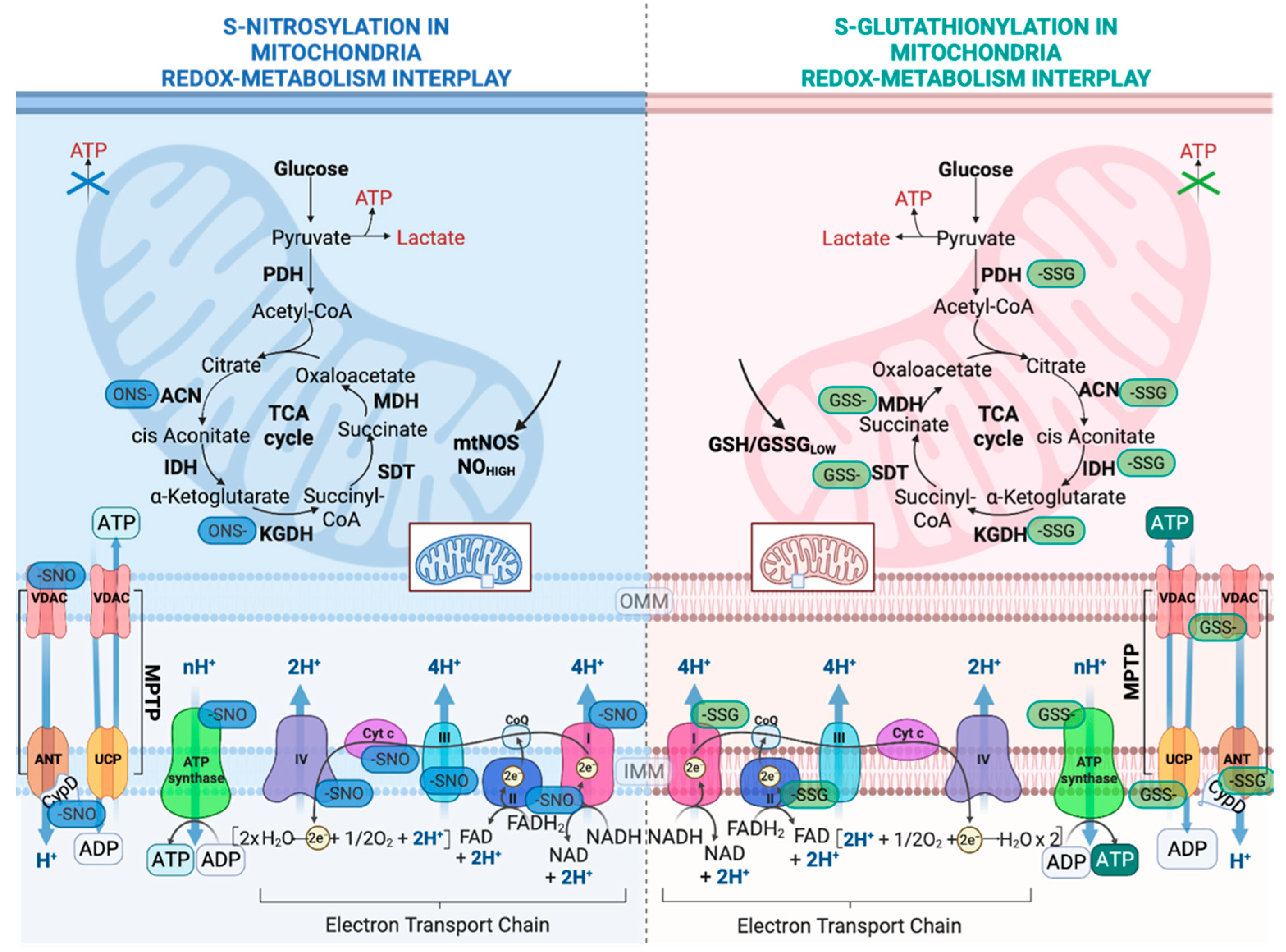
2.2. Regulation of Nutrient Metabolism by S-Glutathionylation

The tricarboxylic acid cycle (TCA) is the most central mechanism for both anaerobic and aerobic organisms. In aerobic organisms, the TCA cycle provides the essential building blocks for macromolecules and strips electrons from nutrients in order to provide ATP. The impact of TCA cycle enzyme S-glutathionylation is enzyme-dependent (Figure 3). Aconitase (ACN) can be reversibly S-glutathionylated at its 4FE–4S cluster upon H2O2 bursts [55]. However, this modification does not disassemble its 4FE–4S cluster due to its interaction with the Frataxin protein [55]. Only upon excessive stress is ACN irreversibly deactivated due to the 4FE–4S cluster’s disassembly [28]. NADP-dependent isocitrate dehydrogenase (IDH), which contributes to the NADPH pool in mitochondria, can be inactivated by S-glutathionylation and that might hamper its reducing power, which is necessary in order to preserve the antioxidant’s defense system [56]. As mentioned above, KGDH can be glutathionylated and reversible oxidation might have a dual role, which includes the modulation of the flow of metabolites via the TCA cycle and amino acid metabolism and the accumulation of 2-oxoglutarate, which in turn quenches H2O2 and minimizes H2O2 production [57]. Pyruvate dehydrogenase (PDH) can be also regulated by reversible S-glutathionylation on its E2 subunit in a similar manner to KGDH [58]. It has been reported that excessive S-glutathionylation inhibits lipid oxidation, thus decreasing fatty-acid-supported OXPHOS [59]. More specifically, mitochondrial long-chain fatty acid oxidation demands the mitochondrial import of long-chain fatty acyl carnitine esters. CACT is an antiporter that is impeded in the mitochondrial inner membrane, which exchanges acyl-carnitines for L-carnitine from the matrix [60]. When the ratio of GSH/GSSG is low, the capacity of CACT for exchange is inhibited by S-glutathionylation [61]. Collectively, both KGDH and PDH are supposed to be redox sensors, and their capacities to commit carbon to further oxidation are redox-regulated. In a similar manner, malate dehydrogenase (MDH) and succinate-CoA translocase (SDT) are both inactivated by S-glutathionylation [62]. These modifications in the OXPHOS system and TCA cycles link the modulation of nutrient delivery and oxidation in mitochondria to S-glutathionylation reactions (Figure 3).

2.3. Regulation of Mitochondrial Permeability Transition Pore and Apoptosis by S-Glutathionylation

It has been reported that uncoupling proteins (UCPs) modulate ROS release via proton leaks in mitochondria, supporting the hypothesis that they assist in diminishing oxidative stress and modulating the relationship between ROS and fuel metabolism [63][64]. Several studies demonstrated that UCP2 and UCP3 (mostly expressed in muscle) are modified by S-glutathionylation, which affects ROS levels [62][64]. The S-glutathionylation of UCP2 and UCP3 leads to increased ROS production, whereas deglutathionylation increases mitochondrial respiration, diminishes ROS production and lowers mitochondrial membrane potential [65]. In the particular case of UCP3, the importance of its glutathionylation was exemplified by the discovery that GRX2 is required for its modification upon its increase in membrane potential and high levels of mitochondrial ROS [43]. Mitochondria tightly regulate the cell’s fate by harboring several death factors, including the apoptogenic factor cytochrome c [66]. Amongst the many ways that mitochondria can facilitate cell death, one involves the opening of mitochondrial permeability transition pores (MPTPs). MPTP opening is induced by many stressors and high ROS, serving as a redox sensor, for which its modulation can be attributed to the S-glutathionylation of various components of the pore [67]. For instance, the S-glutathionylation of ANT has been shown to prevent MPTP opening, whereas the decreased S-glutathionylation of ANT is associated with MPTP opening and apoptosis in neurological tissues [68]. Similarly, the S-glutathionylation of Cyclophilin D (CYPD) prevents mitoptosis by blocking the pore’s opening. Mitochondria suicide (mitoptosis), described in detail by Skulachev in the early 21st century, defines the multiple ways in which mitochondria are degraded without extramitochondrial components [69]. The initial triggers of mitoptosis are excessive ROS, potent inducers of MPTP opening, which prime mitochondria for mitoptosis and degradation by inner or outer mitochondria swelling [69]. In addition, the S-glutathionylation of CYPD might prevent MPTP openings during mitochondria-to-cell communication, and it connects redox buffering with cell signaling in this manner [70]. Interestingly, not many studies have characterized the glutathionylation events inside the mitochondria’s inter-membrane space (IMS) in humans. On the one hand, the import of proteins to the mitochondrial matrix relies on a disulfide-exchange relay system; on the other hand, GRX1 has been identified to reside in IMS, pinpointing the observation that S-glutathionylation may play a crucial role in IMS [70]. However, a recent study showed that the human S-glutathionylation of MIA40, an IMS import receptor, facilitates the maintenance of ROS levels and the optimal function of CIII and CIV [71]. This mechanism highlights the interconnection between ROS levels, protein S-glutathionylation and mitochondrial respiration (Figure 3).

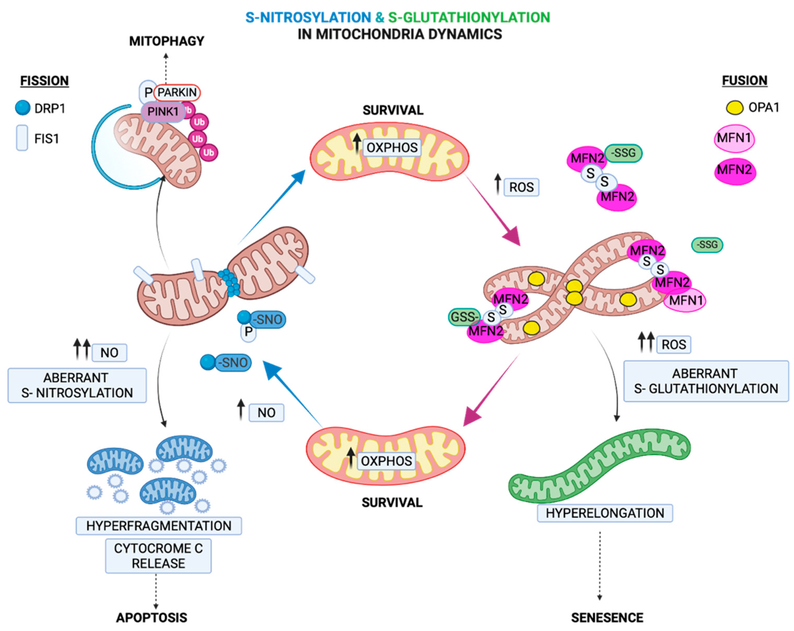
2.4. Regulation of Mitochondria Morphology by S-Glutathionylation

Fusion and fission processes tightly regulate mitochondria morphology and cell fate and are mediated by GTPases located at the outer mitochondrial membrane (OMM). Fusion is regulated by the GTPases mitofusins (MFNs) 1 and 2 and the autosomal dominant optic atrophy protein 1 (OPA1), which also modulate mitochondria cristae [72]. Fission is primarily initiated by the dynamin family of GTPases (DRP1) [72]. Under chronic oxidative and nutrient stresses, fission is triggered, leading to mitochondrial fragmentation. In contrast, under acute stress and glutathione depletion, the GSSG-induced S-glutathionylation of MFN2 and OPA1 oligomerization results in mitochondrial hyperfusion [73]. It has been attributed a role in adaptation to an acute stressor in the mitochondrial hyperfusion process, which provides protection from excessive oxidation and at the same time consolidates ATP production capacities [74]. Therefore, the hyperfusion state induced by S-glutathionylation of MFN2 can act as a regulatory mechanism under acute stressors, serving as a transient response that provides a critical window for adaptation while preserving ATP production (Figure 4). Recently, it was shown that S-glutathionylation of MFN2 led to the formation of necrosomes and their recruitment to mitochondria-associated endoplasmic reticulum (ER) membranes (MAMs), and in turn, it induced ER-mitochondria intra-organelle crosstalk-dependent neuronal necroptosis [75]. This was the first identification that MFN2 S-glutathionylation increases sensitivity to the neuronal necroptotic mechanism in vitro and in vivo. This mechanism can therefore describe an effect of mitofusin’s S-glutathionylation that goes beyond mitochondrial deregulation and can affect other organelles with detrimental consequences to cell fate.
Figure 4. S-Glutathionylation and S-Nitrosylation orchestrate mitochondrial dynamics. Under acute stress, the S-glutathionylation of MFN2 and, in turn, the oligomerization of OPA1 trigger hyperfusion as an adaptive mechanism, whereas the excessive hyper-elongation of mitochondria under aberrant S-glutathionylation leads to cell senescence. The NO-mediated S-nitrosylation of DRP1 triggers its activation and phosphorylation, which in turn activates the fission machinery to eliminate damaged mitochondria. Under excessive NO production, the hyper-fragmentation of mitochondria leads to cytochrome c release and apoptosis. Created with BioRender.com.

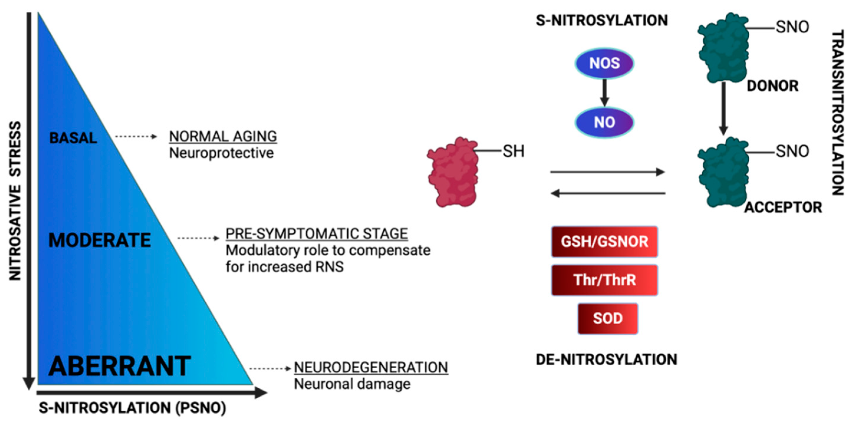
3. Protein S-Nitrosylation

Non-canonical nitric oxide (NO) signaling encompasses covalent post-translational modifications of biomolecules via NO and NO derivatives. These modifications include the S-nitrosylation of protein thiols, metal nitrosylation of transition metals and oxidative nitration or hydroxylation of various molecules such as tyrosine, amines, fatty acids, etc. [76]. Concerning tyrosine nitration, the formation of peroxynitrite (ONOO) and nitrogen dioxide (NO2), are associated with this irreversible modification that can impact protein function. In the case of cysteine residues, peroxynitrite triggers the formation of oxygenated forms such as sulfenic acid (-SOH), sulfinic acid (-SO2H) and sulfonic acid (-SO3H) [77]. S-thiolation forms including S-glutathionylation are induced by peroxinitrite and nitrosothiol formation [77]. Nitrosylation is the reaction in which a nitrosyl moiety of NO is incorporated into another molecule, and when this affects the thiol group of Cys, the reaction is termed S-nitrosylation [78]. S-nitrosylation can propagate beyond the boundaries of its localization via transnitrosylation [33], and in recent years, it has emerged as a prototype redox-induced cell-signaling mechanism that protects cells against oxidative stress and diminishes ROS levels [79]. NO is not an antioxidant and cannot strongly react with protein thiols. Therefore, in order for the majority of S-nitrosylation reactions to take place, NO needs to react with oxygen to increase its oxidation state and then react with protein thiols [80]. For this reaction, many mechanisms have been proposed [76].
Many factors can modulate S-nitrosylation levels including the redox state. More specifically, the decreased levels of antioxidants, which increase the oxidation potential of the cells, trigger the S-nitrosylation of proteins, whereas increased levels of antioxidants have the opposite effect [81]. In particular, under glutathione depletion, the S-nitrosylation of mitochondrial proteins is elevated in order to protect cells against thiol oxidation, membrane permeabilization and apoptosis [82]. Another factor that modulates S-nitrosylation is the denitrosylation process. While the S-nitrosylation process is, in general, a non-enzymatic process (with an exception for prokaryotes), the opposite reaction, denitrosylation, can be enzymatic or non-enzymatic [83]. The spontaneous cleavage of the S-nitrosyl group can be performed by metal ions, nucleophiles, reducing agents, heat, ROS and UV [83]. Enzymatically driven denitrosylation can be processed by two major denitrosylase systems in cells: S-nitrosoglutathione reductase (GSNOR) and thioredoxin reductase (TRXR). Of particular importance for cell homeostasis is the balance between S-nitrosylation and denitrosylation. For example, the over-expression of GSNOR in the brain has been associated with cognitive impairment [84]. Transnitrosylation on the other hand, which is the transfer of the nitrosyl moiety of a nitrosylated protein to the reactive Cys of the interacting protein, allows for signal transmissions, even in sites that are distant from the initial NO source [33]) (Figure 5).
Figure 5. Proposed mechanisms for S-Nitrosylation and Denitrosylation. While NO-mediated protein S-nitrosylation is generally a non-enzymatic reaction, mediated by RNS and ROS species and NOS-mediated NO production, denitrosylation can be both enzymatic and non-enzymatic. The major denitrosylase systems in cells are the Thr/ThrR and glutathione GSH/GSNOR. Moreover, SOD can have denitrosylase functions. The transnitrosylation of a protein can be directly performed by an already S-nitrosylated protein in close proximity. During aging, a subset of proteins is S-nitrosylated, promoting cell signaling cascades that aim for neuroprotection. Upon increased levels of RNS, potentially at early stages of neurodegeneration, increased levels of S-nitrosylation modulate multiple cellular processes and trigger the adjustment of signaling cascades in highly nitrosative environments. Aberrant S-nitrosylation in neurodegenerative disorders exacerbates disease pathology, ultimately leading to neuronal damage. Created with BioRender.com.
In mitochondria bioactivities and quality control, S-nitrosylation and denitrosylation have an integral role [85][86]. The S-nitrosylation of mitochondrial thiols is influenced by fluctuations in mitochondrial respiration and redox states [87]. Intriguingly, the source of NO inside the mitochondria is still controversial. One hypothesis is that mitochondria harbor a particular mitochondria NOS isoform (mtNOS) [88], while another suggests that NO enters mitochondria from cytosol via transnitrosylase-mediated transportation [35]. In the following sections, multiple mitochondrial targets of S-nitrosylation will be discussed, which when modified by NO, mostly inhibits their activity to modulate ROS production, O2 consumption and mitophagy, while simultaneously providing protection against cell death [88].

3.1. Regulation of the OXPHOS System by S-Nitrosylation

All mitochondrial complexes CI-V can be S-nitrosylated and, in turn, inhibited in order to minimize ROS production under oxidizing conditions [89]; moreover, it scavenges NO, prohibiting it from reacting with superoxides to produce peroxynitrite [90] (Figure 3). In hypoxic conditions, NO production is elevated, which could again lead to the S-nitrosylation and inhibition of CI-V enzymatic activities [89]. Under this condition of diminished O2 consumption and the redistribution of intracellular oxygen, the stabilization of hypoxia-inducible factor-1α (HIF1A) can be prevented during hypoxia [91]. The inactivation of CI-V ultimately impedes mitochondrial respiration, which in turn causes mitochondrial depolarization [92]. Consequently, the PINK1/PARKIN pathway (described in detail later) is triggered and induces mitophagy to protect cells from oxidative stress by eliminating defective mitochondria [93]. Additionally, relative to CI-V, cytochrome c, which transfers electrons from CIII to CIV, is also a target of S-nitrosylation. Specifically, cytochrome c is nitrosylated at the iron metal center of the heme moiety, which then transfers the nitrosyl group to glutathione, resulting in the formation of the endogenously abundant transnitrosylase GSNO [94]. While S-nitrosylated cytochrome c is released outside mitochondria during apoptosis, it has been proposed that it can suppress apoptosis by retaining its capability to synthesize GSNO [95].

3.2. Regulation of Metabolic Enzymes by S-Nitrosylation

The fatty acid β-oxidation and aerobic oxidation of pyruvate (TCA cycle) to CO2 take place at the inner mitochondrial membrane and matrix. These processes not only generate ROS by themselves [96][97] but also produce the source of electrons and cofactors that are utilized by the ETC, which in turn also produces ROS [96][98]. Because of this, under oxidative stress, S-nitrosylation inhibits multiple enzymes in these two catabolic processes to inhibit their activities in order to minimize ROS production [87] (Figure 3). Concerning the ETC and TCA cycle, these enzymes include aconitase, the α-ketoglutarate dehydrogenase complex and succinate dehydrogenase, while in the case of fatty acid catabolism, long-chain and short-chain acyl-CoA dehydrogenase and enoyl-CoA hydratase, carnitine palmitoyl transferase 2 and flavoprotein dehydrogenase belong to the enzymes that are inhibited [87]. A recent study revealed a novel mechanism with which RNS generated by macrophages can render inactive α-ketoacid dehydrogenases (including pyruvate dehydrogenase complex and α-ketoglutarate dehydrogenase complex) [99]. Herein, it was shown that the lipoid arm of α-ketoacid dehydrogenase can be modified by RNS-mediated thiol modifications, and in turn, the acyl-transfer activity of the lipoid arm is blocked leading to the inhibition of pyruvate dehydrogenase complex and α-ketoglutarate dehydrogenase complex.

3.3. Regulation of Apoptosis by S-Nitrosylation

Under oxidative conditions, the S-nitrosylation of ETC complexes and the TCA cycle inhibits their activities and the consequent production of ROS, while under the same conditions, S-nitrosylation inhibits pro- and anti-apoptotic or antinecrotic proteins to protect cells from death. This bidirectional S-nitrosylation-mediated inhibition assures that upon decreased energy production, death pathways are impeded (Figure 3). The mitochondrial pro- and anti-apoptotic proteins include the following:
  • Mitochondrial permeability transition pores (MPTPs) are formed under oxidative stress and modulate the redox potential. Under oxidative stress, MPTP increases the permeability relative to high molecular weight macromolecules, leading to mitochondria swelling and necroptosis [100]. Under the basal levels of NO, cyclophilin D (CYPD), critical for an MPTP opening, can be S-nitrosylated [101]. This inhibits its interaction with MPTPs, preventing pore opening and protecting cells during stress. Conversely, under the increased production of NO, peroxynitrite is being produced by excessive ROS, which leads to the opening of MPTPs, the ablation of ATP production and necrosis [102].
  • VDAC has multiple roles. Its localization at the mitochondrial outer membrane allows VDAC to regulate the fluxes of metabolites. At the same time, VDAC modulates cytochrome c, which activates caspase-dependent apoptotic cell death [103]. Similarly to MPTP regulation by NO, VDAC S-nitrosylation at basal levels of NO inhibits its function, protecting cells from apoptosis, whereas elevated levels of NO upregulate VDAC functions [104].
  • Crucial to their role in apoptosis, caspase-3 and caspase-9 are S-nitrosylated in the absence of apoptotic triggers, while their denitrosylation leads to their activation upon the activation of the Fas receptor [105].
  • Another component of the outer mitochondrial membrane with an anti-apoptotic role involves BCL2 family proteins, which interfere with apoptosis by regulating the release of cytochrome c [106]. Upon apoptotic signaling, the S-nitrosylation of BCL-2 inhibits its ubiquitination and degradation, allowing it to exert anti-apoptotic protection [107].

3.4. Regulation of Fission by S-Nitrosylation

A recent discovery in neurons showed that the mitochondrial fission protein DRP1 is regulated by NO. It was shown that the NO-mediated S-nitrosylation of DRP1 triggers its GTPase activity and fission [108]. This discovery is of particular importance for neurons, where the nNOS-mediated regulation of DRP1 might contribute to mitochondrial fragmentation, followed by synaptic loss and neuronal cell death, which are observed in neurodegenerative disorders such as AD [109]. This mechanism of the S-nitrosylation-mediated regulation of fission contrasts the S-glutathionylated regulation of fusion, underlying the importance of these two redox PTMs in modulating mitochondria homeostasis (Figure 4).

This entry is adapted from the peer-reviewed paper 10.3390/ijms232415849

References

  1. Weidinger, A.; Kozlov, A.V. Biological Activities of Reactive Oxygen and Nitrogen Species: Oxidative Stress versus Signal Transduction. Biomolecules 2015, 5, 472–484.
  2. Santos, A.L.; Lindner, A.B. Protein Posttranslational Modifications: Roles in Aging and Age-Related Disease. Oxidative Med. Cell Longev. 2017, 2017, 5716409.
  3. Beckhauser, T.F.; Francis-Oliveira, J.; De Pasquale, R. Reactive Oxygen Species: Physiological and Physiopathological Effects on Synaptic Plasticity. J. Exp. Neurosci. 2016, 10, 23–48.
  4. Jones, D.P. Redefining oxidative stress. Antioxid. Redox. Signal. 2006, 8, 1865–1879.
  5. Ray, P.D.; Huang, B.W.; Tsuji, Y. Reactive oxygen species (ROS) homeostasis and redox regulation in cellular signaling. Cell Signal. 2012, 24, 981–990.
  6. Murphy, M.P. How mitochondria produce reactive oxygen species. Biochem. J. 2009, 417, 1–13.
  7. Lambeth, J.D.; Neish, A.S. Nox enzymes and new thinking on reactive oxygen: A double-edged sword revisited. Annu. Rev. Pathol. 2014, 9, 119–145.
  8. Ma, M.W.; Wang, J.; Zhang, Q.; Wang, R.; Dhandapani, K.M.; Vadlamudi, R.K.; Brann, D.W. NADPH oxidase in brain injury and neurodegenerative disorders. Mol. Neurodegener. 2017, 12, 7.
  9. Di Meo, S.; Reed, T.T.; Venditti, P.; Victor, V.M. Role of ROS and RNS Sources in Physiological and Pathological Conditions. Oxid. Med. Cell Longev. 2016, 2016, 1245049.
  10. Birben, E.; Sahiner, U.M.; Sackesen, C.; Erzurum, S.; Kalayci, O. Oxidative stress and antioxidant defense. World Allergy Organ. J. 2012, 5, 9–19.
  11. Moldogazieva, N.T.; Mokhosoev, I.M.; Feldman, N.B.; Lutsenko, S.V. ROS and RNS signalling: Adaptive redox switches through oxidative/nitrosative protein modifications. Free Radic. Res. 2018, 52, 507–543.
  12. Paul, B.D.; Sbodio, J.I.; Snyder, S.H. Cysteine Metabolism in Neuronal Redox Homeostasis. Trends. Pharm. Sci. 2018, 39, 513–524.
  13. Sbodio, J.I.; Snyder, S.H.; Paul, B.D. Redox Mechanisms in Neurodegeneration: From Disease Outcomes to Therapeutic Opportunities. Antioxid. Redox. Signal. 2019, 30, 1450–1499.
  14. Stadtman, E.R.; Levine, R.L. Free radical-mediated oxidation of free amino acids and amino acid residues in proteins. Amino. Acids. 2003, 25, 207–218.
  15. Davies, M.J. Protein oxidation and peroxidation. Biochem. J. 2016, 473, 805–825.
  16. Wall, S.B.; Oh, J.Y.; Diers, A.R.; Landar, A. Oxidative modification of proteins: An emerging mechanism of cell signaling. Front. Physiol. 2012, 3, 369.
  17. Wani, R.; Nagata, A.; Murray, B.W. Protein redox chemistry: Post-translational cysteine modifications that regulate signal transduction and drug pharmacology. Front. Pharmacol. 2014, 5, 224.
  18. Halloran, M.; Parakh, S.; Atkin, J.D. The role of s-nitrosylation and s-glutathionylation of protein disulphide isomerase in protein misfolding and neurodegeneration. Int. J. Cell Biol. 2013, 2013, 797914.
  19. Chung, H.S.; Wang, S.B.; Venkatraman, V.; Murray, C.I.; Van Eyk, J.E. Cysteine oxidative posttranslational modifications: Emerging regulation in the cardiovascular system. Circ. Res. 2013, 112, 382–392.
  20. Fra, A.; Yoboue, E.D.; Sitia, R. Cysteines as Redox Molecular Switches and Targets of Disease. Front. Mol. Neurosci. 2017, 10, 167.
  21. Davies, M.J. The oxidative environment and protein damage. Biochim. Biophys. Acta 2005, 1703, 93–109.
  22. Pajares, M.; Jimenez-Moreno, N.; Dias, I.H.K.; Debelec, B.; Vucetic, M.; Fladmark, K.E.; Basaga, H.; Ribaric, S.; Milisav, I.; Cuadrado, A. Redox control of protein degradation. Redox. Biol. 2015, 6, 409–420.
  23. Song, H.; Liu, B.; Huai, W.; Yu, Z.; Wang, W.; Zhao, J.; Han, L.; Jiang, G.; Zhang, L.; Gao, C.; et al. The E3 ubiquitin ligase TRIM31 attenuates NLRP3 inflammasome activation by promoting proteasomal degradation of NLRP3. Nat. Commun. 2016, 7, 13727.
  24. Muronetz, V.I.; Medvedeva, M.V.; Sevostyanova, I.A.; Schmalhausen, E.V. Modification of Glyceraldehyde-3-Phosphate Dehydrogenase with Nitric Oxide: Role in Signal Transduction and Development of Apoptosis. Biomolecules 2021, 11, 1656.
  25. Cha, S.J.; Kim, H.; Choi, H.J.; Lee, S.; Kim, K. Protein Glutathionylation in the Pathogenesis of Neurodegenerative Diseases. Oxid. Med. Cell Longev. 2017, 2017, 2818565.
  26. Montagna, C.; Cirotti, C.; Rizza, S.; Filomeni, G. When S-Nitrosylation Gets to Mitochondria: From Signaling to Age-Related Diseases. Antioxid. Redox. Signal. 2020, 32, 884–905.
  27. Nakamura, T.; Tu, S.; Akhtar, M.W.; Sunico, C.R.; Okamoto, S.; Lipton, S.A. Aberrant protein s-nitrosylation in neurodegenerative diseases. Neuron 2013, 78, 596–614.
  28. Mailloux, R.J.; Treberg, J.R. Protein S-glutathionlyation links energy metabolism to redox signaling in mitochondria. Redox. Biol. 2016, 8, 110–118.
  29. Rizza, S.; Cardaci, S.; Montagna, C.; Di Giacomo, G.; De Zio, D.; Bordi, M.; Maiani, E.; Campello, S.; Borreca, A.; Puca, A.A.; et al. S-nitrosylation drives cell senescence and aging in mammals by controlling mitochondrial dynamics and mitophagy. Proc. Natl. Acad. Sci. USA 2018, 115, E3388–E3397.
  30. Zahid, S.; Khan, R.; Oellerich, M.; Ahmed, N.; Asif, A.R. Differential S-nitrosylation of proteins in Alzheimer’s disease. Neuroscience 2014, 256, 126–136.
  31. Sultana, R.; Butterfield, D.A. Oxidative modification of brain proteins in Alzheimer’s disease: Perspective on future studies based on results of redox proteomics studies. J. Alzheimers Dis. 2013, 33 (Suppl. S1), S243–S251.
  32. Sabens Liedhegner, E.A.; Gao, X.H.; Mieyal, J.J. Mechanisms of altered redox regulation in neurodegenerative diseases--focus on S--glutathionylation. Antioxid. Redox. Signal. 2012, 16, 543–566.
  33. Nakamura, T.; Lipton, S.A. Emerging role of protein-protein transnitrosylation in cell signaling pathways. Antioxid. Redox. Signal. 2013, 18, 239–249.
  34. Moosmann, B.; Behl, C. Antioxidants as treatment for neurodegenerative disorders. Expert Opin. Investig. Drugs 2002, 11, 1407–1435.
  35. Mailloux, R.J.; Jin, X.; Willmore, W.G. Redox regulation of mitochondrial function with emphasis on cysteine oxidation reactions. Redox. Biol. 2014, 2, 123–139.
  36. Dalle-Donne, I.; Rossi, R.; Giustarini, D.; Colombo, R.; Milzani, A. S-glutathionylation in protein redox regulation. Free Radic. Biol. Med. 2007, 43, 883–898.
  37. Kang, P.T.; Zhang, L.; Chen, C.L.; Chen, J.; Green, K.B.; Chen, Y.R. Protein thiyl radical mediates S-glutathionylation of complex I. Free Radic. Biol. Med. 2012, 53, 962–973.
  38. Ziegler, D.M. Role of reversible oxidation-reduction of enzyme thiols-disulfides in metabolic regulation. Annu. Rev. Biochem. 1985, 54, 305–329.
  39. Gallogly, M.M.; Starke, D.W.; Mieyal, J.J. Mechanistic and kinetic details of catalysis of thiol-disulfide exchange by glutaredoxins and potential mechanisms of regulation. Antioxid. Redox. Signal. 2009, 11, 1059–1081.
  40. Stroher, E.; Millar, A.H. The biological roles of glutaredoxins. Biochem. J. 2012, 446, 333–348.
  41. Mitra, S.; Elliott, S.J. Oxidative disassembly of the cluster of human Grx2 and redox regulation in the mitochondria. Biochemistry 2009, 48, 3813–3815.
  42. Beer, S.M.; Taylor, E.R.; Brown, S.E.; Dahm, C.C.; Costa, N.J.; Runswick, M.J.; Murphy, M.P. Glutaredoxin 2 catalyzes the reversible oxidation and glutathionylation of mitochondrial membrane thiol proteins: Implications for mitochondrial redox regulation and antioxidant DEFENSE. J. Biol. Chem. 2004, 279, 47939–47951.
  43. Mailloux, R.J.; Xuan, J.Y.; Beauchamp, B.; Jui, L.; Lou, M.; Harper, M.E. Glutaredoxin-2 is required to control proton leak through uncoupling protein-3. J. Biol. Chem. 2013, 288, 8365–8379.
  44. Klaus, A.; Zorman, S.; Berthier, A.; Polge, C.; Ramirez, S.; Michelland, S.; Seve, M.; Vertommen, D.; Rider, M.; Lentze, N.; et al. Glutathione S-transferases interact with AMP-activated protein kinase: Evidence for S-glutathionylation and activation in vitro. PLoS ONE 2013, 8, e62497.
  45. Raza, H. Dual localization of glutathione S-transferase in the cytosol and mitochondria: Implications in oxidative stress, toxicity and disease. FEBS J. 2011, 278, 4243–4251.
  46. Park, J.W.; Mieyal, J.J.; Rhee, S.G.; Chock, P.B. Deglutathionylation of 2-Cys peroxiredoxin is specifically catalyzed by sulfiredoxin. J. Biol. Chem. 2009, 284, 23364–23374.
  47. Aon, M.A.; Cortassa, S.; Maack, C.; O’Rourke, B. Sequential opening of mitochondrial ion channels as a function of glutathione redox thiol status. J. Biol. Chem. 2007, 282, 21889–21900.
  48. Hurd, T.R.; Requejo, R.; Filipovska, A.; Brown, S.; Prime, T.A.; Robinson, A.J.; Fearnley, I.M.; Murphy, M.P. Complex I within oxidatively stressed bovine heart mitochondria is glutathionylated on Cys-531 and Cys-704 of the 75-kDa subunit: Potential role of CYS residues in decreasing oxidative damage. J. Biol. Chem. 2008, 283, 24801–24815.
  49. Chen, C.L.; Zhang, L.; Yeh, A.; Chen, C.A.; Green-Church, K.B.; Zweier, J.L.; Chen, Y.R. Site-specific S-glutathiolation of mitochondrial NADH ubiquinone reductase. Biochemistry 2007, 46, 5754–5765.
  50. Diotte, N.M.; Xiong, Y.; Gao, J.; Chua, B.H.; Ho, Y.S. Attenuation of doxorubicin-induced cardiac injury by mitochondrial glutaredoxin 2. Biochim. Biophys. Acta 2009, 1793, 427–438.
  51. Taylor, E.R.; Hurrell, F.; Shannon, R.J.; Lin, T.K.; Hirst, J.; Murphy, M.P. Reversible glutathionylation of complex I increases mitochondrial superoxide formation. J. Biol. Chem. 2003, 278, 19603–19610.
  52. Tretter, L.; Adam-Vizi, V. Inhibition of Krebs cycle enzymes by hydrogen peroxide: A key role of -ketoglutarate dehydrogenase in limiting NADH production under oxidative stress. J. Neurosci. 2000, 20, 8972–8979.
  53. Garcia, J.; Han, D.; Sancheti, H.; Yap, L.P.; Kaplowitz, N.; Cadenas, E. Regulation of mitochondrial glutathione redox status and protein glutathionylation by respiratory substrates. J. Biol. Chem. 2010, 285, 39646–39654.
  54. Korshunov, S.S.; Skulachev, V.P.; Starkov, A.A. High protonic potential actuates a mechanism of production of reactive oxygen species in mitochondria. FEBS Lett. 1997, 416, 15–18.
  55. Bulteau, A.L.; Lundberg, K.C.; Ikeda-Saito, M.; Isaya, G.; Szweda, L.I. Reversible redox-dependent modulation of mitochondrial aconitase and proteolytic activity during in vivo cardiac ischemia/reperfusion. Proc. Natl. Acad. Sci. USA 2005, 102, 5987–5991.
  56. Kil, I.S.; Park, J.W. Regulation of mitochondrial NADP+-dependent isocitrate dehydrogenase activity by glutathionylation. J. Biol. Chem. 2005, 280, 10846–10854.
  57. McLain, A.L.; Szweda, P.A.; Szweda, L.I. alpha-Ketoglutarate dehydrogenase: A mitochondrial redox sensor. Free Radic. Res. 2011, 45, 29–36.
  58. O’Brien, M.; Chalker, J.; Slade, L.; Gardiner, D.; Mailloux, R.J. Protein S-glutathionylation alters superoxide/hydrogen peroxide emission from pyruvate dehydrogenase complex. Free Radic. Biol. Med. 2017, 106, 302–314.
  59. Mailloux, R.J.; Xuan, J.Y.; McBride, S.; Maharsy, W.; Thorn, S.; Holterman, C.E.; Kennedy, C.R.; Rippstein, P.; de Kemp, R.; da Silva, J.; et al. Glutaredoxin-2 is required to control oxidative phosphorylation in cardiac muscle by mediating deglutathionylation reactions. J. Biol. Chem. 2014, 289, 14812–14828.
  60. Muoio, D.M. Metabolic inflexibility: When mitochondrial indecision leads to metabolic gridlock. Cell 2014, 159, 1253–1262.
  61. Giangregorio, N.; Palmieri, F.; Indiveri, C. Glutathione controls the redox state of the mitochondrial carnitine/acylcarnitine carrier Cys residues by glutathionylation. Biochim. Biophys. Acta 2013, 1830, 5299–5304.
  62. Zhang, J.; Ye, Z.W.; Singh, S.; Townsend, D.M.; Tew, K.D. An evolving understanding of the S-glutathionylation cycle in pathways of redox regulation. Free Radic. Biol. Med. 2018, 120, 204–216.
  63. Cadenas, S. Mitochondrial uncoupling, ROS generation and cardioprotection. Biochim. Biophys. Acta Bioenerg. 2018, 1859, 940–950.
  64. Bouillaud, F.; Alves-Guerra, M.C.; Ricquier, D. UCPs, at the interface between bioenergetics and metabolism. Biochim. Biophys. Acta 2016, 1863, 2443–2456.
  65. Mailloux, R.J.; Seifert, E.L.; Bouillaud, F.; Aguer, C.; Collins, S.; Harper, M.E. Glutathionylation acts as a control switch for uncoupling proteins UCP2 and UCP3. J. Biol. Chem. 2011, 286, 21865–21875.
  66. Nunnari, J.; Suomalainen, A. Mitochondria: In sickness and in health. Cell 2012, 148, 1145–1159.
  67. Kowaltowski, A.J.; Netto, L.E.; Vercesi, A.E. The thiol-specific antioxidant enzyme prevents mitochondrial permeability transition. Evidence for the participation of reactive oxygen species in this mechanism. J. Biol. Chem. 1998, 273, 12766–12769.
  68. Queiroga, C.S.; Almeida, A.S.; Martel, C.; Brenner, C.; Alves, P.M.; Vieira, H.L. Glutathionylation of adenine nucleotide translocase induced by carbon monoxide prevents mitochondrial membrane permeabilization and apoptosis. J. Biol. Chem. 2010, 285, 17077–17088.
  69. Skulachev, V.P. Bioenergetic aspects of apoptosis, necrosis and mitoptosis. Apoptosis 2006, 11, 473–485.
  70. Itani, H.A.; Dikalova, A.E.; McMaster, W.G.; Nazarewicz, R.R.; Bikineyeva, A.T.; Harrison, D.G.; Dikalov, S.I. Mitochondrial Cyclophilin D in Vascular Oxidative Stress and Hypertension. Hypertension 2016, 67, 1218–1227.
  71. Thiriveedi, V.R.; Mattam, U.; Pattabhi, P.; Bisoyi, V.; Talari, N.K.; Krishnamoorthy, T.; Sepuri, N.B.V. Glutathionylated and Fe-S cluster containing hMIA40 (CHCHD4) regulates ROS and mitochondrial complex III and IV activities of the electron transport chain. Redox. Biol. 2020, 37, 101725.
  72. Liesa, M.; Shirihai, O.S. Mitochondrial dynamics in the regulation of nutrient utilization and energy expenditure. Cell Metab. 2013, 17, 491–506.
  73. Willems, P.H.; Rossignol, R.; Dieteren, C.E.; Murphy, M.P.; Koopman, W.J. Redox Homeostasis and Mitochondrial Dynamics. Cell Metab. 2015, 22, 207–218.
  74. Shutt, T.; Geoffrion, M.; Milne, R.; McBride, H.M. The intracellular redox state is a core determinant of mitochondrial fusion. EMBO Rep. 2012, 13, 909–915.
  75. Che, L.; Yang, C.L.; Chen, Y.; Wu, Z.L.; Du, Z.B.; Wu, J.S.; Gan, C.L.; Yan, S.P.; Huang, J.; Guo, N.J.; et al. Mitochondrial redox-driven mitofusin 2 S-glutathionylation promotes neuronal necroptosis via disrupting ER-mitochondria crosstalk in cadmium-induced neurotoxicity. Chemosphere 2021, 262, 127878.
  76. Martinez-Ruiz, A.; Araujo, I.M.; Izquierdo-Alvarez, A.; Hernansanz-Agustin, P.; Lamas, S.; Serrador, J.M. Specificity in S-nitrosylation: A short-range mechanism for NO signaling? Antioxid. Redox. Signal. 2013, 19, 1220–1235.
  77. Martinez-Ruiz, A.; Lamas, S. Signalling by NO-induced protein S-nitrosylation and S-glutathionylation: Convergences and divergences. Cardiovasc. Res. 2007, 75, 220–228.
  78. Fernando, V.; Zheng, X.; Walia, Y.; Sharma, V.; Letson, J.; Furuta, S. S-Nitrosylation: An Emerging Paradigm of Redox Signaling. Antioxidants 2019, 8, 404.
  79. Sun, J.; Steenbergen, C.; Murphy, E. S-nitrosylation: NO-related redox signaling to protect against oxidative stress. Antioxid. Redox. Signal. 2006, 8, 1693–1705.
  80. Lancaster, J.R., Jr. Nitric oxide: A brief overview of chemical and physical properties relevant to therapeutic applications. Future Sci. OA 2015, 1, FSO59.
  81. Beltran, B.; Orsi, A.; Clementi, E.; Moncada, S. Oxidative stress and S-nitrosylation of proteins in cells. Br. J. Pharm. 2000, 129, 953–960.
  82. Whiteman, M.; Chua, Y.L.; Zhang, D.; Duan, W.; Liou, Y.C.; Armstrong, J.S. Nitric oxide protects against mitochondrial permeabilization induced by glutathione depletion: Role of S-nitrosylation? Biochem. Biophys. Res. Commun. 2006, 339, 255–262.
  83. Stomberski, C.T.; Hess, D.T.; Stamler, J.S. Protein S-Nitrosylation: Determinants of Specificity and Enzymatic Regulation of S-Nitrosothiol-Based Signaling. Antioxid. Redox. Signal. 2019, 30, 1331–1351.
  84. Zhang, Y.; Wu, K.; Su, W.; Zhang, D.F.; Wang, P.; Qiao, X.; Yao, Q.; Yuan, Z.; Yao, Y.G.; Liu, G.; et al. Increased GSNOR Expression during Aging Impairs Cognitive Function and Decreases S-Nitrosation of CaMKIIalpha. J. Neurosci. 2017, 37, 9741–9758.
  85. Nakamura, T.; Lipton, S.A. Cell death: Protein misfolding and neurodegenerative diseases. Apoptosis 2009, 14, 455–468.
  86. Ozawa, K.; Komatsubara, A.T.; Nishimura, Y.; Sawada, T.; Kawafune, H.; Tsumoto, H.; Tsuji, Y.; Zhao, J.; Kyotani, Y.; Tanaka, T.; et al. S-nitrosylation regulates mitochondrial quality control via activation of parkin. Sci. Rep. 2013, 3, 2202.
  87. Piantadosi, C.A. Regulation of mitochondrial processes by protein S-nitrosylation. Biochim. Biophys. Acta 2012, 1820, 712–721.
  88. Ghafourifar, P.; Cadenas, E. Mitochondrial nitric oxide synthase. Trends. Pharm. Sci. 2005, 26, 190–195.
  89. Zhang, J.; Jin, B.; Li, L.; Block, E.R.; Patel, J.M. Nitric oxide-induced persistent inhibition and nitrosylation of active site cysteine residues of mitochondrial cytochrome-c oxidase in lung endothelial cells. Am. J. Physiol. Cell Physiol. 2005, 288, C840–C849.
  90. Brown, G.C.; Borutaite, V. Inhibition of mitochondrial respiratory complex I by nitric oxide, peroxynitrite and S-nitrosothiols. Biochim. Biophys. Acta 2004, 1658, 44–49.
  91. Hagen, T.; Taylor, C.T.; Lam, F.; Moncada, S. Redistribution of intracellular oxygen in hypoxia by nitric oxide: Effect on HIF1alpha. Science 2003, 302, 1975–1978.
  92. Chang, A.H.; Sancheti, H.; Garcia, J.; Kaplowitz, N.; Cadenas, E.; Han, D. Respiratory substrates regulate S-nitrosylation of mitochondrial proteins through a thiol-dependent pathway. Chem. Res. Toxicol. 2014, 27, 794–804.
  93. Zorova, L.D.; Popkov, V.A.; Plotnikov, E.Y.; Silachev, D.N.; Pevzner, I.B.; Jankauskas, S.S.; Babenko, V.A.; Zorov, S.D.; Balakireva, A.V.; Juhaszova, M.; et al. Mitochondrial membrane potential. Anal. Biochem. 2018, 552, 50–59.
  94. Basu, S.; Keszler, A.; Azarova, N.A.; Nwanze, N.; Perlegas, A.; Shiva, S.; Broniowska, K.A.; Hogg, N.; Kim-Shapiro, D.B. A novel role for cytochrome c: Efficient catalysis of S-nitrosothiol formation. Free Radic. Biol. Med. 2010, 48, 255–263.
  95. Schonhoff, C.M.; Gaston, B.; Mannick, J.B. Nitrosylation of cytochrome c during apoptosis. J. Biol. Chem. 2003, 278, 18265–18270.
  96. Speijer, D.; Manjeri, G.R.; Szklarczyk, R. How to deal with oxygen radicals stemming from mitochondrial fatty acid oxidation. Philos. Trans. R. Soc. Lond. B Biol. Sci. 2014, 369, 20130446.
  97. Quijano, C.; Trujillo, M.; Castro, L.; Trostchansky, A. Interplay between oxidant species and energy metabolism. Redox. Biol. 2016, 8, 28–42.
  98. Quinlan, C.L.; Perevoshchikova, I.V.; Hey-Mogensen, M.; Orr, A.L.; Brand, M.D. Sites of reactive oxygen species generation by mitochondria oxidizing different substrates. Redox. Biol. 2013, 1, 304–312.
  99. Seim, G.L.; John, S.V.; Arp, N.L.; Fang, Z.; Pagliarini, D.J.; Fan, J. Nitric oxide-driven modifications of lipoic arm inhibit alpha-ketoacid dehydrogenases. Nat. Chem. Biol. 2022. Online ahead of print.
  100. Lemasters, J.J.; Theruvath, T.P.; Zhong, Z.; Nieminen, A.L. Mitochondrial calcium and the permeability transition in cell death. Biochim. Biophys. Acta 2009, 1787, 1395–1401.
  101. Nguyen, T.T.; Stevens, M.V.; Kohr, M.; Steenbergen, C.; Sack, M.N.; Murphy, E. Cysteine 203 of cyclophilin D is critical for cyclophilin D activation of the mitochondrial permeability transition pore. J. Biol. Chem. 2011, 286, 40184–40192.
  102. Ohtani, H.; Katoh, H.; Tanaka, T.; Saotome, M.; Urushida, T.; Satoh, H.; Hayashi, H. Effects of nitric oxide on mitochondrial permeability transition pore and thiol-mediated responses in cardiac myocytes. Nitric. Oxide. 2012, 26, 95–101.
  103. Shoshan-Barmatz, V.; De Pinto, V.; Zweckstetter, M.; Raviv, Z.; Keinan, N.; Arbel, N. VDAC, a multi-functional mitochondrial protein regulating cell life and death. Mol. Asp. Med. 2010, 31, 227–285.
  104. Cheng, Q.; Sedlic, F.; Pravdic, D.; Bosnjak, Z.J.; Kwok, W.M. Biphasic effect of nitric oxide on the cardiac voltage-dependent anion channel. FEBS Lett. 2011, 585, 328–334.
  105. Mannick, J.B.; Schonhoff, C.; Papeta, N.; Ghafourifar, P.; Szibor, M.; Fang, K.; Gaston, B. S-Nitrosylation of mitochondrial caspases. J. Cell Biol. 2001, 154, 1111–1116.
  106. Crow, M.T.; Mani, K.; Nam, Y.J.; Kitsis, R.N. The mitochondrial death pathway and cardiac myocyte apoptosis. Circ. Res. 2004, 95, 957–970.
  107. Azad, N.; Vallyathan, V.; Wang, L.; Tantishaiyakul, V.; Stehlik, C.; Leonard, S.S.; Rojanasakul, Y. S-nitrosylation of Bcl-2 inhibits its ubiquitin-proteasomal degradation. A novel antiapoptotic mechanism that suppresses apoptosis. J. Biol. Chem. 2006, 281, 34124–34134.
  108. Gu, Z.; Nakamura, T.; Lipton, S.A. Redox reactions induced by nitrosative stress mediate protein misfolding and mitochondrial dysfunction in neurodegenerative diseases. Mol. Neurobiol. 2010, 41, 55–72.
  109. Nakamura, T.; Cieplak, P.; Cho, D.H.; Godzik, A.; Lipton, S.A. S-nitrosylation of Drp1 links excessive mitochondrial fission to neuronal injury in neurodegeneration. Mitochondrion 2010, 10, 573–578.
More
This entry is offline, you can click here to edit this entry!
ScholarVision Creations