Heme Metabolism: History
Please note this is an old version of this entry, which may differ significantly from the current revision.
Contributor: ,

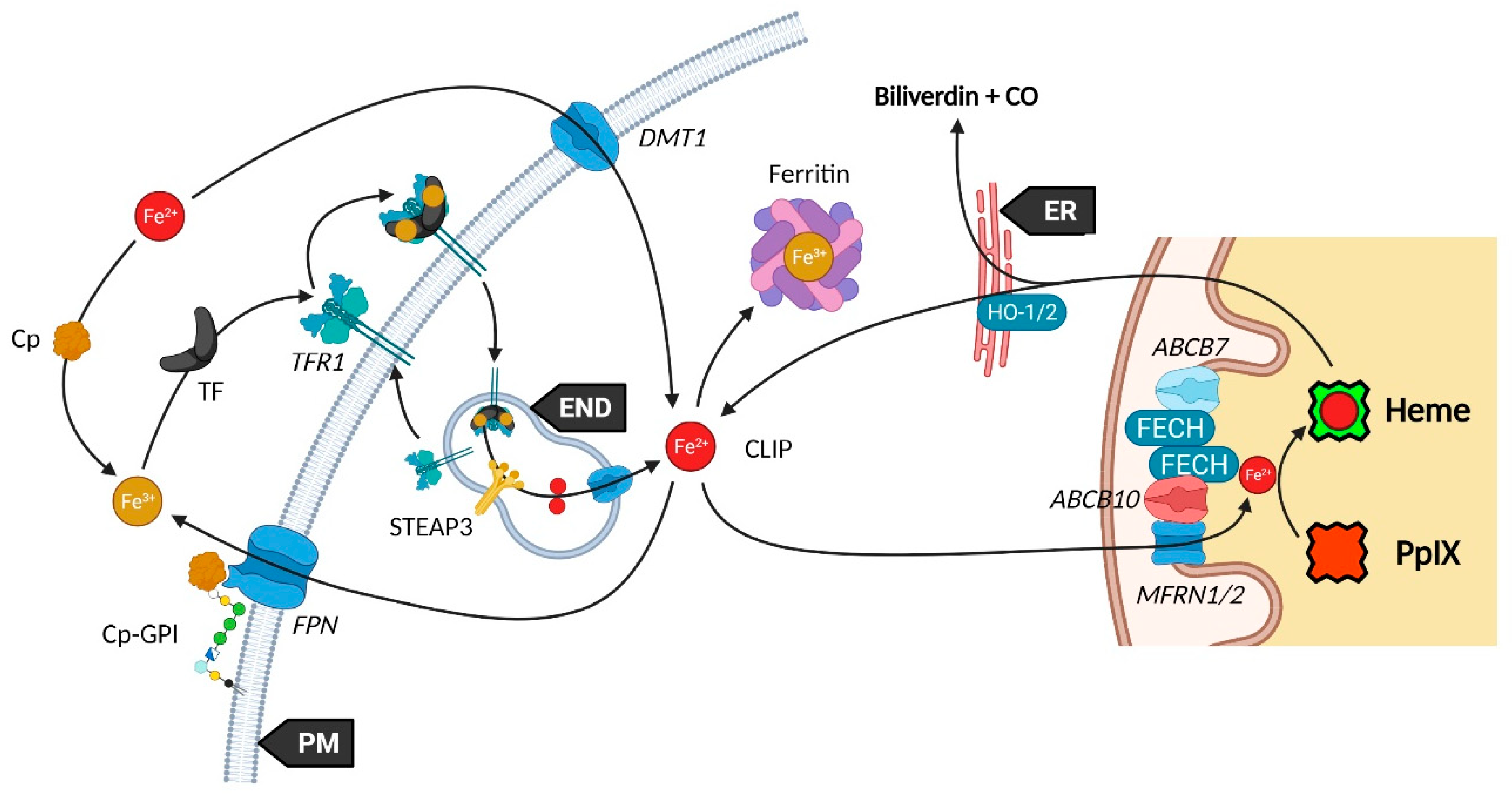
Mitochondria are essential organelles of mammalian cells, often emphasized for their function in energy production, iron metabolism and apoptosis as well as heme synthesis. The heme is an iron-loaded porphyrin behaving as a prosthetic group by its interactions with a wide variety of proteins. These complexes are termed hemoproteins and are usually vital to the whole cell comportment, such as the proteins hemoglobin, myoglobin or cytochromes, but also enzymes such as catalase and peroxidases. The building block of porphyrins is the 5-aminolevulinic acid, whose exogenous administration is able to stimulate the entire heme biosynthesis route. In neoplastic cells, this methodology repeatedly demonstrated an accumulation of the ultimate heme precursor, the fluorescent protoporphyrin IX photosensitizer, rather than in healthy tissues.

  • aminolevulinic acid
  • protoporphyrin IX
  • cancer
  • photodynamic diagnosis

1. Introduction

Since its discovery in 1987 by Malik and Lugaci [1], the selective accumulation of the photosensitizer protoporphyrin IX (PpIX) in neoplastic cells upon administration of 5-aminolevulinic acid (5-ALA) has been extensively reported [2][3][4][5]. However, the mechanisms underlying this crucial feature are still unclear. This is partially due to incomplete knowledge of the heme metabolism, to an obvious insufficient hindsight, but also because of recurrent discrepancies from one study to another.
Unlike other molecules used in photodynamic therapy (PDT) and diagnosis (PDD), 5-ALA is the only naturally occurring agent. Considered present in all aerobic cells, it is a prime player in heme biosynthesis, whose penultimate by-product is PpIX. This pathway is highly regulated by eight enzymes, evenly distributed between mitochondria and the cytoplasm (Figure 1). Although the occurrence of a change in their expression can lead to severe disorders termed porphyria, often associated with skin photosensitivity, the control of this cycle can turn it into a potent tool. Since the conversion of 5-ALA allows to differentiate a normal from a cancerous cell by fluorescence, numerous 5-ALA derivatives have been synthetized to offer an optimized way to perform fluorescence-guided surgery of a wide variety of tumors [6].
Figure 1. Current vision of the major players of heme metabolism and their interactions. Endoplasmic Reticulum (ER), Golgi Apparatus (GA), Outer/Inner Mitochondrial Membrane (OMM/IMM), Inner Mitochondrial Space (IMS), Endosome (END), and Lysosome (Lyso). Porphobilinogen (PBG), Hydroxymethylbilane (HMB), Uroporphyrinogen (UROgen), Coproporphyrinogen (COPROgen), and Protoporphyrinogen (PROTOgenIX). Created with BioRender.com

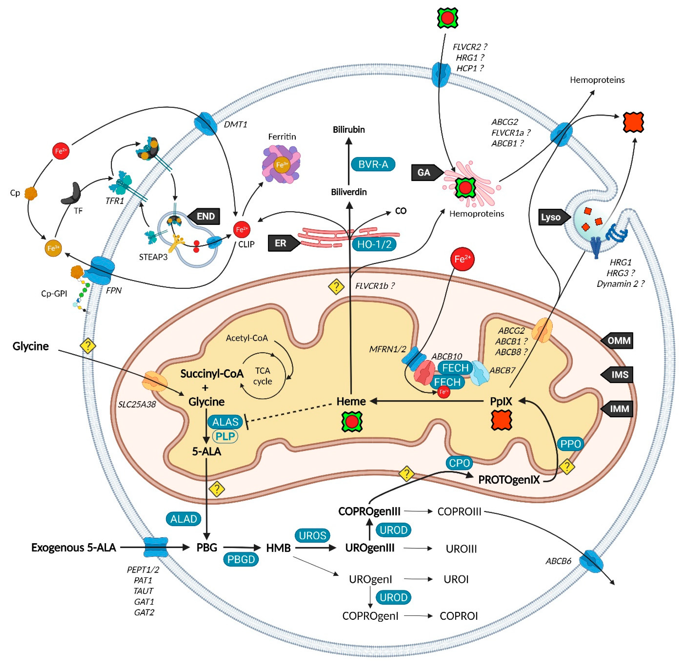
2. Heme Metabolism—A Complex Network Tightly Regulated by Enzymes, Transporters and Other Metabolites

2.1. Biosynthesis of Iron Protoporphyrin IX

Branches surrounding the pathway of 5-ALA conversion into heme have become more and more exhaustive since Shemin and Rittenberg discovered the initial requirement of glycine in 1946 [7].
Heme, protoheme or iron protoporphyrin IX are a prosthetic complex of ferrous iron and PpIX that comes into play in major processes such as electron transfer chains, respiratory complexes, oxygen transport and storage of metal ions.
While 5-ALA exogenous administration leads to protoheme synthesis, the building block of tetrapyrroles is naturally formed in the mitochondrial matrix through the so-called “Shemin pathway” or C-4 [8][9]. Indeed, succinyl-CoA previously obtained by the tricarboxylic acid (TCA) or Krebs cycle is condensed with glycine into 5-ALA thanks to the enzyme ALA-synthase (ALAS, E.C. 2.3.1.37) and its cofactor pyridoxal-5′-phosphate (PLP), before export to the cytosol [10]. This single stage explains why the PpIX-mediated retro-negative feedback that inhibits ALAS can be circumvented by a straight 5-ALA uptake. Then, a second enzyme, ALA-dehydratase (ALAD, E.C. 4.2.1.24) converts two cytosolic 5-amino-4-oxopentanoic acids into the pyrrole derivative porphobilinogen via an asymmetric condensation. The association of four porphobilinogen catalyzed by the porphobilinogen deaminase (PBGD, E.C. 2.5.1.61) subsequently leads to the formation of a linear tetrapyrrolic structure, the 1-hydroxymethylbilane or pre-uroporphyrinogen. Uroporphyrinogen III cosynthase (UROS, E.C. 4.2.1.75) works in tandem with PBGD to close the cycle and enable the formation of the uroporphyrinogen III tetrapyrrolic cycle. This very first cycle is actually composed of eight 5-ALA molecules, the only source of carbon and nitrogen all along the pathway. From this point, decarboxylations are monitored along two stages. Primarily, the four acetate groups are decarboxylated into methyls by uroporphyrinogen decarboxylase (UROD, E.C. 4.1.1.37), turning uroporphyrinogen III into coproporphyrinogen III. Secondly, coproporphyrinogen III oxidase (CPO, E.C. 1.3.3.3) that localizes to the intermembrane space converts two out of four propionate residues into vinyls. The generated protoporphyrinogen IX undergoes further oxidation at the inner mitochondrial membrane by the protoporphyrinogen oxidase (PPO, E.C. 1.3.3.4). The hydrophobic PpIX obtained at this stage is the ultimate heme precursor. Its light-related features of fluorescence and photosensitization finally disappear in heme, when ferrous iron is inserted inside the aromatic structure by ferrochelatase (FECH, E.C. 4.99.1.1) localized at the inner flank of the inner mitochondrial membrane [11].
The synthesis of additional products of the heme biosynthesis pathway was recently explained. Indeed, the hydroxymethylbilane intermediary can spontaneously cyclize and form uroporphyrinogen I, albeit the UROS reaction leading to uroporphyrinogen III overrides it [12]. Similar to its isomer, uroporphyrinogen I can either be oxidized into uroporphyrin I, potentially by various cytochrome P450 isoenzyme catalyzation, or decarboxylated by UROD into coproporphyrinogen I that can in turn undergo spontaneous oxidation into coproporphyrin I. On the main path, uroporphyrinogen III can be transformed to uroporphyrin III, and coproporphyrinogen III can be turned into its coproporphyrin III oxidized counterpart. Both hydrophilic uroporphyrins and coproporphyrins display fluorescence properties very close to PpIX.

2.2. Utilization and Degradation of Iron Protoporphyrin IX

Heme is a ubiquitous component of every eukaryotic cell that plays a crucial role in their survival and behavior. However, an excess of this hydrophobic molecule is toxic to cells. It leads to the direct generation of reactive oxygen species (ROS) that in turn induce lipid peroxidation, DNA damage as well as protein damage and aggregation. Consequently, heme must be degraded by cells to prevent such deleterious effects. This role is endorsed by the heme oxygenase system. Heme oxygenase 1 and 2 (HO-1 and HO-2, E.C. 1.14.14.18) are famous for their role in tetrapyrrolic cycle opening and Fe2+ extraction that produces carbon monoxide (CO) and biliverdin. Located to the endoplasmic reticulum, HO-1 is expressed in most tissues and considered as a protective enzyme due to its inducibility by a profusion of factors. In contrast, HO-2 that contains heme regulatory motifs (HRM) is not induced by such factors but under oxidative stress conditions. Biliverdin reductase-A (BVR-A, E.C. 1.3.1.24) localizes to the cytosol and subsequently transforms biliverdin into bilirubin, that both, as well as HO-1 and CO, display important antioxidant properties [13][14][15]. The fate of the highly lipophilic bilirubin in cells, other than its absorption by hepatocytes, is not well described. The glucuronidation reaction by UDP-glucuronosyltransferase 1A1 (UGT1A1, E.C. 2.4.1.17) is known to transform such lipophilic substrates into hydrophilic metabolites that can subsequently move from hepatocytes to the bile and be excreted [16]. UGT1A1 and other members of its family where recently confirmed to be expressed in keratinocytes, suggesting that a similar elimination mechanism takes place in other cell types [17]. Additionally, the whole balance between heme and bilirubin might be controlled by a double negative feedback inhibition from bilirubin that would inhibit BVR-A [18] and from biliverdin that would inhibit heme oxygenase [19].

2.3. Transporters of the Heme Metabolism: Where Are We Now?

The functioning of the heme biosynthesis pathway is far from the full understanding, as attested by the bunch of inconsistencies on the involvement and role of transporters. Nevertheless, several of them are now unanimously accepted as heme metabolism milestones.
It was recently discovered that, in order to produce 5-ALA, cells import glycine through the mitochondrial glycine transporter localized at the inner mitochondrial membrane (IMM) and encoded by the SLC25A38 solute carrier gene [20]. The newly synthetized 5-ALA then moves out of the mitochondrion by a still obscure way. Previous studies suggested that SLC25A38 facilitates the exchange of glycine and 5-ALA through the mitochondrial membrane [21] and that the ATP-binding cassette subfamily B member 10 (ABCB10) is essential for 5-ALA export to the cytoplasm [22]. In contrast, it was more recently shown that silencing of ABCB10 in zebrafish and murine Friend erythroleukemia cells did not reduce the 5-ALA cytoplasmic concentration and thus rejected a direct interaction of ABCB10 with 5-ALA to export the latter [23].
Concerning the exogenous 5-ALA administration, due to its high hydrophilicity, crossing the cell barrier toughly occurs through passive diffusion, as this is the case for some derivatives (e.g., containing an ester moiety). Active transport and facilitated diffusion are more likely, then many intermediates have been proposed for this uptake, notably based on the idea of a structural similarity between the δ-amino acid 5-ALA, GABA and β-amino acids β-alanine and Taurine. Mainly, PEPT1 (SLC15A1), PEPT2 (SLC15A2), PAT1 (SLC36A1) and GABA neurotransmitter transporters TAUT (Taurine transporter SLC6A6), GAT1 (SLC6A1) and GAT2 (SLC6A13) demonstrated the ability to transport 5-ALA [24][25][26][27][28][29][30].
Once in the cytoplasm, it has been pointed that the ABCB6 transporter inserted inside the outer mitochondrial membrane (OMM) facilitates the mitochondrial crossing by the sub-product coproporphyrinogen III and subsequently acts as a checkpoint that enhances or prevents further heme production [31][32]. Nonetheless, the mitochondrial localization of ABCB6 is controversial and more and more attributed to lysosomes as well as to the plasma membrane [33][34][35]. In fact, ABCB6 is thought to be a potent activatable tetrapyrrole transporter that enables the cells to remove the cytosolic coproporphyrin III excess [31][36] and potentially of other porphyrins COPROI, UROI and UROIII [37].
While the last stages are still not fully resolved, recent studies substantiated the role of many actors in PpIX synthesis and iron metabolism as part of major protein complexes, which would most probably speed-up this last all-in-one stage completed by heme production. TMEM14C was described as essential to the synthesis of PpIX by facilitating the transport of protoporphyrinogen IX, but not of its precursors, into the mitochondria of erythroid cells, while no evidence was found in other cell types [38].
At this stage, the photoactive PpIX may be driven to the cytoplasm and outside of cells by an ABCG2 dimer localized in both mitochondrial and plasma membranes [39]. This multidrug efflux pump, while non-specific toward PpIX, is currently the most described way to regulate its level [40]. Recently, a study also described the involvement of exocytosis and showed that inhibition of dynamin 2, a major player in exocytosis, led to less extracellular and more intracellular PpIX levels [41].
Another transporter, ABCB1 (MDR1), is thought to transport PpIX through the mitochondrial and plasma membranes [42]. Here, researchers demonstrated that inhibition of the oncogenic Ras/MEK pathway restrained the efflux of PpIX due to ABCB1 level reduction in colon cancer cells. This was recently confirmed in a mouse malignant glioma where downregulation of MDR1 increased the cellular 5-ALA-induced PpIX level [43]. Researches on ABCB1 are very limited, hence its role remains to confirm.
It was recently found that ferrochelatase (FECH) homodimerizes to bind ABCB7 on one side and ABCB10 on the other side [44]. Furthermore, ABCB10 is known to bind and stabilize mitoferrin-1 (MFRN1, SLC25A37) an iron importer [45], suggesting a collaboration or close relationship between MFRN1-ABCB10 and ABCB10-FECH-FECH-ABCB7 to import iron and insert it in protoporphyrin IX through ferrochelatase. It is then hypothesized that ABCB7 enables ATP-driven ferrochelatase opening and subsequent iron protoporphyrin IX release [11][44].
ABCB8, another IMM actor, is becoming more and more attractive in heme, iron and cancer fields. It was recently proposed as a renal cell carcinoma prognostic marker. Knockdown of the corresponding gene reduced the migration and viability of renal cancer cell lines in vitro, suggesting a role in tumor progression [46]. It was also shown to mediate iron export in mitochondria of cardiomyocyte cells and to play an essential role in the maturation of iron-sulfur clusters (ISC) [47].
Neuropilin-1 (NRP1) is a transmembrane or cytosolic protein that acts on several signaling pathways, such as angiogenesis through VEGF and HGF in endothelial cells [48], and is a tumor promotor overexpressed in several tumor tissues [49][50][51]. Issit et al. revealed a physical interaction between NRP1 and ABCB8. In addition, their study indicated that NRP1 downregulation reduced ABCB8 expression and increased mitochondrial iron accumulation in endothelial cells. Analysis of iron transporters revealed that NRP1 knockdown also increased the level of the iron exporter FPN1 and the iron importer MFRN1. In contrast, TFR1 and MFRN2 iron importers were not deregulated [52].
Iron availability (see Figure 2) is tightly controlled by diverse mechanisms in cells that have been comprehensively reviewed [14][53][54]. Such control is mandatory to prevent Fe2+ accumulation that leads, through the Fenton reaction, to toxic hydroxyl radical production. Elementarily, ferrous iron (Fe2+) from the extracellular matrix binds to the transferrin (TF) carrier as ferric iron (Fe3+). Transferrin subsequently links to the plasma membrane transferrin receptor 1 (TFR1), forming a complex that will be endocytosed. Endosomal reduction of Fe3+ is handled by STEAP3, the six-transmembrane epithelial antigen of prostate 3 followed by translocation of free Fe2+ into the cytosol by the divalent metal transporter 1 (DMT1). DMT1 was also found to mediate Fe2+ intracellular level by acting as a plasma membrane importer. Similar to the Fe-TF-TFR1 complex, Fe-DMT1 can be internalized in endosomes and act on the cytosolic labile iron pool (CLIP) or deliver Fe to mitochondria. Additionally, DMT1 was found to be expressed in the outer mitochondrial membrane (OMM) and to act as both a mitochondrial iron importer and exporter [55]. A more direct use of the CLIP might take place throughout a “kiss and run” process that may operate by the sole touch of the endosome with the mitochondrion. The CLIP excess is controlled by the iron efflux protein ferroportin (FPN) that dispatches Fe2+ back in the cytoplasm, as well as by the iron storing complex ferritin [56]. Iron from the cytosolic pool may be incorporated in mitochondria through the MFRN1-ABCB10 complex, as mentioned above, in order to act as a substrate of PpIX.
Figure 2. Utilization and regulation of the cytosolic labile iron pool. Created with BioRender.com
Ceruloplasmin (Cp) is a potent copper sequestering protein that circulates in the blood flow. It shows a ferroxidase activity that is essential to the ferrous to ferric iron oxidation, a necessary step for transferrin (TFR1) loading and iron delivery inside cells [53][57]. Cp also mediates iron efflux by cooperating with ferroportin (FPN, IREG1) but must be glycosylphosphatidylinositol (GPI)-anchored to the plasma membrane next to FPN to activate the latter [58].
Heme subsequently moves to the cytoplasm to exert different functions. From this statement, it was proposed that heme cytoplasm homeostasis is controlled by mitochondrial exporters and cell membrane heme importers and exporters. The topic has been reviewed by Ponka et al. who blame a lack of evidence leading to several extrapolations [59].
Starting with mitochondrial export, FLVCR1b (Feline Leukemia Virus Subgroup C Cellular Receptors) is thought to make a way for heme to the cytoplasm considering that its expression is directly correlated to the cytoplasmic heme level [60]. However, no study corroborated this hypothesis and the localization on the mitochondrial membrane is not described yet. Additionally, it was recently noticed that FLVCR1b overexpression does not modify myoglobin heme-insertion [61]. Whether FLVCR1b directly transports heme is a mere assumption, but a secondary role for this protein is likely.
From the cytoplasm, heme might be either further expelled out to the extracellular matrix through FLVCR1a and ABCG2, or degraded into biliverdin and bilirubin while releasing its iron content and carbon monoxide (CO). The heme export role of FLVCR1a was proposed by Quigley et al. who showed that overexpression of FLVCR1a decreases the cytoplasmic heme level of rat epithelial cells incubated with the heme analog ZnMP [62]. Other studies reported a similar role, however, Destefanis et al. recently showed that FLVCR1a silencing did not lead to heme accumulation, both without and under 5-ALA treatment, in the SNU-407 colorectal adenocarcinoma cell line [63]. Nonetheless, a clear upregulation of the ABCG2 mRNA, a PpIX exporter, was found when FLVCR1a was silenced. Thus, it seems an equilibrium needs to be implemented as the lack of FLVCR1a could induce a porphyrin accumulation, even though this is not observed in the study.
FLVCR2 was suggested to import heme across the plasma membrane of mammalian cells [64]. Unlike its FLVCR1 homolog, an export role was not found for FLVCR2 (named FLVCRL14q) in vitro [62]. In contrast, no import feature was noticed in a Saccharomyces Cerevisiae yeast model [65].
Lately, Li et al. reported a new mode of action for FLVCR2 (named MFSD7C9) in an extensive study [66]. They first confirmed that FLVCR2 displays three binding sites to heme. Then, immunoprecipitation, subcellular fractionation coupled to Western blotting, FLVCR2-GFP and MitoTracker analysis confirmed a major localization to mitochondria, a transient interaction with energy transfer chain (ETC) complexes III, IV and V, and the endoplasmic reticulum ATPase SERCA2b implied in thermogenesis. Based on their results, they make the hypothesis that heme binding to FLVCR2 dissociates the latter from the ETC and SERCA2b that induces a mitochondrial respiration switch from ATP synthesis to thermogenesis.
In addition, they report an interaction between FLVCR2 and the heme catabolism proteins HO-1 and TFR1 [66]. Thus, FLVCR2 might be important for heme opening by HO-1 and Fe2+ homeostasis.
Heme-responsive gene 1 (HRG1) protein was shown to bind and transport heme while locating in endosomes and lysosomes [67]. In 2019, the same group demonstrated in a mice HRG1 KO model that heme accumulated in phagolysosomes of the reticuloendothelial system macrophages [68]. An over 10-fold accumulation in heme was observed, and the size of the lysosomes was enlarged from 10 to 100-fold compared to normal mice. This storage was explained by a crystallization of heme into hemozoin, potentially to avoid heme toxicity [68].
In 2013, a team related the HRG1 plasma membrane localization with a high degree of cell invasive and migratory features. They hypothesize that HRG1 induces the vacuolar-(H+) ATPase that co-expresses to the plasma membrane, whose role in pH regulation, glucose metabolism and metalloproteinases activities may regulate the metastatic ability of cancer cells [69]. Altogether, this suggests a direct role of heme metabolism regulation into cancer cells behavior.
Heme carrier protein 1 (HCP1), a transmembrane proton-coupled folate transporter (PCFT) was found to be a potent intestinal heme transporter [70]. More recently, an in vivo study displayed a role in hepatocyte iron regulation [71]. HCP1 gene (SLC46A1) silencing was associated with a reduced liver iron level and increased TFR1 and FPN protein expressions. They confirmed that HCP1 in hepatocytes was able to transport heme, however heme treatment decreased HCP1 expression in hepatic cell line [71].
Progesterone receptor membrane component 1 (PGRMC1) is another regulator of the heme metabolism. Its role in heme regulation through FECH interaction was described in human cells by Piel et al. who also suggested a direct interaction between PGRMC1 and heme [72]. More recently, human PGRMC1 was confirmed to link to both ferrous and ferric forms of heme [73] and to bind to various cytochromes P450 in a heme-independent fashion [74].
Some players and their attributed role can now be trusted; however, many others are either disputed or not sufficiently examined. The overall comprehension of the transport mechanisms of heme-related actors is still faraway and extrapolations should be handled with great care.

This entry is adapted from the peer-reviewed paper 10.3390/ijms23147974

References

  1. Malik, Z.; Lugaci, H. Destruction of erythroleukaemic cells by photoactivation of endogenous porphyrins. Br. J. Cancer 1987, 56, 589–595.
  2. D’Hallewin, M.-A.; Bezdetnaya, L.; Guillemin, F. Fluorescence Detection of Bladder Cancer: A Review. Eur. Urol. 2002, 42, 417–425.
  3. Collaud, S.; Juzeniene, A.; Moan, J.; Lange, N. On the selectivity of 5-aminolevulinic acid-induced protoporphyrin IX formation. Curr. Med. Chem. Anticancer Agents 2004, 4, 301–316.
  4. Diez Valle, R.; Hadjipanayis, C.G.; Stummer, W. Established and emerging uses of 5-ALA in the brain: An overview. J. Neuro-Oncol. 2019, 141, 487–494.
  5. Kim, H.I.; Wilson, B.C. Photodynamic Diagnosis and Therapy for Peritoneal Carcinomatosis from Gastrointestinal Cancers: Status, Opportunities, and Challenges. J. Gastric Cancer 2020, 20, 355–375.
  6. Fotinos, N.; Campo, M.A.; Popowycz, F.; Gurny, R.; Lange, N. 5-Aminolevulinic Acid Derivatives in Photomedicine: Characteristics, Application and Perspectives. Photochem. Photobiol. 2006, 82, 994–1015.
  7. Shemin, D.; Rittenberg, D. The biological utilization of glycine for the synthesis of the protoporphyrin of hemoglobin. J. Biol. Chem. 1946, 166, 621–625.
  8. Bali, S.; Palmer, D.J.; Schroeder, S.; Ferguson, S.J.; Warren, M.J. Recent advances in the biosynthesis of modified tetrapyrroles: The discovery of an alternative pathway for the formation of heme and heme d 1. Cell. Mol. Life Sci. 2014, 71, 2837–2863.
  9. Heinemann, I.U.; Jahn, M.; Jahn, D. The biochemistry of heme biosynthesis. Arch. Biochem. Biophys. 2008, 474, 238–251.
  10. Hunter, G.A.; Ferreira, G.C. Molecular enzymology of 5-aminolevulinate synthase, the gatekeeper of heme biosynthesis. Biochim. Biophys. Acta 2011, 1814, 1467–1473.
  11. Medlock, A.E.; Shiferaw, M.T.; Marcero, J.R.; Vashisht, A.A.; Wohlschlegel, J.A.; Phillips, J.D.; Dailey, H.A. Identification of the Mitochondrial Heme Metabolism Complex. PLoS ONE 2015, 10, e0135896.
  12. Fortian, A.; Castaño, D.; Gonzalez, E.; Laín, A.; Falcon-Perez, J.M.; Millet, O. Structural, thermodynamic, and mechanistical studies in uroporphyrinogen III synthase: Molecular basis of congenital erythropoietic porphyria. Adv. Protein Chem. Struct. Biol. 2011, 83, 43–74.
  13. Kumar, S.; Bandyopadhyay, U. Free heme toxicity and its detoxification systems in human. Toxicol. Lett. 2005, 157, 175–188.
  14. Yanatori, I.; Richardson, D.R.; Toyokuni, S.; Kishi, F. How iron is handled in the course of heme catabolism: Integration of heme oxygenase with intracellular iron transport mechanisms mediated by poly (rC)-binding protein-2. Arch. Biochem. Biophys. 2019, 672, 108071.
  15. Stocker, R.; Yamamoto, Y.; McDonagh, A.F.; Glazer, A.N.; Ames, B.N. Bilirubin Is an Antioxidant of Possible Physiological Importance. Science 1987, 235, 1043–1046.
  16. Thomas, M.; Hardikar, W.; Greaves, R.F.; Tingay, D.G.; Loh, T.P.; Ignjatovic, V.; Newall, F.; Rajapaksa, A.E. Mechanism of bilirubin elimination in urine: Insights and prospects for neonatal jaundice. Clin. Chem. Lab. Med. 2021, 59, 1025–1033.
  17. Sumida, K.; Kawana, M.; Kouno, E.; Itoh, T.; Takano, S.; Narawa, T.; Tukey, R.H.; Fujiwara, R. Importance of UDP-glucuronosyltransferase 1A1 expression in skin and its induction by UVB in neonatal hyperbilirubinemia. Mol. Pharmacol. 2013, 84, 679–686.
  18. Kutty, R.K.; Maines, M.D. Purification and characterization of biliverdin reductase from rat liver. J. Biol. Chem. 1981, 256, 3956–3962.
  19. Kutty, R.K.; Maines, M.D. Hepatic heme metabolism: Possible role of biliverdin in the regulation of heme oxygenase activity. Biochem. Biophys. Res. Commun. 1984, 122, 40–46.
  20. Lunetti, P.; Damiano, F.; De Benedetto, G.; Siculella, L.; Pennetta, A.; Muto, L.; Paradies, E.; Marobbio, C.M.; Dolce, V.; Capobianco, L. Characterization of Human and Yeast Mitochondrial Glycine Carriers with Implications for Heme Biosynthesis and Anemia. J. Biol. Chem. 2016, 291, 19746–19759.
  21. Guernsey, D.L.; Jiang, H.; Campagna, D.R.; Evans, S.C.; Ferguson, M.; Kellogg, M.D.; Lachance, M.; Matsuoka, M.; Nightingale, M.; Rideout, A.; et al. Mutations in mitochondrial carrier family gene SLC25A38 cause nonsyndromic autosomal recessive congenital sideroblastic anemia. Nat. Genet. 2009, 41, 651–653.
  22. Bayeva, M.; Khechaduri, A.; Wu, R.; Burke, M.A.; Wasserstrom, J.A.; Singh, N.; Liesa, M.; Shirihai, O.S.; Langer, N.B.; Paw, B.H.; et al. ATP-binding cassette B10 regulates early steps of heme synthesis. Circ. Res. 2013, 113, 279–287.
  23. Seguin, A.; Takahashi-Makise, N.; Yien, Y.Y.; Huston, N.C.; Whitman, J.C.; Musso, G.; Wallace, J.A.; Bradley, T.; Bergonia, H.A.; Kafina, M.D.; et al. Reductions in the mitochondrial ABC transporter Abcb10 affect the transcriptional profile of heme biosynthesis genes. J. Biol. Chem. 2017, 292, 16284–16299.
  24. Frolund, S.; Marquez, O.C.; Larsen, M.; Brodin, B.; Nielsen, C.U. Delta-aminolevulinic acid is a substrate for the amino acid transporter SLC36A1 (hPAT1). Br. J. Pharmacol. 2010, 159, 1339–1353.
  25. Tran, T.T.; Mu, A.; Adachi, Y.; Adachi, Y.; Taketani, S. Neurotransmitter transporter family including SLC6A6 and SLC6A13 contributes to the 5-aminolevulinic acid (ALA)-induced accumulation of protoporphyrin IX and photodamage, through uptake of ALA by cancerous cells. Photochem. Photobiol. 2014, 90, 1136–1143.
  26. Iwaki, K.; Fujiwara, T.; Ito, T.; Suzuki, C.; Sasaki, K.; Ono, K.; Saito, K.; Fukuhara, N.; Onishi, Y.; Yokoyama, H.; et al. Flow Cytometry-Based Photodynamic Diagnosis with 5-Aminolevulinic Acid for the Detection of Minimal Residual Disease in Multiple Myeloma. Tohoku J. Exp. Med. 2019, 249, 19–28.
  27. Rodriguez, L.; Batlle, A.; Di Venosa, G.; MacRobert, A.J.; Battah, S.; Daniel, H.; Casas, A. Study of the mechanisms of uptake of 5-aminolevulinic acid derivatives by PEPT1 and PEPT2 transporters as a tool to improve photodynamic therapy of tumours. Int. J. Biochem. Cell Biol. 2006, 38, 1530–1539.
  28. Ning, S.; Kang, Q.; Fan, D.; Liu, J.; Xue, C.; Zhang, X.; Ding, C.; Zhang, J.; Peng, Q.; Ji, Z. Protein 4.1R is Involved in the Transport of 5-Aminolevulinic Acid by Interaction with GATs in MEF Cells. Photochem. Photobiol. 2018, 94, 173–178.
  29. Bermudez Moretti, M.; Correa Garcia, S.; Perotti, C.; Batlle, A.; Casas, A. Delta-Aminolevulinic acid transport in murine mammary adenocarcinoma cells is mediated by beta transporters. Br. J. Cancer 2002, 87, 471–474.
  30. Rud, E.; Gederaas, O.; Høgset, A.; Berg, K. 5-Aminolevulinic Acid, but not 5-Aminolevulinic Acid Esters, is Transported into Adenocarcinoma Cells by System BETA Transporters. Photochem. Photobiol. 2000, 71, 640–647.
  31. Krishnamurthy, P.C.; Du, G.; Fukuda, Y.; Sun, D.; Sampath, J.; Mercer, K.E.; Wang, J.; Sosa-Pineda, B.; Murti, K.G.; Schuetz, J.D. Identification of a mammalian mitochondrial porphyrin transporter. Nature 2006, 443, 586–589.
  32. Zhao, S.G.; Chen, X.F.; Wang, L.G.; Yang, G.; Han, D.Y.; Teng, L.; Yang, M.C.; Wang, D.Y.; Shi, C.; Liu, Y.H.; et al. Increased expression of ABCB6 enhances protoporphyrin IX accumulation and photodynamic effect in human glioma. Ann. Surg. Oncol. 2013, 20, 4379–4388.
  33. Bergam, P.; Reisecker, J.M.; Rakvacs, Z.; Kucsma, N.; Raposo, G.; Szakacs, G.; van Niel, G. ABCB6 Resides in Melanosomes and Regulates Early Steps of Melanogenesis Required for PMEL Amyloid Matrix Formation. J. Mol. Biol. 2018, 430, 3802–3818.
  34. Kiss, K.; Brozik, A.; Kucsma, N.; Toth, A.; Gera, M.; Berry, L.; Vallentin, A.; Vial, H.; Vidal, M.; Szakacs, G. Shifting the paradigm: The putative mitochondrial protein ABCB6 resides in the lysosomes of cells and in the plasma membrane of erythrocytes. PLoS ONE 2012, 7, e37378.
  35. Paterson, J.K.; Shukla, S.; Black, C.M.; Tachiwada, T.; Garfield, S.; Wincovitch, S.; Ernst, D.N.; Agadir, A.; Li, X.; Ambudkar, S.V.; et al. Human ABCB6 Localizes to Both the Outer Mitochondrial Membrane and the Plasma Membrane. Biochemistry 2007, 46, 9443–9452.
  36. Matsumoto, K.; Hagiya, Y.; Endo, Y.; Nakajima, M.; Ishizuka, M.; Tanaka, T.; Ogura, S. Effects of plasma membrane ABCB6 on 5-aminolevulinic acid (ALA)-induced porphyrin accumulation in vitro: Tumor cell response to hypoxia. Photodiagn. Photodyn. Ther. 2015, 12, 45–51.
  37. Fukuda, Y.; Cheong, P.L.; Lynch, J.; Brighton, C.; Frase, S.; Kargas, V.; Rampersaud, E.; Wang, Y.; Sankaran, V.G.; Yu, B.; et al. The severity of hereditary porphyria is modulated by the porphyrin exporter and Lan antigen ABCB6. Nat. Commun. 2016, 7, 12353.
  38. Yien, Y.Y.; Robledo, R.F.; Schultz, I.J.; Takahashi-Makise, N.; Gwynn, B.; Bauer, D.E.; Dass, A.; Yi, G.; Li, L.; Hildick-Smith, G.J.; et al. TMEM14C is required for erythroid mitochondrial heme metabolism. J. Clin. Investig. 2014, 124, 4294–4304.
  39. Kobuchi, H.; Moriya, K.; Ogino, T.; Fujita, H.; Inoue, K.; Shuin, T.; Yasuda, T.; Utsumi, K.; Utsumi, T. Mitochondrial localization of ABC transporter ABCG2 and its function in 5-aminolevulinic acid-mediated protoporphyrin IX accumulation. PLoS ONE 2012, 7, e50082.
  40. Partha, K.; John, D.S. The Role of ABCG2 and ABCB6 in Porphyrin Metabolism and Cell Survival. Curr. Pharm. Biotechnol. 2011, 12, 647–655.
  41. Kitajima, Y.; Ishii, T.; Kohda, T.; Ishizuka, M.; Yamazaki, K.; Nishimura, Y.; Tanaka, T.; Dan, S.; Nakajima, M. Mechanistic study of PpIX accumulation using the JFCR39 cell panel revealed a role for dynamin 2-mediated exocytosis. Sci. Rep. 2019, 9, 8666.
  42. Yoshioka, E.; Chelakkot, V.S.; Licursi, M.; Rutihinda, S.G.; Som, J.; Derwish, L.; King, J.J.; Pongnopparat, T.; Mearow, K.; Larijani, M. Enhancement of Cancer-Specific Protoporphyrin IX Fluorescence by Targeting Oncogenic Ras/MEK Pathway. Theranostics 2018, 8, 2134–2146.
  43. Shono, K.; Mizobuchi, Y.; Yamaguchi, I.; Nakajima, K.; Fujiwara, Y.; Fujihara, T.; Kitazato, K.; Matsuzaki, K.; Uto, Y.; Sampetrean, O.; et al. Elevated cellular PpIX potentiates sonodynamic therapy in a mouse glioma stem cell-bearing glioma model by downregulating the Akt/NF-kappaB/MDR1 pathway. Sci. Rep. 2021, 11, 15105.
  44. Maio, N.; Kim, K.S.; Holmes-Hampton, G.; Singh, A.; Rouault, T.A. Dimeric ferrochelatase bridges ABCB7 and ABCB10 homodimers in an architecturally defined molecular complex required for heme biosynthesis. Haematologica 2019, 104, 1756–1767.
  45. Chen, W.; Paradkar, P.N.; Li, L.; Pierce, E.L.; Langer, N.B.; Takahashi-Makise, N.; Hyde, B.B.; Shirihai, O.S.; Ward, D.M.; Kaplan, J.; et al. Abcb10 physically interacts with mitoferrin-1 (Slc25a37) to enhance its stability and function in the erythroid mitochondria. Proc. Natl. Acad. Sci. USA 2009, 106, 16263–16268.
  46. Blajan, I.; Miersch, H.; Schmidt, D.; Kristiansen, G.; Perner, S.; Ritter, M.; Ellinger, J.; Klumper, N. Comprehensive Analysis of the ATP-binding Cassette Subfamily B Across Renal Cancers Identifies ABCB8 Overexpression in Phenotypically Aggressive Clear Cell Renal Cell Carcinoma. Eur. Urol. Focus 2021, 7, 1121–1129.
  47. Ichikawa, Y.; Bayeva, M.; Ghanefar, M.; Potini, V.; Sun, L.; Mutharasan, R.K.; Wu, R.; Khechaduri, A.; Jairaj, N.T.; Ardehali, H. Disruption of ATP-binding cassette B8 in mice leads to cardiomyopathy through a decrease in mitochondrial iron export. Proc. Natl. Acad. Sci. USA 2012, 109, 4152–4157.
  48. Sulpice, E.; Plouet, J.; Berge, M.; Allanic, D.; Tobelem, G.; Merkulova-Rainon, T. Neuropilin-1 and neuropilin-2 act as coreceptors, potentiating proangiogenic activity. Blood 2008, 111, 2036–2045.
  49. Stasikowska-Kanicka, O.; Wągrowska-Danilewicz, M.; Danilewicz, M. Immunohistochemical study on neuropilin 1 (NRP1) immunoexpression in oral squamous cell carcinoma. Folia Histochem. Cytobiol. 2018, 56, 98–105.
  50. Stephenson, J.M.; Banerjee, S.; Saxena, N.K.; Cherian, R.; Banerjee, S.K. Neuropilin-1 is differentially expressed in myoepithelial cells and vascular smooth muscle cells in preneoplastic and neoplastic human breast: A possible marker for the progression of breast cancer. Int. J. Cancer 2002, 101, 409–414.
  51. Hong, T.M.; Chen, Y.L.; Wu, Y.Y.; Yuan, A.; Chao, Y.C.; Chung, Y.C.; Wu, M.H.; Yang, S.C.; Pan, S.H.; Shih, J.Y.; et al. Targeting neuropilin 1 as an antitumor strategy in lung cancer. Clin. Cancer Res. 2007, 13, 4759–4768.
  52. Issitt, T.; Bosseboeuf, E.; De Winter, N.; Dufton, N.; Gestri, G.; Senatore, V.; Chikh, A.; Randi, A.M.; Raimondi, C. Neuropilin-1 Controls Endothelial Homeostasis by Regulating Mitochondrial Function and Iron-Dependent Oxidative Stress. iScience 2019, 11, 205–223.
  53. Litwack, G. Micronutrients (Metals and Iodine). Hum. Biochem. 2018, 591–643.
  54. Dietz, J.V.; Fox, J.L.; Khalimonchuk, O. Down the Iron Path: Mitochondrial Iron Homeostasis and Beyond. Cells 2021, 10, 2198.
  55. Wolff, N.A.; Ghio, A.J.; Garrick, L.M.; Garrick, M.D.; Zhao, L.; Fenton, R.A.; Thevenod, F. Evidence for mitochondrial localization of divalent metal transporter 1 (DMT1). FASEB J. 2014, 28, 2134–2145.
  56. Paul, B.T.; Manz, D.H.; Torti, F.M.; Torti, S.V. Mitochondria and Iron: Current questions. Expert Rev./Hematol. 2017, 10, 65–79.
  57. Osaki, S.; Johnson, D.A.; Frieden, E. The Possible Significance of the Ferrous Oxidase Activity of Ceruloplasmin in Normal Human Serum. J. Biol. Chem. 1966, 241, 2746–2751.
  58. Jeong, S.Y.; David, S. Glycosylphosphatidylinositol-anchored ceruloplasmin is required for iron efflux from cells in the central nervous system. J. Biol. Chem. 2003, 278, 27144–27148.
  59. Ponka, P.; Sheftel, A.D.; English, A.M.; Scott Bohle, D.; Garcia-Santos, D. Do Mammalian Cells Really Need to Export and Import Heme? Trends Biochem. Sci. 2017, 42, 395–406.
  60. Chiabrando, D.; Marro, S.; Mercurio, S.; Giorgi, C.; Petrillo, S.; Vinchi, F.; Fiorito, V.; Fagoonee, S.; Camporeale, A.; Turco, E.; et al. The mitochondrial heme exporter FLVCR1b mediates erythroid differentiation. J. Clin. Investig. 2012, 122, 4569–4579.
  61. Tupta, B.; Stuehr, E.; Sumi, M.P.; Sweeny, E.A.; Smith, B.; Stuehr, D.J.; Ghosh, A. GAPDH is involved in the heme-maturation of myoglobin and hemoglobin. FASEB J. 2022, 36, e22099.
  62. Quigley, J.G.; Yang, Z.; Worthington, M.T.; Phillips, J.D.; Sabo, K.M.; Sabath, D.E.; Berg, C.L.; Sassa, S.; Wood, B.L.; Abkowitz, J.L. Identification of a human heme exporter that is essential for erythropoiesis. Cell 2004, 118, 757–766.
  63. Destefanis, F.; Fiorito, V.; Altruda, F.; Tolosano, E. Investigating the Connection Between Endogenous Heme Accumulation and COX2 Activity in Cancer Cells. Front. Oncol. 2019, 9, 162.
  64. Duffy, S.P.; Shing, J.; Saraon, P.; Berger, L.C.; Eiden, M.V.; Wilde, A.; Tailor, C.S. The Fowler syndrome-associated protein FLVCR2 is an importer of heme. Mol. Cell. Biol. 2010, 30, 5318–5324.
  65. Yuan, X.; Protchenko, O.; Philpott, C.C.; Hamza, I. Topologically conserved residues direct heme transport in HRG-1-related proteins. J. Biol. Chem. 2012, 287, 4914–4924.
  66. Li, Y.; Ivica, N.A.; Dong, T.; Papageorgiou, D.P.; He, Y.; Brown, D.R.; Kleyman, M.; Hu, G.; Chen, W.W.; Sullivan, L.B.; et al. MFSD7C switches mitochondrial ATP synthesis to thermogenesis in response to heme. Nat. Commun. 2020, 11, 4837.
  67. Rajagopal, A.; Rao, A.U.; Amigo, J.; Tian, M.; Upadhyay, S.K.; Hall, C.; Uhm, S.; Mathew, M.K.; Fleming, M.D.; Paw, B.H.; et al. Haem homeostasis is regulated by the conserved and concerted functions of HRG-1 proteins. Nature 2008, 453, 1127–1131.
  68. Pek, R.H.; Yuan, X.; Rietzschel, N.; Zhang, J.; Jackson, L.; Nishibori, E.; Ribeiro, A.; Simmons, W.; Jagadeesh, J.; Sugimoto, H.; et al. Hemozoin produced by mammals confers heme tolerance. Elife 2019, 8, e49503.
  69. Fogarty, F.M.; O’Keeffe, J.; Zhadanov, A.; Papkovsky, D.; Ayllon, V.; O’Connor, R. HRG-1 enhances cancer cell invasive potential and couples glucose metabolism to cytosolic/extracellular pH gradient regulation by the vacuolar-H(+) ATPase. Oncogene 2014, 33, 4653–4663.
  70. Shayeghi, M.; Latunde-Dada, G.O.; Oakhill, J.S.; Laftah, A.H.; Takeuchi, K.; Halliday, N.; Khan, Y.; Warley, A.; McCann, F.E.; Hider, R.C.; et al. Identification of an intestinal heme transporter. Cell 2005, 122, 789–801.
  71. Li, H.; Wang, D.; Wu, H.; Shen, H.; Lv, D.; Zhang, Y.; Lu, H.; Yang, J.; Tang, Y.; Li, M. SLC46A1 contributes to hepatic iron metabolism by importing heme in hepatocytes. Metabolism 2020, 110, 154306.
  72. Piel, R.B., 3rd; Shiferaw, M.T.; Vashisht, A.A.; Marcero, J.R.; Praissman, J.L.; Phillips, J.D.; Wohlschlegel, J.A.; Medlock, A.E. A Novel Role for Progesterone Receptor Membrane Component 1 (PGRMC1): A Partner and Regulator of Ferrochelatase. Biochemistry 2016, 55, 5204–5217.
  73. Kaluka, D.; Batabyal, D.; Chiang, B.Y.; Poulos, T.L.; Yeh, S.R. Spectroscopic and mutagenesis studies of human PGRMC1. Biochemistry 2015, 54, 1638–1647.
  74. McGuire, M.R.; Mukhopadhyay, D.; Myers, S.L.; Mosher, E.P.; Brookheart, R.T.; Kammers, K.; Sehgal, A.; Selen, E.S.; Wolfgang, M.J.; Bumpus, N.N.; et al. Progesterone receptor membrane component 1 (PGRMC1) binds and stabilizes cytochromes P450 through a heme-independent mechanism. J. Biol. Chem. 2021, 297, 101316.
More
This entry is offline, you can click here to edit this entry!
Video Production Service