The biologically active form of vitamin D3, 1α,25-dihydroxyvitamin D3 (1,25(OH)2D3), modulates innate and adaptive immunity via genes regulated by the transcription factor vitamin D receptor (VDR). In order to identify the key vitamin D target genes involved in these processes, transcriptome-wide datasets were compared, which were obtained from a human monocytic cell line (THP-1) and peripheral blood mononuclear cells (PBMCs) treated in vitro by 1,25(OH)2D3, filtered using different approaches, as well as from PBMCs of individuals supplemented with a vitamin D3 bolus. The led to the genes ACVRL1, CAMP, CD14, CD93, CEBPB, FN1, MAPK13, NINJ1, LILRB4, LRRC25, SEMA6B, SRGN, THBD, THEMIS2 and TREM1. Public epigenome- and transcriptome-wide data from THP-1 cells were used to characterize these genes based on the level of their VDR-driven enhancers as well as the level of the dynamics of their mRNA production.
1. Introduction
Vitamin D3 is known as a micronutrient that is essential for calcium homeostasis and bone formation
[1][2]. However, vitamin D3 is also a pre-hormone that is endogenously produced when skin is exposed to sufficient amounts of UV-B
[3]. Vitamin D3 affects gene regulation after it is converted via hydroxylation at carbon 25 (providing 25-hydroxyvitamin D3) and at carbon 1 to 1α,25-dihydroxyvitamin D3 (1,25(OH)2D3), which is the high affinity ligand of the transcription factor vitamin D receptor (VDR)
[4]. Thus, ligand-activated VDR binds to accessible genomic sites in the vicinity of its target genes and modulates their transcription
[5].
Interestingly, a fully potent VDR protein evolved some 550 million years ago in a boneless vertebrate, i.e., at a time when there was no need for calcium homeostasis and bone formation
[6][7][8]. VDR’s first evolutionary function was the control of metabolism, in order to support the evolving immune system of ancestral vertebrates with energy
[9]. Thus, VDR and its ligand first specialized in the modulation of innate and adaptive immunity, such as fighting against bacterial and viral infections
[10][11] and preventing autoimmune diseases, such as multiple sclerosis and rheumatoid arthritis
[12][13], before they took on the additional task of regulating bone metabolism. Thus, vitamin D deficiency is causing an increase in bone disease, such as rickets
[14], and it may also be one of the reasons for increased vulnerability, particularly in elderly persons, against viral infections, such as the recent coronavirus (COVID-19) outbreak
[15].
Since monocytes are the most vitamin D responsive cell type in the immune system, the human monocytic leukemia cell line THP-1 is often used as a model system for the description of vitamin D signaling
[16]. Peripheral blood mononuclear cells (PBMCs), which are a mixture of lymphocytes and monocytes isolated from blood, are an interesting and easily accessible primary vitamin D-responsive tissue
[17]. In this study, we will use data from both cellular systems, in order to analyze the regulation of vitamin D target genes in innate and adaptive immunity.
The key regulatory regions of a gene are its transcription start site (TSS) and enhancer(s). The latter are stretches of genomic DNA that bind one or several signal-responsive transcription factors
[18]. Single enhancers have a dominant transcription factor binding site, while super-enhancers are formed by multiple single enhancers
[19]. Genomic DNA is always embedded in chromatin, the major function of which is to control the access of transcription factors to enhancer and TSS regions
[20][21]. Accordingly, the epigenome of a cell is represented by covalent and structural modifications of its chromatin
[22]. For example, active chromatin is detected via acetylated histone H3 proteins at position lysine 27 (H3K27ac)
[23], while tri-methylated histone H3 protein at position lysine 4 (H3K4me3) indicates active TSS regions
[24].
The next-generation sequencing method, chromatin immunoprecipitation followed by sequencing (ChIP-seq) is used for the genome-wide detection of transcription factor binding and histone modifications
[25], while formaldehyde-assisted identification of regulatory elements followed by sequencing (FAIRE-seq) determines genome-wide chromatin accessibility
[26]. Interestingly, a number of attributes of the epigenome are vitamin D sensitive, such as the binding of transcription factors like VDR and pioneer factors, and the accessibility of chromatin and changes in histone modifications
[4]. A VDR ChIP-seq meta-analysis indicated that there are on average more than 10,000 VDR binding loci per cell type
[27]. However, only a few hundred of these sites are persistently occupied by VDR and some 2000 are bound transiently, while the majority of them are only found at later points in time
[28]. Based on the chromatin model of vitamin D signaling
[4], the looping of these VDR-bound enhancers to TSS regions modulates the activity of hundreds of vitamin D target genes in VDR expressing tissues. These vitamin D-induced shifts in the epigenome and transcriptome represent the molecular basis of vitamin D signaling
[5].
2. Key Vitamin D Target Genes with Functions in the Immune System
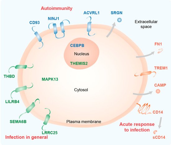
As expected for immune-related genes, most of the proteins encoded by these genes are located in or at the plasma membrane (ACVRL1, CD14, CD93, LILRB4, LRRC25, NINJ1, SEMA6B, THBD, TREM1) or are even secreted (CAMP, FN1 and SRGN) (Figure 5). Furthermore, the transcription factor CEBPB and the Toll-like receptor (TLR) signaling scaffold protein THEMIS2 act in the nucleus and the kinase MAPK13 acts in the cytoplasm.
Figure 5. Functional profile of key immune-related vitamin D target genes. Schematic picture of a cell indicating the main location of the proteins encoded by the 15 key genes. The information is based on GeneCards (
www.genecards.org) and publications cited in the text. The classification of the proteins (group 1: orange, group 2: green, group 3: blue) is based on their transcriptome profile. The main immune-related function of the protein groups is indicated in red.
The majorly increased production of the anti-microbial peptide CAMP is a well-known example of the action of vitamin D in supporting innate immunity in the fight against bacteria, such as Mycobacterium tuberculosis
[29][30]. The glycoprotein CD14 shows highest expression in monocytes and macrophages. It is anchored via glycosylphosphatidylinositol on the surface of the plasma membrane and is also found in a secreted form (sCD14). CD14 acts as a co-receptor for the pattern recognition receptors TLR1-4, 6, 7 and 9
[31]. It delivers the pathogen-associated molecule lipopolysaccharide (LPS), which is produced exclusively by gram-negative bacteria, to TLR4 resulting in pro-inflammatory responses
[32]. The transmembrane glycoprotein TREM1, which is found in monocytes, macrophages and neutrophils, is also heavily involved in TLR4 signaling, i.e., it promotes inflammation in response to bacterial infection
[33]. TREM1 is already known as a vitamin D target gene
[34]. Similarly, the gene encoding for the extracellular matrix protein FN1 has long been known as a vitamin D target
[35]. The protein is secreted by a large variety of cell types, such as macrophages, fibroblasts and epithelial cells. FN1 functions in cell adhesion and wound healing but also participates in LPS/TLR4 signaling, i.e., in the inflammatory responses
[36]. Thus, the proteins encoded by the highly vitamin D-responsive genes of group 1 are all involved in acute responses to infection.
THBD is a transmembrane protein expressed in endothelial cells, monocytes and macrophages
[37]. The traditional role of THBD is to bind thrombin and turn its pro-coagulative action to anti-coagulative, i.e., reducing blood clots. However, THBD can also bind LPS and induces its binding to the complex of CD14 and TLR4, i.e., it prevents the pro-inflammatory consequences of NF-ĸB signaling
[38]. The THBD gene is known as a key vitamin D target both in monocytes
[39] and in PBMCs
[40]. LILRB4 is an inhibitory immuno-regulatory receptor that acts on antigen-presenting cells like dendritic cells, macrophages, monocytes and microglia. It affects TNF production and bactericidal activity
[41] as well as the modulation of the differentiation of regulatory T cells
[42]. The LILRB4 gene is known as a vitamin D target gene in PBMCs
[43]. The transmembrane protein SEMA6B belongs to a the semaphorin protein family, members of which have immune functions related to the control of cell movements and cell-cell communication
[44]. Interestingly, the family member SEMA3B is known as a vitamin D target gene in bone
[45]. The transmembrane protein LRRC25 is found in monocytes, dendritic cells, granulocytes and T lymphocytes. It acts as a negative regulator of the signaling pathways of NF-ĸB
[46] and interferon
[47], i.e., it suppresses the production of inflammatory cytokines and modulates the response to viral infections. The latter is a long overlooked effect of vitamin D on the immune system
[48][49] and a reason why vitamin D deficiency may lead to high vulnerability against viral infections in the elderly, but also in school children
[15][50][51].
The kinase MAPK13 is involved in LPS/TLR4 signaling and together with MAPK11 it regulates cytokine-induced inflammatory responses
[52]. The MAPK13 gene is expressed in a large variety of cell types and it has already been described as a vitamin D target in skeletal muscle
[53]. The TLR signal transduction modulatory protein THEMIS2 is expressed in B lymphocytes, macrophages and dendritic cells and regulates LPS-induced TNF production downstream of TLR4
[54]. Moreover, the protein is involved in the development of T lymphocytes and serves as a marker of monocytic differentiation
[55]. Thus, the proteins encoded by the mid vitamin D responsive genes of group 2 are primarily involved in general responses to infection.
CD93 is a transmembrane glycoprotein that is expressed primarily in endothelial cells but also in granulocytes, monocytes, platelets and stem cells
[56]. The protein is involved in several processes of innate immunity, such as adhesion, phagocytosis and inflammation
[57]. In the context of the latter, CD93 may act as a plasma membrane receptor for DNA for delivery to endosomal TLR9. Moreover, CD93 boosts the inflammatory response of monocytes by increasing LPS recognition by TLR4. CD93 may have a protective role in autoimmune encephalomyelitis via the control of the severity of inflammation, apoptosis and bystander neuronal injury
[58]. NINJ1 is a transmembrane protein expressed primarily in myeloid and endothelial cells
[59] but it was first described in the peripheral nervous system inducing neurite extension. In the latter context, NINJ1 was found to be involved in the immune pathogenesis of multiple sclerosis
[60]. NINJ1 functions in cell adhesion and inflammation, such as leukocyte migration to sites of inflammation in the endothelium. CEBPB is a transcription factor that plays diverse roles in inflammation, e.g., through T helper cell 17 (TH17)-dependent regulation of inflammation in models of multiple sclerosis
[61]. The CEBPB gene has already been described as a vitamin D target in myeloid leukemia cells
[62]. ACVRL1 is a transmembrane protein of the transforming growth factor beta superfamily, which mediates the bone morphogenetic protein (BMP) 9- and BMP10-induced signaling that orchestrates the development of blood vessels
[63]. This relates to the control of monocyte to macrophage differentiation
[64]. SRGN is a secreted proteoglycan of endothelial cells, monocytes, mast cells and lymphocytes
[65]. LPS up-regulates SRGN expression in macrophages, suggesting the protein plays a role of in storage and secretion of inflammatory mediators
[66]. Thus, the proteins encoded by the low vitamin D responsive genes of group 3 are primarily involved in autoimmunity.
This entry is adapted from the peer-reviewed paper 10.3390/nu12041140

