Bioactive Antimicrobial Peptides for Infected Diabetic Foot Ulcers: History
Please note this is an old version of this entry, which may differ significantly from the current revision.

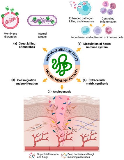
Diabetic foot ulcer (DFU) is a devastating complication, affecting 15% of diabetic patients and representing the leading cause of non-traumatic amputations. Notably, the risk of mixed bacterial–fungal infection is elevated and highly associated with wound necrosis and poor clinical outcomes. Antimicrobial peptides (AMPs) are endogenous peptides that are naturally abundant in several organisms, such as bacteria, amphibians and mammals. These molecules have shown broad-spectrum antimicrobial activity and some of them even have wound-healing activity, establishing themselves as ideal candidates for treating multi-kingdom infected wounds.

  • antimicrobial peptides
  • chronic non-healing wounds
  • diabetic foot ulcers
  • wound healing
  • bacterial and fungal infections
  • biofilms

1. Introduction

Diabetes mellitus (DM) is a chronic disease, with a continuously increasing worldwide prevalence, that affected 463 million adults globally in 2019 [1,2,3]. In Europe alone, the DM prevalence was about 59 million adults in 2019, and it is estimated to rise to over 68 million by 2045, representing an increase of 15% [1,3]. Similarly, the DM-associated complications are also expected to increase [1,2,3,4]. Indeed, diabetic foot and lower limb complications affect between 40 to 60 million people globally, representing an important source of morbidity in people with DM [1,3]. About 15% of patients with DM will develop foot ulcers in their life time, requiring prolonged hospitalizations and amputations in 85% of the cases [1,2,3,4,5,6].
A diabetic foot ulcer (DFU) is a devastating and costly complication of diabetes, consisting of deep tissue lesions associated with both peripheral neuropathy and peripheral vascular disease [7,8]. DFU represents a severe public health problem with an urgent need for new effective treatments, which are crucial to reduce the associated high morbidity and mortality rates, as well as to reduce the economic and social burden [2,6,8]. The persistent hyperglycemia, chronic inflammation, hypoxia, peripheral neuropathy, impaired angiogenesis, and difficulty to fight infections in diabetes are factors that impair the wound healing progress. Importantly, around 60% of DFUs become infected, predominantly with bacterial colonies of S. aureus and C. striatum, and fungal colonies of C. albicans [9,10,11,12,13,14,15,16].
Antimicrobial peptides (AMPs) are endogenous peptides found in different organisms, particularly in the skin, that act as a first line of defense against infection [21,22]. Furthermore, these molecules not only play key roles in fighting infection through broad-spectrum antimicrobial activity against Gram-positive and Gram-negative bacteria, viruses, and fungi, but they also play important roles in wound healing [23,24,25,26].

2. Diabetic Foot Infection

Wound healing comprises a complex and dynamic series of cellular and biochemical events which consists of the following four overlapping phases: hemostasis, inflammation, proliferation, and remodeling [5,27,28,29]. The hemostasis phase begins with constriction of the injured blood vessels and activation of platelets to form a fibrin clot to stop the bleeding [27,30]. Subsequently, the inflammatory phase initiates with the recruitment of neutrophils to the clot as a first line of defense against pathogens to remove debris, in order to provide a propitious environment for wound healing [27,28,30]. Neutrophils reach their peak population between 24 and 48 h after injury, after which they reduce greatly in number, and macrophages, in turn, arrive at the wound site and continue clearing debris [27,28]. Macrophages secrete growth factors and proteins that attract adaptive immune system cells to the wound site, such as Langerhans cells, dermal dendritic cells and T cells, which are involved either in the clearance of cellular debris or in the combat of infection [27,28]. Once the wound has been cleaned out and the inflammation decrease, the proliferative phase occurs with the following three different stages: filling of the wound with granulation tissue, contraction of the wound margins, and covering of the wound with epithelial cells, also called re-epithelization [27,28,30]. Finally, the remodeling phase takes place with collagen fiber reorganization, tissue remodeling and maturation, and an overall increase in the tensile strength can be observed [27,28,30].
However, besides its complexity, the healing process is also susceptible to interruption or delay, due to impairment of local and systemic factors that are important in the healing process. Chronic non-healing wounds often develop in people with diabetes. If wounds do not heal within 12 weeks, they are defined as chronic wounds according to the Food and Drug Administration [29,31,32]. Furthermore, in the presence of conditions such as hyperglycemia, chronic inflammation, hypoxia, peripheral neuropathy, impaired angiogenesis, and infection, the wound healing progress in diabetes can become stalled [31,32].
Diabetic foot infections (DFIs) are defined by invasion and multiplication of microorganisms in diabetic non-healing wounds and are associated with tissue destruction and/or alterations in the host’s inflammatory response [9,33,34]. DFIs are among the most serious and frequent complications in people with diabetes. They are estimated to develop in about 60% of all DFU cases and represent an important source of morbidity in these patients [9,35,36,37]. Several aspects of the wound microbiology influence the development of DFI, including the microbial load, the microbe diversity, the existence of pathogenic microorganisms, and the synergistic association amongst microbial species [9,33,35,38]. Among the most predominantly identified bacteria in DFUs are not only Gram-positive bacteria, such as S. aureus (MSSA—methicillin-susceptible Staphylococcus aureus, and MRSA—methicillin-resistant Staphylococcus aureus), Streptococcus β-hemolytic and C. striatum, but also Gram-negative bacteria, such as P. aeruginosa, E. coli, A. baumannii, Proteus spp., Enterobacter spp., and Citrobacter spp., in addition to some anaerobes deeper in the wound bed, such as Bacteroides spp., Prevotella spp., Clostridium spp., and Peptostreptococcus spp. (Table 1).
Table 1. Most predominantly identified microorganisms in DFUs, comprising both Gram-positive and Gram-negative bacteria, as well as anaerobic bacteria and fungi. All microorganisms are presented in order of the greatest abundance in DFUs.


Gram-Positive

BACTERIA

Gram-Negative


Anaerobes

S. aureus (MSSA and MRSA) [8,9,10,14,15,17,32,34,35,36,37,38]

C. striatum [10,32,34,43]

Streptococcus β-hemolytic [8,9,17,18,32]

P. aeruginosa [8,9,10,14,17,35,36,37,38,44]

Proteus spp. [8,9,17,37]

Enterobacter spp. [8,17,37]

Citrobacter spp. [8,17,42]

E. coli [8,17,37]

A. baumannii [8,17,38,43]

Bacteroides spp. [9,17,18]

Prevotella spp. [9,10,17]

Peptostreptococcus spp. [9]

Clostridium spp. [9]


FUNGI


C. albicans [11,12,13,14,19,20,39,40]

C. tropicalis [12,14,20,39,40]

C. glabrata [12,39,40]

C. parapsilosis [14,19,20,39,40]

T. rubrum [12,13,44]

T. mentagrophytes [12,13,40]

A. fumigatus [12,14,20]

T. asahii [14,19,20]

C. herbarum [19,20,40]

Furthermore, DFUs have a polymicrobial basis, and the risk for the diabetic foot syndrome development is mostly associated with mycotic infections [11,12,13,14,33]. However, few studies have considered the prevalence of fungal colonies in DFUs. Indeed, more than a quarter of DFUs undergo fungal infection, but remain undetected or undiagnosed by regular and standard microbiology laboratory protocols in the DFU clinics, in most cases, as it also happens with anaerobic bacteria [14,20,39,40]. It has also been demonstrated that patients with higher systemic glycosylated hemoglobin levels, such as diabetic patients, have significantly more fungal infections, which contribute to delayed wound healing [14]. Importantly, the mycobiome represents a scaffold for bacterial attachment and provides additional protection from external threats, promoting the formation of multi-kingdom biofilms [19,20]. Moreover, increased fungal pathogens in DFUs have been highly associated with wound necrosis and poor clinical outcomes [19,20]. The fungi most commonly isolated are Candida spp., Trichophyton spp., Aspergillus spp., Trichosporon spp., and Cladosporium herbarum (Table 1).
The formation of microbial biofilm in DFUs, defined as a structured arrangement of microorganisms in a self-produced polysaccharide matrix with transformed phenotype and growth patterns, has been related to wound chronicity and infection [9,35,38,41]. Biofilms may be explained by the organization of these microorganisms into functionally equivalent pathogroups (FEP) in DFUs, where pathogenic and commensal microorganisms co-aggregate symbiotically in a pathogenic biofilm for more efficient nutrient cycling and enhanced protection from external threats, further promoting chronic infection [17,35,42,43]. Additionally, it is noteworthy that biofilm-forming microbial colonies are 10 to 1000 times more resistant to antimicrobials, including both antibiotics and antiseptics, in comparison with planktonic ones, which consists of free-floating microorganisms. Therefore, it is urgent to find effective treatments for chronic infected DFUs with a polymicrobial basis. The combination of multidisciplinary treatment approaches should help to overcome some of the DFI-related hurdles [17,35,38]. As a result, the role of AMPs with antifungal activity in wound management needs to be considered and further investigated, in association with antibacterial agents, such as antibiotics and AMPs with antibacterial activity, or alternatively the application of a broad-spectrum antimicrobial agent that targets both bacteria and fungi.

3. Antimicrobial Peptides

Antimicrobial peptides (AMPs), also known as endogenous host defense peptides, are naturally abundant peptides found in bacteria, plants, insects, amphibians, reptiles, and mammals. These peptides play essential roles in the innate immune response and contribute to the first line of defense against infection [21,22,26,45]. Upon injury and infection, the innate immune system is activated and leads to the production of these small molecules by different resident cells of the skin such as keratinocytes, the predominant cell type of the epidermis [26,41,42,43]. Indeed, pathogen-associated molecular patterns (PAMPs), such as lipoarabinomannan, lipopolysaccharides and proinflammatory cytokines, are recognized by the innate immune system, leading to the up-regulation and overexpression of AMPs to promote a fast and effective response to injury and infection [23,29,45,46].
AMPs are composed of 15 to 50 amino acids, are generally positively charged, form amphipathic structures, and are classified into different categories according to their primary structures and topologies, including human endogenous β-defensins (hBDs) 1–3, cathelicidin antimicrobial peptide (LL-37) and dermcidins [41,45,46,47,48,49]. The two most predominant types of AMPs in human skin include hBDs and cathelicidins, particularly hBDs 1–3 and LL-37, with their primary, secondary and tertiary structures, and their related physicochemical properties presented in Table 2 [30,46,49,50]. These physicochemical properties, including length, molecular weight (MW), isoelectric point (pI), net charge, and hydrophobicity, are important to predict their antimicrobial potential for further clinical application.
Table 2. Main endogenous AMP primary, secondary, and tertiary structures, and their related physicochemical properties, including length, molecular weight (MW), isoelectric point (pI), net charge, and hydrophobicity. PBD codes were obtained from the Protein Data Bank: www.rcsb.org (accessed on 1 December 2021). The physicochemical properties were obtained from www.pepdraw.com (accessed on 5 December 2021) and confirmed in other similar software, whereas the secondary and tertiary structures were obtained from www.compbio.dundee.ac.uk/jpred4/index.html (accessed on 7 December 2021) and www.rcsb.org/structure/ (accessed on 1 December 2021), respectively. N.F. — Not found.

AMP

Primary

Structure

Length

(aa)

PDB

Code

Secondary

Structure

Tertiary

Structure

MW

(Da)

pI

Net

Charge

Hydrophob.

(kcal/mol)

hBD-1

DHYNCVSSGG QCLYSACPIF TKIQGTCYRG KAKCCK

36

1IJU

α-helix

+

β-strand

three antiparallel β-sheets stabilized by three disulfide bridges and flanked by an α-helix segment, together stabilized by a disulfide bridge

3932

8.55

+4

+28.98

hBD-2

GIGDPVTCLK SGAICHPVFC PRRYKQIGTC GLPGTKCCKKP

41

1FD4

4331

9.26

+6

+32.25

hBD-3

GIINTLQKYY CRVRGGRCAV LSCLPKEEQI GKCSTRGRKC CRRKK

45

N.F.

5158

10.47

+11

+45.26

LL-37

LLGDFFRKSK EKIGKEFKRI VQRIKDFLRN LVPRTES

37

2K6O

α-helix

one α-helical conformation

4491

11.15

+6

+41.03

The hBDs 1–3 and LL-37 have a peptide length below 50 amino acids (aa) and a relatively similar MW, thereby being referred to as AMPs, but also as small peptides (Table 2). The small length and low MW of these peptides can promote their insertion of the peptide into the microbial membrane, contributing to their higher antimicrobial activity [51]. Moreover, all four of these endogenous AMPs exhibit a high positive net charge, ranging from +4 to +11, and a relatively similar isoelectric point, ranging from 8.55 to 11.15 (Table 2). This net positive charge is a requirement for their antimicrobial potential, in order to permeabilize the negatively charged membranes of microbes [22,46,47,52]. In addition, the hydrophobicity properties are also crucial for partial or total insertion of AMPs into the membrane’s hydrophobic core [51]. This AMP membrane insertion will enable the destabilization of the bilayer and/or promote the cell depolarization, denoting the importance of a high AMP hydrophobicity for antimicrobial potential [51]. All four peptides (hBDs 1–3 and LL-37) exhibit a high hydrophobicity value ranging from +28.98 to +45.26 kcal/mol, another important property highlighting their antimicrobial potential (Table 2). Furthermore, the secondary and tertiary structure properties are another key feature influencing the biological function of these small peptides [51]. Regarding the secondary structure, hBDs 1–3 present a mixed α-helix + β-strand conformation, whereas LL-37 exhibits only an α-helix arrangement (Table 2) [30,49], which are the most common conformations in AMPs [53]. In regard to their tertiary structure, hBDs 1–3 present a relatively similar “defensin-like” topology, i.e., a core consisting of three antiparallel β-sheets interconnected with three intramolecular disulfide bridges flanked by an α-helix segment, all together stabilized by a disulfide bridge, making them members of the defensin family. On the other hand, LL-37 presents a predominant α-helical conformation, making it the only human member of the cathelicidin family (Table 2) [30,52,54]. As a result, these physicochemical properties greatly influence the activity and the potential of AMPs, therefore highlighting the need for the inclusion of such parameters when evaluating AMPs and selecting them for further clinical application.
Besides their well-known broad-spectrum antimicrobial activity against Gram-positive and Gram-negative bacteria, viruses, and fungi, some AMPs also play key roles in wound healing by promoting cell migration and proliferation, angiogenesis, chemokine and cytokine production, and wound closure (Figure 1) [23,24,25,26,50]. Therefore, these aforementioned AMPs are usually referred to as peptides with dual antimicrobial and wound-healing properties [46].
Figure 1. Mechanisms of action of AMPs supporting their therapeutic application for treating infected non-healing wounds—unraveled antimicrobial and wound-healing properties: (a) direct killing of microbes; (b) modulation of the host’s immune system; (c) promotion of cell migration and proliferation; (d) induction of angiogenesis; and (e) enhancement of extracellular matrix synthesis. Figure created in BioRender.com (accessed on 9 November 2021).
AMPs can achieve direct eradication of microbes by disrupting microbial membranes through pore formation and by interacting with intracellular targets, such as hBD-2, hBD-3 and LL-37 (Figure 1) [22,47,49]. This antimicrobial mechanism of action by the disruption of microbial membranes is based on the permeabilization of negatively charged membranes of the microbes followed by microbial lysis, due to the positive charge of AMPs [22,46,47]. On the other hand, AMPs can also modulate the host immune system by the recruitment and activation of immune cells through the induction of chemokine and cytokine production, and, therefore, enhancing indirect pathogen killing and clearance and controlling inflammation, namely hBD-2, hBD-3, LL-37 and dermcidin-1L (Figure 1) [25,30,47,49].
Importantly, some AMPs are also able to promote re-epithelization and wound closure through activation of receptor-signaling mechanisms responsible for cell proliferation and migration, such as hBD-2, hBD-3 and LL-37 (Figure 1) [54]. In addition, they can also support angiogenesis by the induction of endothelial cell tube formation and up-regulation of angiogenic proteins, namely LL-37 (Figure 1) [54]. Furthermore, they can enhance extracellular matrix synthesis, promote the contraction capacity of fibroblasts by inducing fibroblast-to-myofibroblast differentiation, and enhance wound healing by increasing α-smooth muscle actin expression by fibroblasts (Figure 1) [23,47,49,55].
Therefore, AMPs need to be further investigated as promising alternatives to conventional antibiotics to overcome the emergence of multidrug-resistant (MDR) microorganisms and as an attractive strategy for polymicrobial-infected DFUs, due to their dual antimicrobial and wound-healing properties [21,25,47,57].

4. Changes of Endogenous AMPs in DFUs

Wound healing and infection control are efficiently carried out in the skin by AMPs and other molecules, such as growth factors. Important endogenous AMPs participating in these events include hBDs, LL-37, and dermcidins, which are naturally abundant in different organisms, particularly in the skin [23,53,58]. However, their expression levels and/or activity may be altered under certain conditions, including diabetes, leading to inadequate infection control, and contributing to impaired wound healing.
Lan et al. have shown that when human keratinocytes isolated from normal adult foreskin are cultured in vitro under a high glucose environment for 7 days, hBD-2 expression is reduced through the downregulation of signal transducer and activator of transcription 1 (STAT-1) signaling [59]. Indeed, STAT-1 is a transcription factor that is involved in the upregulation of many genes, due to a signal by either type I, II or III interferons, suggesting that functional STAT-1 signaling is required to achieve optimal hBD-2 transcription. In addition, the skin of streptozotocin (STZ)-induced diabetic rats showed inadequate β-defensin expression after wounding compared with skin from control rats, contributing to poor diabetic wound healing [59]. Moreover, Gonzalez-Curiel et al. have determined that patients with type 2 diabetes express lower levels of CAMP (LL-37) and DEFB4 (hBD-2) genes in peripheral blood cells, which could explain the higher susceptibility to infectious diseases [23]. Moreover, Galkowska et al. have revealed that chronic wounds, grade 2–4 DFUs according to the Wagner’s classification and venous calf ulcers, present underexpression of hBD-2 in comparison to normal skin, which may point to the involvement of this peptide in the chronicity of ulcers [60]. Conversely, Rivas-Santiago et al. have demonstrated that hBDs were overexpressed in biopsies from grade 3 DFUs according to the Wagner’s classification, whereas LL-37 is under expressed or absent in comparison with biopsies from healthy skin donors [50]. Although Rivas-Santiago et al. found that hBDs are expressed in DFUs, their activity seems to be inefficient to fight infection and promote proper wound healing [50].
All together, these results suggest that though some endogenous AMPs are expressed in DFU, their expression level and activity is not appropriate, highlighting the need to restore the expression level and enhance the activity of these peptides at the wound site. When doing this, one needs to bear in mind factors that weaken their function, such as those found in the diabetic microenvironment, protease degradation and serum inactivation.
Nonetheless, neither the increase of PAMPs to induce up-regulation and overexpression of AMPs nor the increase of AMPs itself should be used as therapeutic approaches, due to undesirable side effects in patients that PAMPs may induce and due to the potential toxicity of free AMPs, respectively [23,48]. Therefore, a suitable strategy may rely on the performance of chemical modifications and/or the use of delivery systems, in order to increase the stability of these peptides in the DFU microenvironment, reduce their toxicity, enhance their dual antimicrobial and wound-healing activities, and improve their targeting and prolonged delivery, including deeper in the wound bed [41,48].

5. Endogenous and Synthetic AMPs as Promising Therapeutic Agents for Infected Wounds

Several AMPs are being studied as promising therapies to combat infected non-healing wounds, and some of them are even in clinical trials as formerly referred, either free or loaded onto a delivery system. Table 3 and Table 4 summarize the AMPs being explored as promising therapies for chronic non-healing wounds and their respective roles in antimicrobial and wound-healing activities, either free or loaded on a delivery system, respectively. All AMPs are presented according to the following criteria: (1) free vs. chemically modified or loaded on a delivery system vs. chemically modified and loaded on a delivery system; (2) endogenous vs. synthetic; and (3) alphabetic order.
Table 3. Free AMPs being applied as promising therapies for infected chronic wounds and their respective roles in antimicrobial and wound-healing activities. All AMPs are presented according to the following criteria: (1) free vs. chemically modified; (2) endogenous vs. synthetic; and (3) alphabetic order. AMP sequences are presented using the one-letter amino acid code, as per the IUPAC-IUBMB Joint Commission on Biochemical Nomenclature rules. ↑ — increase; ↓ — decrease. 1 AMPs that were tested against fungi.

AMP

Sequence

Source

Delivery Method

Role in Antimicrobial and Wound-Healing Activities

Ref.

hBD-2


LL-37

GIGDPVTCLK SGAICHPVFC PRRYKQIGTC GLPGTKCCKKP

LLGDFFRKSK EKIGKEFKRI VQRIKDFLRN LVPRTES

Endogenous

(human)

Endogenous

(human)

Free


Free

↑ antimicrobial activity (E. coli)

↑ keratinocyte migration

[23]

CW49

APFRMGICTTN

Synthetic

(frog skin)

Free

↑ angiogenic ability

↑ anti-inflammatory effect

little effect on re-epithelialization

[61]

IDR-1018

VRLIVAVRIWRR-NH2

Synthetic

Free

↓ in vitro toxicity compared to LL-37

↑ wound healing in S. aureus infected porcine and non-diabetic but not in diabetic murine wounds

[62]

IDR-1018

VRLIVAVRIWRR-NH2

Synthetic

Free

↑ angiogenic ability

↑ anti-inflammatory effect

↑ migration of endothelial cells

[57]

Pexiganan

GIGKFLKKAK KFGKAFVKILKK

Synthetic

(analogue of magainin II—frog skin)

Free

↑ antimicrobial activity (E. coli, E. cloacae, Citrobacter spp., P. vulgaris, M. morganii, K. pneumoniae, S. marcescens, P. aeruginosa, A. baumannii, S. agalactiae, S. pyogenes, E. faecium, MSSA and MRSA)

[58]

3.1-PP4

KKLLKWLLKL LKTTKS

Synthetic

Free

(chemically modified)

↓ toxicity to HFF-1 human fibroblasts

↑ antimicrobial activity (E. coli, P. aeruginosa, and K. pneumoniae, including MDR isolates)

↓ formation of K. pneumoniae biofilms

[66]

PP4-3.11

KTTKSKKLLK WLLKLL

Synthetic

Free

(chemically modified)

↑ antimicrobial activity (Gram-positive and Gram-negative bacteria, including MDR isolates, as well as against relevant Candida spp.)

[67]

A-hBD-2

APKAMVTCLK SGAICHPVFC PRRYKQIGTC GLPGTKCCKKP

Synthetic

Free

(chemically modified)

↑ structural stability

↓ toxicity to keratinocytes

↑ antimicrobial activity (S. aureus)

↑ migration and proliferation of keratinocytes

↓ terminal differentiation of keratinocytes

↑ mobilization of intracellular Ca2+

↑ wound healing in vivo

[26]

LFcinB

FKCRRWQWRM KKLGAPSITC VRRAF

Synthetic

(derived from bLF)

Free

(chemically modified)

↑ keratinocyte migration in vitro and ex vivo

↑ wound healing

↑ antimicrobial activity (B. pumilus and S. aureus)

↑ angiogenesis and collagen deposition

↓ inflammation

[68]

SHAP1

APKAMKLLKK LLKLQKKGI

Synthetic

Free

(chemically modified)

↓ toxicity to human erythrocytes and keratinocytes

↑ stability to proteases exposure

↑ wound closure compared to LL- 37 in vitro

↑ healing in vivo full-thickness excisional wounds

↑ antimicrobial activity (S. aureus)

↑ healing in S. aureus-infected murine wounds

[69]

SR-03791

MLKLIFLHRL KRMRKRLDLysRK

Synthetic

Free

(chemically modified)

↑ proliferation of human dermal fibroblasts

↑ antimicrobial activity (bacteria, including drug-resistant, and also fungi, namely: E. coli, P. aeruginosa, S. aureus, C. krusei, T. mentagrophytes, T. rubrum, MRSA and A. baumannii (MDR))

↑ accelerated wound healing in two different wound-healing rat models

[47]

Table 4. AMPs loaded on delivery systems being applied as promising therapies for infected chronic wounds and their respective roles in antimicrobial and wound-healing activities. All AMPs are presented according to the following criteria: (1) loaded on a delivery system vs. chemically modified and loaded on a delivery system; (2) endogenous vs. synthetic; and (3) alphabetic order. AMP sequences are presented using the one-letter amino acid code, as per the IUPAC-IUBMB Joint Commission on Biochemical Nomenclature rules. ↑ — increase; ↓ — decrease. 1 AMPs that were tested against fungi; 2 AMPs that were/are under clinical trials.

AMP

Sequence

Source

Delivery Method

Role in Antimicrobial and Wound-Healing Activities

Ref.

hBD-1


HNP-1

GNFLTGLGHR SDHYNCVSSG GQCLYSACPI FTKIQGTCYR GKAKCCK

EPLQARADEV AAAPEQIAAD IPEVVVSLAW DESLAPKHPG SRKNMACYCR IPACIAGERR YGTCIYQGRLWAFCC

Endogenous

(human)


Endogenous

(human)

Niosomal gel


Niosomal gel

↑ antimicrobial activity (MRSA-infected wound in rats and MSSA and MRSA isolated from patients with DFIs)

[70]

Nisin

ITSISLCTPG CKTGALMGCN MKTATCH(or N)CSIHVSK

Endogenous

(bacteria)

Guar gum gel

↑ antimicrobial activity against S. aureus DFU biofilm-producing isolates, including some MDR clinical isolates

[25]

Nisin

ITSISLCTPG CKTGALMGCN MKTATCH(or N)CSIHVSK

Endogenous

(bacteria)

Guar gum gel

↑ antibacterial activity against biofilms formed by DFI S. aureus

[71]

aCT1 2

RQPKIWFPNR RKPWKKRPRP DDLEI-acid

Synthetic

(analogue of Cx43)

Hydroxyethyl cellulose gel

↓ ulcer area in DFU patients

↑ ulcer re-epithelialization in DFU patients

↓ time-to-complete-ulcer closure in DFU patients

[56]

ASP-1

ASP-2

RRWVRRVRRW VRRVVRVVRRWVRR

RWWRWWRRWWRR

Synthetic

Gel, Stratex or PU-based dressings

↑ eradication of mono- and polymicrobial biofilms of MDR pathogens: S. aureus, A. baumannii, K. pneumoniae, P. aeruginosa, and MRSA

↑ BI compared to free ASP-1 and ASP-2

[72]

IKYLSVN

IKYLSVN

Synthetic

GOx-loaded hydrogel

↑ antimicrobial activity (S. aureus)

↓ blood glucose concentration of diabetic patients

[73]

LL-37

LLGDFFRKSK EKIGKEFKRI VQRIKDFLRN LVPRTESC

Synthetic

Gold-nanoscale formulation

↑ phosphorylation of EGFR and ERK1/2

↑ migratory properties of keratinocytes

↑ wound-healing activity in vivo

↑ expression of collagen, IL6 and VEGF

[48]

Pexiganan 2

GIGKFLKKAK KFGKAFVKILKK

Synthetic

(analogue of magainin II—frog skin)

Cream

= clinical outcome, microbiological eradication (S. aureus, E. coli, E. cloacae, S. marcescens, P. aeruginosa, Enterococcus spp., MSSA and MRSA), and wound healing as ofloxacin

↓ bacterial resistance in vivo

[74]

Cys-KR12

CKRIVKRIKKWLR

Synthetic

(originated from LL37)

SF nanofiber membrane

(chemically modified)

↑ antimicrobial activity (S. aureus, S. epidermidis, E. coli, and P. aeruginosa)

↑ proliferation of keratinocytes and fibroblasts

↑ differentiation of keratinocytes

↓ LPS-induced TNF-α expression of monocytes

[75]

K11R-K17R1

DSHAKRHHGY RRKFHERHHSHRGY

Synthetic

(analogue of Hst-5 peptide)

HPMC-based bioadhesive hydrogel

(chemically modified)

↑ antimicrobial activity (C. albicans strains resistant to traditional antifungals)

↑ cell proliferation and migration in human oral keratinocytes

[76]

KSL-W

KKVVFWVKFK

Synthetic

(analogue of KSL peptide)

Pluronic F-127 gel

(chemically modified)

↑ antibiofilm and antimicrobial activity (chronic wound infection biofilm-embedded bacteria, including MRSA, S. epidermidis, CoNS, and A. baumannii)

[77]

TC19

LRCMCIKWWSG KHPK

Synthetic

(derived from human TC-1-derived peptide L3)

HPMC gel

(chemically modified)

↓ toxicity to human fibroblasts

↑ antimicrobial activity (ESKAPE panel in vitro, and MRSA and A. baumannii (MDR) in a murine superficial wound infection model)

↓ bacterial resistance inflammation in vitro

[78]

Tet213

KRWWKWWRRC

Synthetic

(cysteinylated HHC36 peptide)

Alg/HA/Col dressing

(chemically modified)

↑ antimicrobial activity (E. coli, S. aureus, MRSA)

↑ proliferation of NIH 3T3 fibroblast cells

↑ wound healing, re-epithelialization, collagen deposition, and angiogenesis in vivo rat model of partial-thickness mixed-bacterial infected wounds

[79]

6. Conclusion and Future Perspectives

Despite a wealth of research about AMPs and their respective application as potential therapy for non-healing infected wounds, this area needs further investigation. There is evidence that the performance of chemical modifications and the use of delivery systems can greatly improve the characteristics of AMPs to be applied as alternatives to antibiotics and antifungals. Indeed, these strategies can protect AMPs from host diabetic microenvironment, protease degradation and serum inactivation, reduce their inherent toxicity and improve their targeting and prolonged delivery. Accordingly, AMP-based approaches could be a solution for the emergence of antimicrobial resistance or could be applied in association with antibiotics or antifungals to promote a synergistic action for treating chronic wounds. However, few have been developed to treat polymicrobial infections that include anaerobic bacteria, fungi, and biofilms, and consequently to improve the treatment of infected DFUs. Only Gomes et al., Tomioka et al., and Sultan et al. have evaluated the action of PP4-3.1, SR-0379 and K11R-K17R against fungi, respectively, without any study considering the action of AMPs against anaerobic bacteria present in the DFU microenvironment. Therefore, further studies will need to include more models of infection with anaerobic bacteria, fungi, and biofilms, since infected DFUs tend to have a multi-kingdom basis. It is noteworthy that the infection models used in the different studies presented herein include microorganisms that are more pathogenic and predominant in DFUs, such as S. aureus (MSSA and MRSA), P. aeruginosa, E. coli, and A. baumannii, as well as some Candida spp. However, these infection models only include one or two of these microbes, and do not consider the complexity of polymicrobial infections and biofilms in human-infected chronic wounds. Furthermore, more accurate models of infected DFUs need to be included in future research to prove the efficacy of novel AMP delivery systems as therapeutic approaches for treating chronic infected wounds. Indeed, better wound models also need to be implemented to better mimic the human condition, including full-thickness infected wound models. Together, these future improvements could conduct to a greater translation into the clinical practice and consequently to a reduction of clinical trial failure rates, leading to effective management and treatment approaches for multi-kingdom infected DFUs, to enhance the health and the quality of life of these patients.

This entry is adapted from the peer-reviewed paper 10.3390/biom11121894

This entry is offline, you can click here to edit this entry!
Video Production Service