T Cell Immunotherapy in Acute Myeloid Leukemia: History
Please note this is an old version of this entry, which may differ significantly from the current revision.
Subjects: Immunology
Contributor:

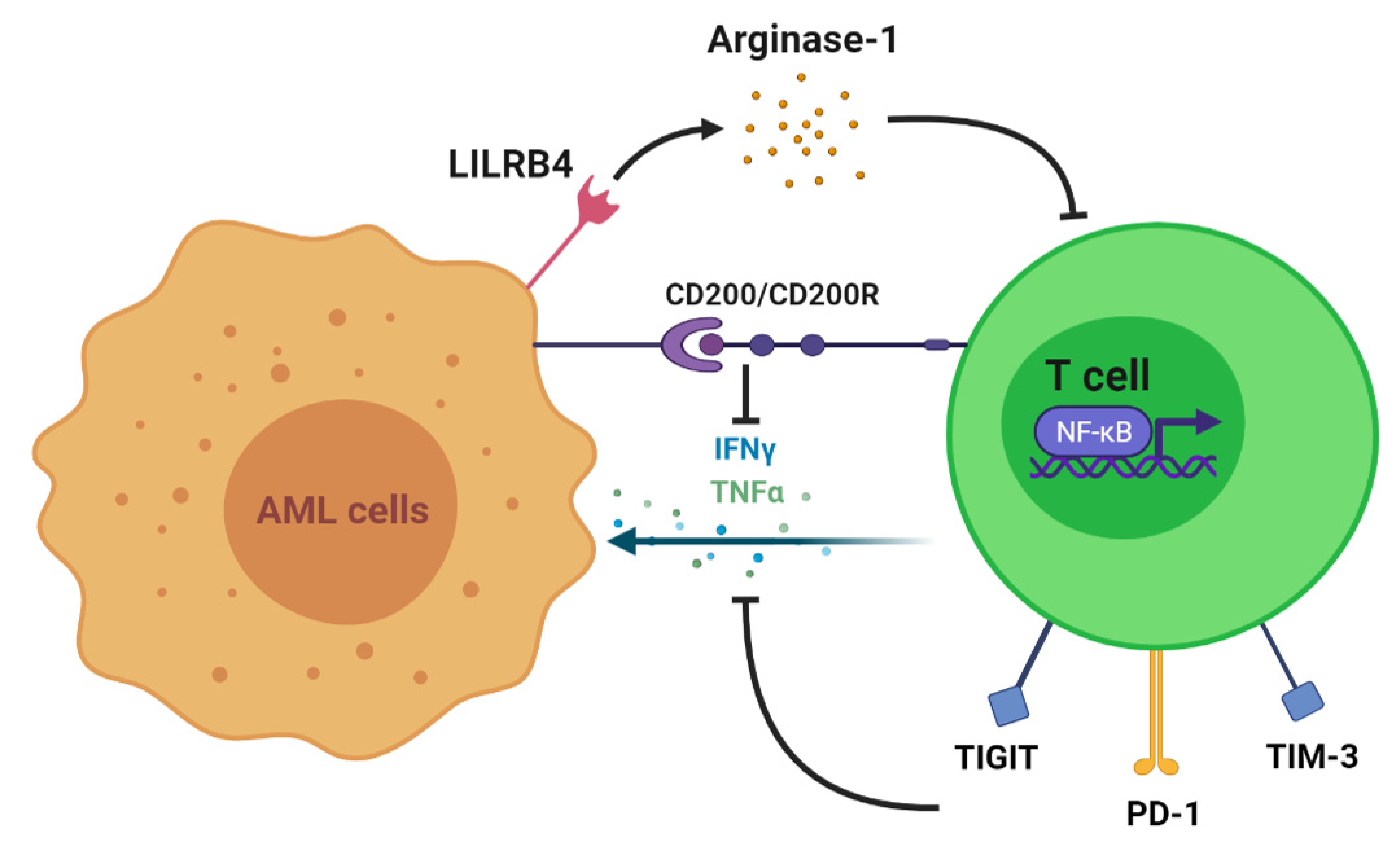
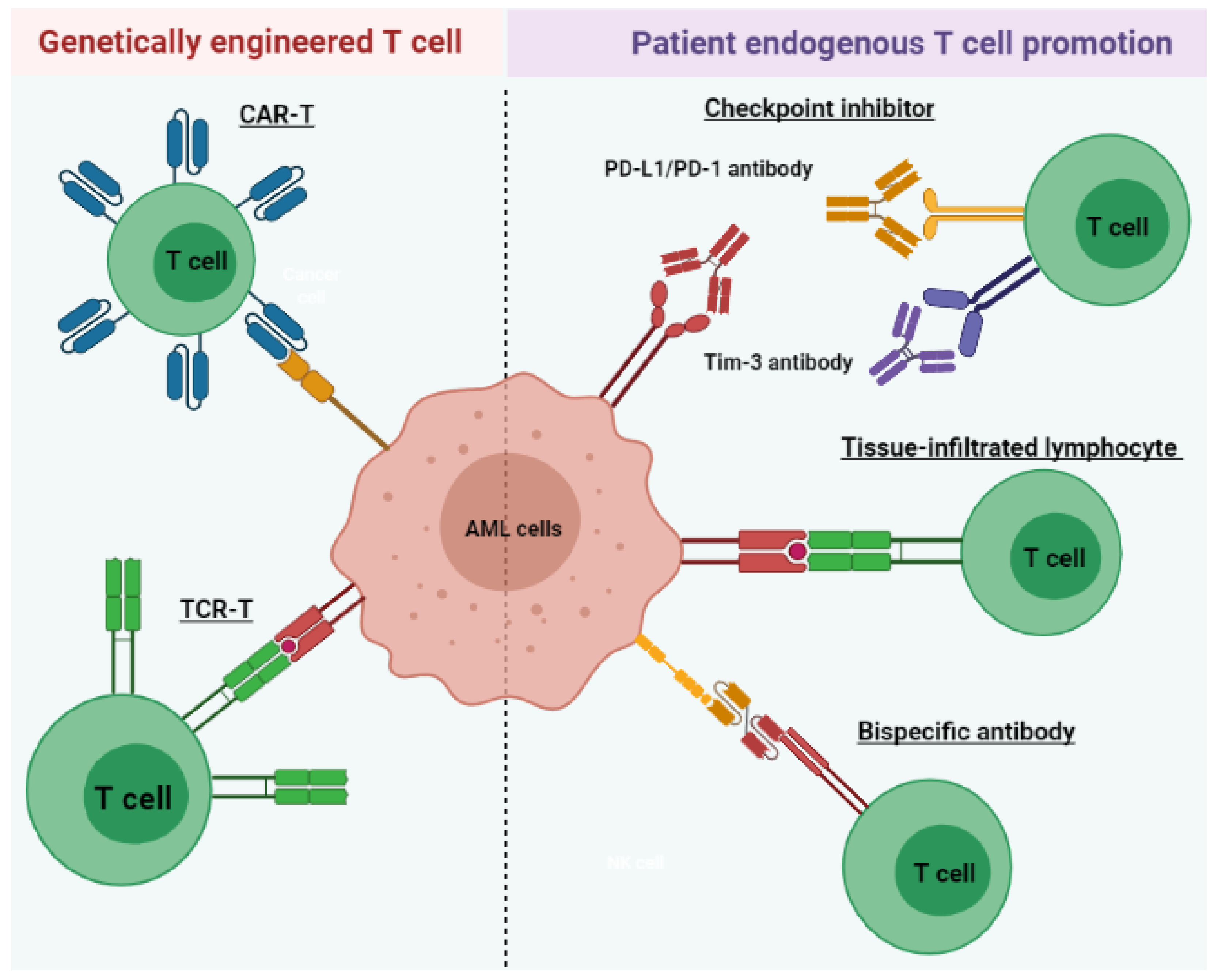
Acute myeloid leukemia (AML) is a heterogeneous disease associated with various alterations in T cell phenotype and function leading to an abnormal cell population, ultimately leading to immune exhaustion. However, restoration of T cell function allows for the execution of cytotoxic mechanisms against leukemic cells in AML patients. Therefore, long-term disease control, which requires multiple therapeutic approaches, includes those aimed at the re-establishment of cytotoxic T cell activity. AML treatments that harness the power of T lymphocytes against tumor cells have rapidly evolved over the last 3 to 5 years through various stages of preclinical and clinical development. These include tissue-infiltrated lymphocytes (TILs), bispecific antibodies, immune checkpoint inhibitors (ICIs), chimeric antigen receptor T (CAR-T) cell therapy, and tumor-specific T cell receptor gene-transduced T (TCR-T) cells.

  • acute myeloid leukemia
  • T cell immunotherapy
  • T cell alteration
  • CAR-T
  • TCR-T

1. Introduction

Acute myeloid leukemia (AML) is a heterogeneous disease characterized by infiltration of immature myelogenous cells in the blood, bone marrow (BM), and other tissues, which ultimately results in ineffective hematopoiesis and various symptoms of leukemic disease. Because the disease is composed of various cytogenic and molecular abnormalities, individual treatment strategies are complex and require multiple approaches [1,2].
Despite numerous studies aimed at harnessing the power of the natural immunologic antileukemic response, there has been no significant advancement made in the standard treatment regimen in over 50 years [3]. Current treatment consists of a high-intensity induction phase wherein cytotoxic chemotherapy, commonly the “7+3 regimen” of cytarabine plus anthracycline, or other target-specific agents are administered based on disease profile as well as patient risk and comorbidities [3,4,5]. Complete remission (CR) may be attained in approximately 70–80% of patients < 60 years of age or in 50% of patients 60 or older [6]. However, due to frequent residual disease and potential for sequential relapse, a consolidation phase often follows that may include additional chemotherapy, specific antigen-targeting therapies, myeloablative conditioning, or either autologous or allogeneic hematopoietic stem cell transplantation (HSCT) depending on the presence of certain molecular and cytogenetic abnormalities [3,4,6,7].
For those with a poor disease prognosis, allogeneic HSCT is the first-line treatment as it can offer disease cure. It is preferred to autologous HSCT, which demonstrates higher rates of relapse due to various mechanisms of immune escape [8,9,10]. The success of allogeneic HSCT is recognized as a graft-versus-leukemia (GvL) effect, which is mainly attributed to the ability of donor T lymphocytes to target recipient leukemia cells [11]. Donor lymphocyte infusions emphasize the role of T cells in GvL as demonstrated by their effectiveness in relapsed chronic myeloid leukemia (CML) and potential to lower relapse incidence in high-risk AML patients [12,13]. However, despite these advantages, significant side effects of allogeneic HSCT exist, namely, graft-versus-host disease (GvHD), which can be fatal or result in chronic health consequences. Thus, various priming strategies are being investigated to preserve the GvL effect of T cells while avoiding complications encompassing GvHD [10,14,15].
The promising role of T lymphocytes in eradicating leukemic disease has led to the recent development of therapeutics that utilize this cytotoxic potential while minimizing risk for treatment-related morbidity and mortality as well as disease relapse.

2. T Cell Alteration in AML

In the presence of AML, multiple clinical studies have demonstrated various disruptions in T cell immunity, including augmented T regulatory cells and reduced T helper cells, T cell exhaustion, and dysregulated activity of transcription factors. To begin with, T cell numbers and functions are altered to allow the progression of myeloid disease. In vitro studies show that coculture with AML cells and incubations with AML plasma inhibit the proliferation of T cells [16,17]. One proven mechanism of T cell growth inhibition is the expression of AML surface receptors that interact with T cells and alter their function. This includes leukocyte immunoglobulin-like receptor B4 (LILRB4), an inhibitory immune checkpoint receptor restrictively expressed on monocytic leukemic cells (M4 and M5 subtypes) (Figure 1). LILRB4 mediates the release of arginase-1, thus directly suppressing T cell proliferation and cytotoxicity in vitro and in vivo [18]. Another immune-suppressive molecule, CD200, is upregulated on the surface of AML cells. CD200 is a type I membrane glycoprotein belonging to the immunoglobulin superfamily whose interaction with the T cell CD200 receptor (CD200R) is associated with both increased regulatory T cell (Treg) populations and reduced memory T cell function [19] (Figure 1). This is one of several mechanisms by which the AML tumor microenvironment may alter subsets of T cells to suppress the immune response. A higher frequency of Tregs impairs the cell-mediated antileukemic immune response [20]. This is supported by an AML mouse model demonstrating an increased frequency of Tregs in vivo in addition to in vitro suppression of effector T cell function. However, with Treg depletion, in vitro effector T cell function was restored, and in vivo treatment outcomes were improved. These results suggest that the expansion of Tregs may be a mechanism of AML persistence [21]. Moreover, increased numbers of Tregs have been identified in AML patients when compared with healthy controls [20]. Shenghui et al. compared the peripheral blood (PB) and BM of 182 patients with newly diagnosed AML with those of 20 healthy age-matched controls [22]. The frequency of Treg cells in PB from patients with AML was significantly higher than that found in the disease-free counterparts (9.2% versus 5.44%, p < 0.001). Furthermore, another T cell subset, helper T cells, has demonstrated reduced numbers in AML. T helper 1 (Th1) cells predominantly secrete interferon γ (IFN-γ) and thus play a role in host defense against intracellular pathogens by promoting macrophage activation at sites of inflammation. Th1 cells exhibit a reduced frequency as well as a decreased expression of IFN-γ in AML patients compared with healthy controls (21.03% versus 11.27%, p < 0.001) [23]. Patients who obtained CR following standard induction chemotherapy exhibited a significant increase in Th1 cells as opposed to newly diagnosed patients. Additionally, increased frequencies of Th17 cells were identified in the PB and BM of AML patients when compared with healthy donors. Th17 cells secrete interleukin-17 (IL-17), which promotes the proliferation of AML cells and inhibits the differentiation of Th1 cells in vitro. Patients with higher frequencies of Th17 cells had poorer survival than those with lower numbers as well [24,25].
Cells 10 03376 g001
Figure 1. Mechanisms of T cell suppression and dysfunction induced by AML. LILRB4 is an inhibitory receptor present in monocytic AML that directly suppresses T cell proliferation. T cell proliferation and activation are also hindered due to the downregulation of transcription factors, including NFκB, by the tumor microenvironment. Upregulated expressions of checkpoint molecules, such as Tim-3, PD-1, and TIGIT, on T cells and interactions between CD200 and CD200R reduce the secretion of IFN-γ and TNF-α, thus promoting immune evasion.

3. T Cell Immunotherapy

Given the effective antileukemic properties of T cells, current immunotherapies aim to restore weakened T cell activity in AML patients through multiple strategies (Figure 2). In this section, we will discuss such therapeutics under clinical investigation for potential AML therapy. These include those that utilize autologous T cells and spare the use of cell modification, including tissue-infiltrating lymphocytes (TILs), bispecific antibodies, and immune checkpoint inhibitors (ICIs). We will also briefly cover the updated information of artificially engineered CAR-T cell therapy and TCR-T cell treatment. Clinical trials of these immunotherapies are summarized in Table 1.
Cells 10 03376 g002
Figure 2. Immunotherapeutic strategies include the promotion of endogenous patient T cells and genetically engineered T cells for AML treatment. Checkpoint inhibitors prevent immune-suppressive signaling through PD-1/PD-L1 and Tim-3. TILs express surface receptors (green) that specifically target AML antigens and exhibit increased cytotoxicity. Bispecific antibodies bind and cross-link leukemic antigens to T cells, facilitating their antileukemic activities. CAR-T or TCR-T provides another strategy to improve leukemia-directed T cell activity via genetic modification of the T cell surface. TCR-T cells express receptors specific for a tumor-associated antigen presented in MHC molecules, similar to TIL. CAR-T cells express receptors (blue) targeting nonspecific, naturally occurring antigens.
Table 1. Clinical trial information of T cell immunotherapies.
Name Target CheckPoint/Antigen Clinical Trial Start Time Trial Number/Phase Disease Status
T cell-recruiting bispecific antibody CD3 and CD33 2015 NCT02520427/Phase 1 Relapsed/Refractory AML Recruiting; estimated completion on 28 February 2023
2017 NCT03224819/Phase 1 AML Active; estimated completion on 21 March 2022
2017 NCT03144245/Phase 1 AML Completed on 5 November 2020; no result posted
2019 NCT03915379/Phase 1 AML and myelodysplastic syndromes Recruiting; estimated completion on 26 October 2022
2018 NCT03516760//Phase 1 Relapsed/Refractory AML Active; estimated completion on 31 December 2020
Immune checkpoint inhibitor PD-1 2015 NCT02397720/Phase 2 AML Recruiting; estimated completion on 30 April 2022
2016 NCT02845297/Phase 2 AML Active; estimated completion in October 2021
2020 NCT04214249/Phase 2 AML Not yet recruiting; estimated completion on 31 July 2024
Chimeric antigen receptor T cell therapy CD123 2015 NCT02159495/Phase 1 AML Recruiting; estimated completion on 15 December 2021
CD33/Lewis Y 2013 NCT01864902/Phase 1 and 2 Relapsed/refractory AML Unknown recruiting status; estimated completion in April 2017
Tumor-specific T cell receptor gene-transduced T cells WT 1 2012 NCT01640301/Phase 1 and 2 Recurrent and secondary AML Active; estimated completion on 1 September 2029
2002 NCT00052520/Phase 1 and 2 AML Completed in June 2013; no result posted

4. Conclusions

In conclusion, significant progress has been made regarding understanding the complexity of the immune biology of AML in addition to noteworthy advancements in the technical development of novel T cell-based AML therapeutics. Despite numerous ongoing trials, T cell immunotherapies for myeloid malignancies remain at an elementary stage. We predict that clinical advancements in immunotherapy will be facilitated by an increased focus on the analysis of potential biomarkers as therapeutic targets. This will aid in elucidating the mechanisms of immune resistance, predicting patient response, and allow for improved management of undesired toxicities. Other than biomarker-driven approaches, the application of novel genomic and cellular techniques, such as single-cell genome sequencing, single-cell cytokine analysis, and mass cytometry, using patient samples will help to reveal additional T cell roles in the tumor microenvironment. These methods may also lead to the discovery of immune components other than cell-mediated that contribute to the immune response as well as escape, ultimately guiding the development of innovative immune therapy strategies. To illustrate, single-cell TCR sequencing generates a TCR repertoire encompassing a wide array of T cell populations in terms of antigen specificities. T cells expressing specific receptors that recognize MHC on tumor cells are likely to characterize TILs. These TCR sequences may identify TIL markers to be used for TIL isolation and amplification. The sequences generated can also be used to identify novel target antigens for personalized immunotherapy and vaccines. Thus, in the near future, we expect promising clinical outcomes for T cell immunotherapies in AML.

This entry is adapted from the peer-reviewed paper 10.3390/cells10123376

This entry is offline, you can click here to edit this entry!
Video Production Service