S1P in Multiple Sclerosis: History
Please note this is an old version of this entry, which may differ significantly from the current revision.
Subjects: Neurosciences

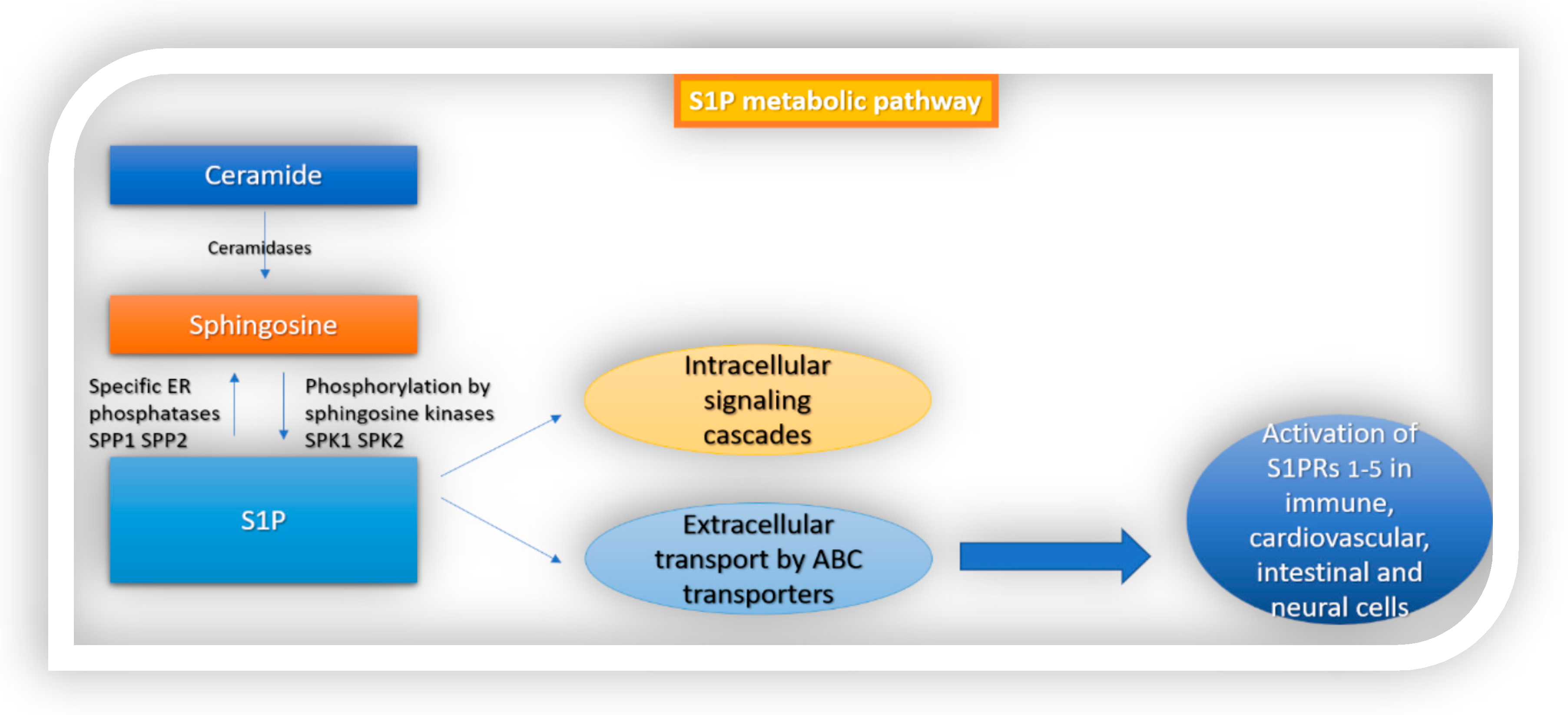
Sphingosine 1-phosphate (S1P) is a signaling molecule with complex biological functions that are exerted through the activation of sphingosine 1-phosphate receptors 1–5 (S1PR1–5). S1PR expression is necessary for cell proliferation, angiogenesis, neurogenesis and, importantly, for the egress of lymphocytes from secondary lymphoid organs. Since the inflammatory process is a key element of immune-mediated diseases, including multiple sclerosis (MS), S1PR modulators are currently used to ameliorate systemic immune responses. 

  • sphingosine 1-phoshate
  • sphingosine 1-phosphate receptors
  • multiple sclerosis

1. Introduction

Sphingolipids, including sphingomyelin and its metabolites, are structural components of all cell membranes and of the myelin sheath in the nervous system. They were discovered by J.Thudichum in 1874 [1] but it was not until 1997, when the specific, high-affinity G-protein-coupled receptors for the sphingomyelin metabolite sphingosine 1-phosphate (S1P) were identified [2][3], that their multiple physiologic roles in the human body started to be recognized. Subsequent molecular and physiologic research studies suggested that the S1P system mediates various intracellular signaling cascades in the CNS, cardiovascular and immune systems [4]. S1P is a bioactive lipid second messenger that has important signaling functions in cell growth, cell proliferation and angiogenesis. It also regulates biological functions in health and disease [5][6]. S1P concentrations are low in intracellular and interstitial fluids and increased within blood and lymph, in the sub-micromolar range, therefore creating a S1P gradient, which is important for regulating physiologic actions such as lymphocyte egress from secondary lymphoid organs. Multiple enzymes are essential for maintaining the S1P gradient between tissues and systemic circulation and a dynamic equilibrium between S1P and sphingosine. S1P is a lipid metabolite of ceramide. Ceramide can be either broken down by ceramidases to sphingosine, or phosphorylated in the Golgi apparatus by ceramide kinase to produce ceramide-1-phosphate (C1P), another sphingolipid metabolite. In contrast with ceramide and sphingosine, which are considered to activate an apoptotic response, S1P is associated with the suppression of apoptosis [7]. S1P is produced intracellularly through the phosphorylation of sphingosine by sphingosine kinases (SphK1, SphK2), or extracellularly through the hydrolysis of sphingosyl phosphorylcholine by autotoxin. SphK1 is mainly responsible for the cytosolic and extracellular S1P, has trophic functions and can be upregulated in response to proinflammatory cytokines. In contrast, SphK2 can translocate into the nucleus and has the capacity to enhance apoptosis [5]. Following its production, S1P interacts with intracellular targets or is transported extracellularly by ABC transporters, in autocrine or paracrine cell targets, to activate the G-protein-coupled receptor named sphingosine 1-phosphate receptors 1–5 (S1PRs 1–5) (Figure 1). S1P is converted to sphingosine by S1P-specific ER phosphatases (SPP1 and SPP2) or degraded by S1P lyase (S1PL) [5][8][9].
Figure 1. S1P metabolic pathway. S1P is formed from ceramide. Ceramidase converts ceramide to sphingosine, which is then phosphorylated by sphingosine kinases 1 and 2 to S1P. Following its production, S1P interacts with intracellular targets or is transported extracellularly by ABC transporters. Finally, it activates sphingosine 1-phosphate receptors 1–5 (S1PRs 1–5) which are ubiquitously expressed in different cell types in the body.

2. S1P Signaling in the Immune System

S1P signaling plays a multifactorial role in immunity. Initially recognized and well-described is the role of S1P−S1PR1 modulation of T-cell trafficking, which has a crucial impact on adaptive immunity [10]. Recent findings suggest, however, additional roles of S1P signaling in B-cell trafficking [7] and also in innate immunity [11]. S1PR1 is widely expressed by T cells, B cells, macrophages, neutrophils, dendritic cells, monocytes, eosinophils, mast cells and NK cells. S1PR2 is expressed on macrophages, monocytes, and eosinophils and mast cells. S1PR3 is expressed on macrophages, neutrophils during inflammation, dendritic cells, monocytes, eosinophils and mast cells. S1PR4 is expressed on macrophages, neutrophils, dendritic cells, monocytes, eosinophils and mast cells. S1PR5 is expressed on patrolling monocytes and NK cells [11][12].

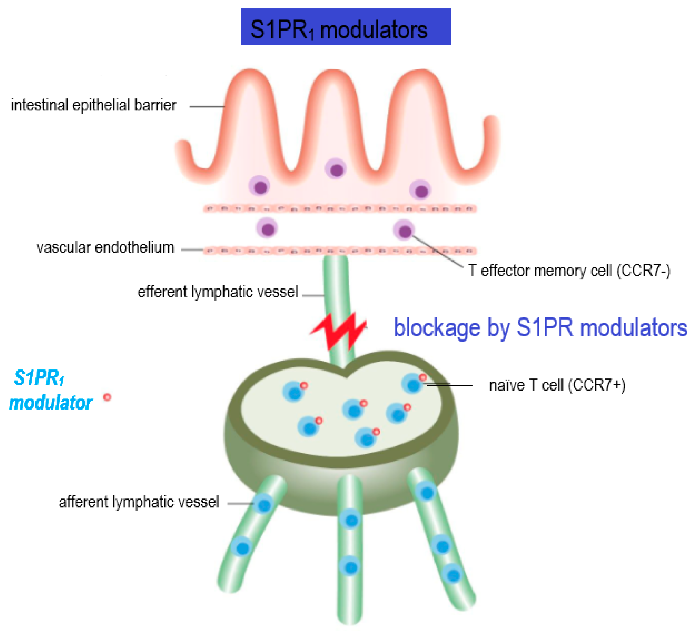
2.1. Lymphocyte Trafficking and Regulation of Adaptive Immune Functions

S1PR1 expression is required for B and T cells to egress from lymph nodes, for the exit of mature T cells from the thymus and for the migration of natural killer T cells from secondary lymphoid organs into the circulation [13]. The circulation of naïve B and T lymphocytes between the blood and lymphatic systems is important for various functions of the immune system and is orchestrated by S1P and S1PR1.
Described above, the positive S1P gradient between lymphoid organs and the lymphatic circulation is utilized by T lymphocytes to egress from thymus and peripheral lymphoid organs into the lymphatic circulation [7][14]. However, the entry into secondary lymphoid organs does not require S1P signaling. Studies of S1PR1 knockout mice in hematopoietic cells reported significant lymphopenia, resulting from intact lymphocyte maturation but the inability of the last to egress from thymus and peripheral lymphoid organs [15]. Similarly, the inhibition of sphingosine kinase 1/2 or S1P lyase results in lymphopenia, by a similar mechanism [14][16].
As mentioned above, not all T cell subpopulations’ trafficking is affected by S1P signaling within the lymph nodes. S1P−S1PR1 action can overcome the retention signals mediated by the chemokine receptor CCR7, which is expressed by naïve T cells, central memory T cells, and B cells. In contrast, terminally differentiated effector T cells and effector memory T cells do not express CCR7 receptors, therefore they have the capacity to egress from lymph nodes independent of S1P−S1PR1 signaling [7][17][18]. This specific property of T effector and T effector memory cells to overcome S1P regulation has crucial implications in clinical practice, where the administration of S1P receptor modulators, such as fingolimod does not inhibit these cells from recirculating to peripheral tissue and performing important immunosurveillance functions [7] (Figure 2).
Figure 2. Mechanism of action of S1PR modulators. The figure illustrates the blockage of T cell egress by S1PR modulators, which is the core of their therapeutic effect in MS. CCR7, a chemokine receptor expressed in naïve and central memory T cells, retains lymphocytes within lymph nodes by overcoming S1P1-mediated immune egress. Following activation and clonal expansion, T cells switch to a state favoring egress over retention by downregulating CCR7. Importantly, effector memory cells do not express CCR7 receptors and have the ability to overcome S1P regulation.

2.2. Regulation of Innate Immune Cell Trafficking

S1P signaling controls trafficking of the other immune cells, including dendritic cells [10][19], natural killer cells [20][21] and hematopoietic stem cells [14][22][23]. S1PR1 is responsible for neutrophil migration [17]. In addition, it mediates neutrophil recruitment, as was demonstrated through a rat model of hyperalgesia [24], and a model of Candida albicans-induced vasculitis, where the administration of an SPR1 agonist, ONO-W061 suppressed neutrophil recruitment [25].
In innate cells, S1PR3 is present on multiple cell subtypes. It promotes dendritic cell maturation. It drives macrophage chemotaxis and killing, as shown by studies in vitro and in vivo where S1PR3-mediated macrophage action caused atherosclerosis via altering smooth muscle cell behavior [26]. S1PR3 also mediates neutrophil and eosinophil recruitment in models of asthma [7]. It also drives leukocyte rolling on endothelial cells [11]. S1PR4 is expressed on most immune cells, and current research suggests a role in plasmacytoid dendritic cell differentiation and activation, as evidenced by studies in S1PR4 knockout mice [27]. It regulates neutrophil migration from blood to tissue [17][23], and has important roles in macrophages as well. Interestingly, it was found that S1PR4 is the only receptor that displays a significant difference in its expression between M1 and M2 states of activated macrophages, suggesting an important role for this receptor in multiple macrophage actions as migration and cytokine release [28]. Last, the S1PR4-mediated regulation of cytokine production by myeloid cells appears to be the central function of this receptor, compared to its less active role in immune cell migration [23]. The co-expression of CXCR4 and S1PR5 is a prerequisite for mature natural killer cells to leave the lymph nodes and bone marrow [11][21]. S1PR5 is also required for the recruitment of NK cells to sites of inflammation [7][11]. Patrolling monocytes, similar to NK cells, require S1PR5 to egress from the bone marrow. However, unlike T cells, they do not require S1P gradient for their trafficking, suggesting that S1PR5-mediated trafficking is controlled by a different mechanism [11][29]. Recent findings have suggested that S1PR1-myeloid cell signaling is considered to impact the initiation and progress of CNS autoimmunity [30]. Studies have focused on unravelling their role in CD14 and CD16 myeloid cells [31]. (Table 1)
Table 1. S1P receptor expression in cells of immune, gastrointestinal and nervous system and associated cell functions.

Receptor

Associated Cell Types

Functions (Described up to Date)

S1PR1

Immune system:

T cells, B cells

Macrophages

Neutrophils

DCs

Monocytes

Eosinophils

Mast cells

NK cells

Egress from lymph nodes, exit of mature T cells from thymus, migration of natural killer T cells from secondary lymphoid organs to circulation, transfer of immature B cells from bone marrow to circulation

Macrophage recruitment

Neutrophil migration, recruitment

Trafficking of DCs

Trafficking of monocytes

Eosinophil recruitment

Mast cell recruitment

GI:

IECs

Upregulation of intestinal barrier proteins (claudin1, occludin)

CNS:

Astrocytes

Oligodendrocytes

Neurons

Microglia

Activation, differentiation, proliferation of astrocytes, astrogliosis

Differentiation of oligodendrocytes, process extension, survival of oligodendrocyte progenitors, myelination

Growth cone formation, enhancement of neurite extension, synaptic transmission

S1PR2

Immune system:

Macrophages

Monocytes

Mast cells

Eosinophils

Enhance antibody-mediated phagocytosis, inhibit phagocytosis of bacteria and fungi

Degranulation of mast cells

GI:

IECs

Upregulation of c-Myc, cyclin D1, E-cadherin and Zona occludin 1, proliferation of IECs, absorption of NaCl, prevention of IECs apoptosis

CNS:

Neurons

Microglia

Growth cone formation, inhibition of neurite extension, control of neural excitability

S1PR3

Immune system:

Macrophages

Monocytes

Neutrophils

DCs

Eosinophils

Mast cells

Leucocyte rolling on endothelial cells

Macrophage’s chemotaxis and killing

Neutrophil recruitment

DC maturation

Eosinophil recruitment

GI:

IECs


CNS:

Astrocytes

Microglia

Activation, differentiation, proliferation of astrocytes, astrogliosis

S1PR4

Immune system:

Macrophages

Monocytes

Neutrophils

DCs

Eosinophils

Mast cells

Macrophage migration and cytokine release

Neutrophil migration

Plasmacytoid DC activation and differentiation

GI:

IECs


CNS:


S1PR5

Immune system:

Patrolling monocytes

NK cells

Egress of mature NK cells from lymph nodes and bone marrow

GI:

IECs


CNS:

Mature oligodendrocytes

Neurons

Cell survival, process retraction, inhibition of OPC migration, myelination

Growth cone formation, inhibition of neurite extension

3. S1P Signaling in the Intestine

The gastrointestinal (GI) tract is the largest epithelial barrier that protects the human body from the external environment. Preservation of an intact intestinal mucosal barrier is important for GI functions including digestion of food, absorption of nutrients, expulsion of waste, and protection against hostile bacteria [32]. The proliferation and migration of intestinal epithelial cells (IECs) must be carefully balanced and regulated [33]. This way, the body can preserve the integrity of the intestinal mucosal barrier and manage to maintain the protection of the host from the environment but also the communication between host and its environment. These functions are regulated by a variety of signaling molecules [34]. The intestinal immune system must preserve its immunological homeostasis. The disturbance of the intestinal immune function causes the outbreak of allergic, inflammatory, and infectious diseases [35][36]. S1P is a biolipid mediator that controls various cell functions, as described in detail above. In the gastrointestinal system, S1P has important roles in maintaining intestinal epithelial cell barrier structural and functional integrity, regulating IEC proliferation, migration and apoptosis. It also mediates immunological actions like immunoglobulin A production and T cell trafficking [32][35][36]. In general, S1P is a byproduct of the catalyzation of sphingomyelin and is produced mainly by platelets, erythrocytes, and endothelial cells in mammals. However, in the intestinal tissue the production of S1P is mainly due to epithelial cell activity.
Interestingly, it has been suggested that the levels of dietary sphingomyelin intake and the activity of the enzymes sphingomyelinase and sphingosine kinase may influence the incidence and severity of intestinal inflammatory processes [35][37][38][39]. Furthermore, cholesterol ingested by food antagonizes the intestinal absorption of sphingolipids. Thus, it is implied that modern Western diets, rich in cholesterol, may promote S1P-mediated intestinal immunity dysfunction and the subsequent genesis of inflammatory disorders, through its impact in reducing the availability of S1P precursors [36][40]. Recent studies have brought up evidence supporting the role of S1P signaling in the activation of two critical transcription factors, nuclear factor kappa B (NF-κB) and signal transducer and activator of transcription 3 (STAT3). These transcription factors are known to regulate key inflammatory signaling pathways that promote colon inflammation and carcinogenesis in the intestinal as well as in other human systems [41].

Implication in Intestinal Barrier Homeostasis

S1P is found abundantly in the human intestine. All five S1PRs are expressed in intestinal epithelial cells, as indicated by the findings from the Human Protein Atlas, but the expression level of each one varies, with the highest being that of S1PR2 [42]. The role of S1P in intestinal epithelial barrier integrity has been investigated, but still not all its functions have been revealed. Greenspon et.al have demonstrated that, following S1P administration in differentiated rat IECs, intestinal barrier function was enhanced. Treatment with S1P led to an increase in expression of an adherens junction protein, E-cadherin, and also in more efficient redistribution of this protein on the barrier. Increased abundance of S1P, resulting from upregulated expression of SphK1 in IECs, was also related to augmented levels of other barrier proteins including claudin-1 and occludin. These effects were attributed to activation of S1PR1 [43]. Chen et al. have shown that S1P also upregulates the expression of c-Myc, cyclin D1, E-cadherin and zona occluden-1 (ZO-1) via activation of S1PR2 [34]. These effects result in maintaining the integrity of the intestinal epithelial cell barrier. The above authors later conducted further research experiments, using an S1PR2-knockout mice model, with the administration of Dextran sulfate sodium (DSS) to induce colitis. The pivotal role of S1P-mediated S1PR2 activation in regulating intestinal barrier structural integrity and function was reaffirmed. It was demonstrated that the S1P/S1PR2 axis in IECs prevented intestinal barrier damage by mediating (a) CD4+T-cell activation via the ERK pathway and (b) MHC-II expression. In addition, they found that IFN-γ, secreted by CD4+T cells, augmented DSS-induced damage of the intestinal barrier function. This was done through downregulation of ZO-1 by IFN-γ. On the other hand, S1P was capable of restricting DSS/IFN-γ-induced damage of intestinal mucosa permeability by increasing ZO-1 [44]. Additionally, S1P has been found to promote intestinal epithelial cell proliferation and migration, in a dose-dependent manner. This is achieved through S1P-mediated activation of ERK1/2 via S1PR2 [34]. Furthermore, S1P-mediated activation of S1PR2 leads to increased expression of down-regulated in adenoma (DRA), the major Cl-/HCO3-exchanger that regulates NaCl absorption in the intestine of mammals [45]. MEK/ERK activity controls JNK activation via regulating MAPK phosphatase (MKP-1), which prevents cell apoptosis. S1P enhances the ERK1/2 and Akt signaling pathways thus preventing intestinal epithelial cells’ apoptosis [46][47] (Table 1).

4. S1P Signaling in the CNS

S1P is widely present in the CNS, where it is released by cerebral sphingosines and acts on S1P receptors. All S1PRs except S1PR4 are expressed in the CNS by neuronal and glial cell populations and also in the cerebral vascular system, to varying degrees. Their activation by S1P mediates numerous physiologic processes involved in neuronal plasticity including myelination, neurogenesis and neuroprotection [48]. Evidence from animal models and studies in vitro suggest that S1P regulates multiple physiologic functions in the CNS and that the expression of S1PRs is dynamic, influenced by temporal and spatial factors and stimuli of the cell environment [4][49][50].
In neurons, S1P signaling has an active role in neural progenitor migration, synaptic activity (=neurotransmission), differentiation, process extension, calcium signaling and survival [51]. S1P stimulates neurogenesis more effectively than fibroblast growth factor (FGF) [52]. S1P binding to S1PR1, S1PR2 and S1PR5 participates in the regulation of growth cone formation, neurite extension and retraction. In vitro experiments of stimulation of neurons with nerve growth factor (NGF) showed the resulting enhancement of neurite extension which is mediated by S1PR1. Opposingly, the activation of S1PR2 and S1PR5 inhibits neurite extension [53]. Several researchers have addressed the role of S1P-mediated circuits in neural progenitor stem cells (NPSCs). The effect of S1P on NPSCs is mediated by increased laminin expression and extracellular matrix (ECM) interactions with progenitor integrins [54]. NPSCs that migrate out of the embryoid body stem cells (ESCs) are considered to upregulate S1PR1 [55]. However, there are important variations between studies concerning the description of S1PR expression in NPSCs [52]. S1PR1 also affects plasma membrane excitability and neurotransmitter release, thus promoting synaptic transmission, as demonstrated in studies with rat hippocampus models [56][57]. Control of neural activity has been attributed to S1PR2 signaling [58], as models of S1PR2-knockout mice have resulted in excess neural excitability and the occurrence of seizures at 3–7 weeks of age [51][59]. The regulation of synaptic activity, neurogenesis and cell survival is such an important factor that these receptors have attracted interest as potential drug targets in memory disorders and neurodegenerative diseases.
Oligodendrocytes and oligodendrocyte precursor cells (OPCs) widely express S1PRs, and the level of each S1PR’s expression depends on the cells’ stage of development and on the myelinating state [49][60][61]. In mature oligodendrocytes, S1PR5 expression is highest and is thought to promote cell survival via the S1PR5-mediated Akt signaling pathway and inhibit OPC migration via the S1PR5-mediated Rho GTPase/Rho kinase pathway [62], whereas it is assumed that early stages of differentiation are mainly mediated by S1PR1 signaling [50]. Indeed, various authors suggest that higher expression level and activation of S1PR1 induce the differentiation of oligodendrocytes along with the survival of oligodendrocyte progenitors [52][63]. Regarding the role of S1P in oligodendrocyte morphology, there is an interesting dual action. Via the modulation of S1PR5, it enhances process retraction [64], while via S1PR1, it promotes process extension, indicating reciprocal effects on cytoskeletal elements. S1PR5 is also considered to have a pivotal role in myelination, although data from S1PR5-null mice showed that myelination was normally developed despite the lack of S1PR5 expression [64]. On the other hand, the targeted deletion of S1PR1 in oligodendrocyte lineage cells led to abnormal formation of myelin and increased susceptibility to cuprizone-induced demyelination [65]. This finding suggests an important role for S1PR1 in normal myelination of the brain. S1P actions on oligodendrocytes and OPCs are also influenced by the action of neurotrophic factors and the lysophosphatidic acid receptors expressed on these cells, such as neurotrophin-3 and platelet-derived growth factor. It seems very probable that the process of myelination is coordinated with the contribution of all the above cellular structures [51][66]. Altogether, interactions between S1P and S1PRs in oligodendrocytes and OPCs control cell survival, migration and differentiation, maintenance of cell morphology, myelination and remyelination.
S1P signaling mediates various functions in astrocytes, including differentiation [52][67], proliferation, migration, gap junction communication, growth factor production and astrogliosis [50][51]. Astrocytes mainly express S1PR1 and S1PR3, with scarce expression of S1PR2. S1PR5 expression by astrocytes was detected during in vitro studies, when exposed to growth factors [68]. Astrocyte proliferation is stimulated by S1P via activation of the extracellular-signal-regulated kinase (ERK) pathway [69]. Observations of activated astrocytes in response to pathogens suggest that they overexpress S1PR1 and S1PR3, pointing to a role of these receptors in astrogliosis [70]. Studies in S1PR-knockout mice also suggest that astrogliosis requires the activation of S1PR1 and S1PR3 [71]. Some research groups support that S1PR3 is overexpressed in astrocytes under proinflammatory conditions [52][67]. Interestingly, S1P antagonism by fingolimod reduces the production of the inflammatory chemokines CXCL5/LIX, C-X-C motif chemokine 10 (CXCL10) and monocyte chemoattractant protein-1 (MCP-1) in astrocytes and microglial cultures [72]. Therefore, S1P signaling in astrocytes seems to mediate both proinflammatory and anti-inflammatory effects.
S1PR1, S1PR2 and S1PR3 are also expressed by microglia. Microglia exist in a nonactivated state and switch to an activated state when stimulated by proinflammatory cytokines. The expression of S1PRs in microglia cells depends on their activation state [73]. Extracellular S1P is a powerful chemoattractant for microglial cells in the brain [52]. S1P levels have been shown to increase at sites of brain damage, where microglia and neural progenitors accumulate. S1PR1 and S1PR3 are downregulated in activated microglia, and S1PR2 is upregulated. In models of experimental autoimmune encephalomyelitis (EAE), S1PR1 deletion was correlated with reduced activation of microglia cells [72]. (Table 1)

Implication in BBB Homeostasis

Plasma S1P-knockout mice display increased morbidity due to increased vascular leak and anaphylaxis following the administration of platelet-activating factor or histamine [74]. Likewise, pharmacologic blockage of S1PR1 or ApoM deficiency leads to compromised vascular integrity and excess inflammation [75]. Brain endothelial SP1 signaling also supports the BBB integrity by arranging the localization of tight junction proteins [76]. Maintenance of vascular integrity requires proper rearrangements of the cytoskeleton as well as the assembly of adherens junctions in endothelial cells. S1P promotes the structural integrity of the actin cytoskeleton, the accumulation of VE-cadherin and α-, β- and γ-catenin at the sites of adjacent cell contact, and adherens junction assembly [77]. The activation of small G proteins Rac and Rho downstream of the S1PR1 and S1PR3 signaling pathways are involved in the processes [77][78]. S1P−S1PR1 signaling is considered to have a pivotal function in regulating the inflammatory status of vascular endothelial cells. Giovani et al. have shown that endothelial S1PR1 abundance was enhanced in regions of vascular inflammation. Additionally, proinflammatory adhesion proteins such as VCAM-1 and ICAM-1 were upregulated in the descending aorta of mice with endothelial cell-specific deletion of S1PR1 and suppressed in mice with endothelial cell-specific overexpression of S1PR1 [79]. These observations indicate that proper S1PR1 localization and signaling are important to maintain vascular homeostasis, and that the impairment of S1PR1 signaling because of receptor internalization predisposes endothelial cells to an inflammatory phenotype [14].

5. Implication for the Role of S1P in Multiple Sclerosis

The ubiquitous presence of S1P and its receptors in the CNS implies that the S1P−S1PR signaling system may be targeted for therapeutic purposes in neurological disorders like MS [80]. MS is a T cell-mediated autoimmune disease of the CNS and its etiology is believed to constitute an interaction between genetic predisposition and environmental factors. MS is classified as follows, according to the clinical course of the disease: relapsing–remitting MS, primary progressive MS, and secondary progressive MS [81].
The activation and clonal expansion of CNS-directed, autoreactive CD8+ T cells, differentiated CD4+ TH1 and TH17 cells, B cells and innate immune cells in the periphery are considered to be the first step in MS pathogenesis [82]. Following this, T cells cross the BBB, with the interference of integrins and, upon entrance, they are reactivated by epitopes on myelin and induce inflammation with the release of cytokines such as tumor necrosis factor alpha (TNFα) and IFNγ. These events lead to increased BBB permeability, demyelination and axonal degeneration [81][83]. After their activation by TNFα and IFNγ, CNS resident microglia and infiltrating macrophages are transferred to the site of inflammation, also contributing to destruction of myelin and prolonging neurodegeneration [84]. The B lymphocytes’ role in the pathogenesis of the disease has attracted vivid interest and thorough research in the last years, and therapeutic agents targeting these cells have shown positive results in limiting disease progress [85]. B cells contribute to MS pathophysiology through multiple mechanisms such as antigen presentation to T cells, transport of antigens from tissues to secondary lymphoid organs and the release of proinflammatory or anti-inflammatory cytokines. Pathogenetic auto-antibodies have been identified in a subgroup of MS patients [86].
Following the acute inflammatory attack, the patient exhibits functional recovery that is attributed to the remyelination of the axons and brain plasticity. However, axonal damage, which is secondary to demyelination and neurotoxic factors released from activated microglia, is not reversible. Accumulating axonal damage leads to neuronal degeneration and brain atrophy, which is the main contributor of progressive irreversible neurological disability [48]. Another residue of the acute inflammatory event is the subsequent chronic inflammation which is restricted within the CNS. This is due to the persistence of activated immune cells in the CNS, despite the absence of infiltrating lymphocytes from the periphery. This chronic inflammatory process affects the whole brain parenchyma, even at sites far from the underlying focal demyelinating lesions. Diffuse chronic CNS inflammation is considered more prevalent in patients with progressive forms of MS [48][87]. Inflammation in MS is thus differentiated both in the space axis (peripheral vs. local CNS inflammation) and in the time axis (acute vs. chronic inflammation). Both inflammatory and neurodegenerative components of the disease are thought to be present from the early stages, and proceed in parallel, but are responsible for different aspects of the clinical phenotype.

This entry is adapted from the peer-reviewed paper 10.3390/cells10113217

References

  1. Thudichum, J.L.W. Treatise on the Chemical Constitution of the Brain: Based Throughout upon Original Researches. Glasgow Med. J. 1884, 22, 363–364.
  2. An, S.; Bleu, T.; Huang, W.; Hallmark, O.G.; Coughlin, S.R.; Goetzl, E.J. Identification of cDNAs encoding two G protein-coupled receptors for lysosphingolipids. FEBS Lett. 1997, 417, 279–282.
  3. Lee, M.J.; van Brocklyn, J.R.; Thangada, S.; Liu, C.H.; Hand, A.R.; Menzeleev, R.; Spiegel, S.; Hla, T. Sphingosine-1-phosphate as a ligand for the G protein-coupled receptor EDG-1. Science 1998, 279, 1552–1555.
  4. Hla, T.; Brinkmann, V. Sphingosine 1-phosphate (S1P): Physiology and the effects of S1P receptor modulation. Neurology 2011, 76, S3–S8.
  5. Rosen, H.; Gonzalez-Cabrera, P.J.; Sanna, M.G.; Brown, S. Sphingosine 1-phosphate receptor signalling. Annu. Rev. Biochem. 2009, 78, 743–768.
  6. Prager, B.; Spampinato, S.F.; Ransohoff, R.M. Sphingosine 1-phosphate signaling at the blood-brain barrier. Trends Mol. Med. 2015, 21, 354–363.
  7. Aoki, M.; Aoki, H.; Ramanathan, R.; Hait, N.C.; Takabe, K. Sphingosine-1-Phosphate Signaling in Immune Cells and Inflammation: Roles and Therapeutic Potential. Mediators Inflamm. 2016, 2016, 8606878.
  8. Marsolais, D.; Rosen, H. Chemical modulators of sphingosine-1-phosphate receptors as barrier-oriented therapeutic molecules. Nat. Rev. Drug Discov. 2009, 8, 297–307.
  9. Mendoza, A.; Bréart, B.; Ramos-Perez, W.R.; Pitt, L.A.; Gobert, M.; Sunkara, M.; Lafaille, J.; Morris, A.J.; Schwab, S.R. The transporter Spns2 is required for secretion of lymph but not plasma sphingosine-1- phosphate. Cell Rep. 2012, 2, 1104–1110.
  10. Cyster, J.G.; Schwab, S.R. Sphingosine-1-Phosphate and Lymphocyte Egress from Lymphoid Organs. Annu. Rev. Immunol. 2012, 30, 69–94.
  11. Bryan, A.M.; del Poeta, M. Sphingosine-1-phosphate receptors and innate immunity. Cell Microbiol. 2018, 20, e12836.
  12. Mao-Draayer, Y.; Sarazin, J.; Fox, D.; Schiopu, E. The sphingosine-1-phosphate receptor: A novel therapeutic target for multiple sclerosis and other autoimmune diseases. Clin. Immunol. 2017, 175, 10–15.
  13. Tsai, H.-C.; Han, M.H. Sphingosine-1-Phosphate (S1P) and S1P Signaling Pathway: Therapeutic Targets in Autoimmunity and Inflammation. Drugs 2016, 76, 1067–1079.
  14. Obinata, H.; Hla, T. Sphingosine 1-phosphate and inflammation. Int. Immunol. 2019, 23, 617–625.
  15. Matloubian, M.; Lo, C.G.; Cinamon, G.; Lesneski, M.J.; Xu, Y.; Brinkmann, V.; Allende, M.L.; Proia, R.L.; Cyster, J.G. Lymphocyte egress from thymus and peripheral lymphoid organs is dependent on S1P receptor 1. Nature 2004, 427, 355.
  16. Schwab, S.R.; Pereira, J.P.; Matloubian, M.; Xu, Y.; Huang, Y.; Cyster, J.G. Lymphocyte sequestration through S1P lyase inhibition and disruption of S1P gradients. Science 2005, 309, 1735–1739.
  17. Allende, M.L.; Bektas, M.; Lee, B.G.; Bonifacino, E.; Kang, J.; Tuymetova, G.; Chen, W.; Saba, J.D.; Proia, R.L. Sphingosine-1-phosphate lyase deficiency produces a pro-inflammatory response while impairing neutrophil trafficking. J. Biol. Chem. 2011, 286, 7348–7358.
  18. Maeda, Y.; Seki, N.; Kataoka, H.; Takemoto, K.; Utsumi, H.; Fukunari, A.; Sugahara, K.; Chiba, K. IL-17-producing Vγ4+ γδ T cells require sphingosine 1-phosphate receptor 1 for their egress from the lymph nodes under homeostatic and inflammatory conditions. J. Immunol. 2015, 195, 1408–1416.
  19. Czeloth, N.; Bernhardt, G.; Hofmann, F.; Genth, H.; Förster, R. Sphingosine-1-phosphate mediates migration of mature dendritic cells. J. Immunol. 2005, 175, 2960.
  20. Walzer, T.; Chiossone, L.; Chaix, J.; Calver, A.; Carozzo, C.; Garrigue-Antar, L.; Jacques, Y.; Baratin, M.; Tomasello, E.; Vivier, E. Natural killer cell trafficking in vivo requires a dedicated sphingosine 1-phosphate receptor. Nat. Immunol. 2007, 8, 1337.
  21. Jenne, C.N.; Enders, A.; Rivera, R.; Watson, S.R.; Bankovich, A.; Pereira, J.; Xu, Y.; Roots, C.M.; Beilke, J.N.; Banerjee, A.; et al. T-bet-dependent S1P5 expression in NK cells promotes egress from lymph nodes and bone marrow. J. Exp. Med. 2009, 206, 2469–2481.
  22. Massberg, S.; Schaerli, P.; Knezevic-Maramica, I.; Köllnberger, M.; Tubo, N.; Moseman, E.A.; Huff, I.V.; Junt, T.; Wagers, A.J.; Mazo, I.B.; et al. Immunosurveillance by hematopoietic progenitor cells trafficking through blood, lymph, and peripheral tissues. Cell 2007, 131, 994.
  23. Olesch, C.; Ringel, C.; Brüne, B.; Weigert, A. Beyond Immune Cell Migration: The Emerging Role of the Sphingosine-1-phosphate Receptor S1PR4 as a Modulator of Innate Immune Cell Activation. Mediators Inflamm. 2017, 2017, 1–12.
  24. Finley, A.; Chen, Z.; Esposito, E.; Cuzzocrea, S.; Sabbadini, R.; Salvemini, D. Sphingosine 1-Phosphate Mediates Hyperalgesia via a Neutrophil-Dependent Mechanism. PLoS ONE 2013, 8, e55255.
  25. Miyabe, C.; Miyabe, Y.; Komiya, T.; Shioya, H.; Miura, N.N.; Takahashi, K.; Ohno, N.; Tsuboi, R.; Luster, A.D.; Kawai, S.; et al. A sphingosine 1-phosphate receptor agonist ameliorates animal model of vasculitis. Inflamm. Res. 2017, 66, 335–340.
  26. Keul, P.; Lucke, S.; Lipinski, K.V.W.; Bode, C.; Gräler, M.; Heusch, G.; Levkau, B. Sphingosine-1-phosphate receptor 3 promotes recruitment of monocyte/macrophages in inflammation and atherosclerosis. Circ. Res. 2011, 108, 314–323.
  27. Dillmann, C.; Mora, J.; Olesch, C.; Brüne, B.; Weigert, A. S1PR4 is required for plasmacytoid dendritic cell differentiation. Biol. Chem. 2015, 396, 775–782.
  28. Müller, J.; von Bernstorff, W.; Heidecke, C.-D.; Schulze, T. Differential S1P Receptor Profiles on M1- and M2-Polarized Macrophages Affect Macrophage Cytokine Production and Migration. BioMed Res. Int. 2017, 2017, 1–10.
  29. Debien, E.; Mayol, K.; Biajoux, V.; Daussy, C.; de Agüero, M.G.; Taillardet, M.; Dagany, N.; Brinza, L.; Henry, T.; Dubois, T.; et al. S1PR5 is pivotal for the homeostasis of patrolling monocytes. Eur. J. Immunol. 2013, 43, 1667–1675.
  30. Tsai, H.-C.; Nguyen, K.; Hashemi, E.; Engleman, E.; Hla, T.; Han, M.H. Myeloid sphingosine-1-phosphate receptor 1 is important for CNS autoimmunity and neuroinflammation. J. Autoimmun. 2019, 105, 102290.
  31. Cohen, J.; Bar-Or, A.; Cree, B.A.C.; Mao-Draayer, Y.; Han, M.H.; Singer, B.; Jannu, A.; Kolodny, S.; Meng, X.; Winger, R.C. The FLUENT study design: Investigating immune cell subset and neurofilament changes in patients with relapsing multiple sclerosis treated with fingolimod. Mult. Scler. J. Exp. Transl. Clin. 2019, 5, 2055217318819245.
  32. Salvo Romero, E.; Alonso Cotoner, C.; Pardo Camacho, C.; Casado Bedmar, M.; Vicario, M. The intestinal barrier function and its involvement in digestive disease. Rev. Esp. Enferm. Dig. 2015, 107, 686–696.
  33. Klunder, L.J.; Faber, K.N.; Dijkstra, G.; van Ijzendoorn, S.C.D. Mechanisms of Cell Polarity-Controlled Epithelial Homeostasis and Immunity in the Intestine. Cold Spring Harb. Perspect Biol. 2017, 9, a027888.
  34. Chen, T.; Huang, Z.; Liu, R.; Yang, J.; Hylemon, P.B.; Zhou, H. Sphingosine-1 phosphate promotes intestinal epithelial cell proliferation via S1PR2. Front. Biosci. 2017, 22, 596–608.
  35. Smith, A.D.; Rao, J.N.; Turner, D.J. Sphingosine-1-Phosphate and the Intestine. Surgery 2012, 2, 1.
  36. Kunisawa, J.; Kiyono, H. Immunological Function of Sphingosine 1-Phosphate in the Intestine. Nutrients 2012, 4, 154–166.
  37. Maines, L.W.; Fitzpatrick, L.R.; French, K.J.; Zhuang, Y.; Xia, Z.; Keller, S.N.; Upson, J.J.; Smith, C.D. Suppression of ulcerative colitis in mice by orally available inhibitors of sphingosine kinase. Dig. Dis. Sci. 2008, 53, 997–1012.
  38. Mazzei, J.C.; Zhou, H.; Brayfield, B.P.; Hontecillas, R.; Bassaganya-Riera, J.; Schmelz, E.M. Suppression of intestinal inflammation and inflammation-driven colon cancer in mice by dietary sphingomyelin: Importance of peroxisome proliferator-activated receptor gamma expression. J. Nutr. Biochem. 2011, 22, 1160–1171.
  39. Fischbeck, A.; Leucht, K.; Frey-Wagner, I.; Bentz, S.; Pesch, T.; Kellermeier, S.; Krebs, M.; Fried, M.; Rogler, G.; Hausman, M.; et al. Sphingomyelin induces cathepsin D-mediated apoptosis in intestinal epithelial cells and increases inflammation in DSS colitis. Gut 2011, 60, 55–65.
  40. Nyberg, L.; Duan, R.D.; Nilsson, A. A mutual inhibitory effect on absorption of sphingomyelin and cholesterol. J. Nutr. Biochem. 2000, 11, 244–249.
  41. Suh, J.H.; Saba, J.D. Sphingosine-1-phosphate in inflammatory bowel disease and colitis-associated colon cancer: The fat’s in the fire. Transl. Cancer Res. 2015, 4, 15.
  42. The Human Protein Atlas, Knut & Alice Wallenberg Foundation. Available online: http://www.proteinatlas.org (accessed on 8 August 2021).
  43. Greenspon, J.; Li, R.; Xiao, L.; Rao, J.N.; Sun, R.; Strauch, E.D.; Shea-Donohue, T.; Wang, J.; Turner, D.J. Sphingosine-1-phosphate regulates the expression of adherens junction protein E-cadherin and enhances intestinal epithelial cell barrier function. Dig. Dis. Sci. 2011, 56, 1342–1353.
  44. Chen, T.; Lin, R.; Jin, S.; Chen, R.; Xue, H.; Ye, H.; Huang, Z. The Sphingosine-1-Phosphate/Sphingosine-1-Phosphate Receptor 2 Axis in Intestinal Epithelial Cells Regulates Intestinal Barrier Function During Intestinal Epithelial Cells–CD4+T-Cell Interactions. Cell Physiol. Biochem. 2018, 48, 1188–1200.
  45. Anbazhagan, A.N.; Priyamvada, S.; Alakkam, A.; Kumar, A.; Borthakur, A.; Saksena, S. Transcriptional Modulation of SLC26A3 (DRA) by Sphingosine-1-Phosphate. Am. J. Physiol. Gastrointest. Liver Physiol. 2016, 310, G1028–G1035.
  46. Greenspon, J.; Li, R.; Xiao, L.; Rao, J.N.; Marasa, B.S.; Strauch, E.D.; Wang, J.; Turner, D.J. Sphingosine-1-phosphate protects intestinal epithelial cells from apoptosis through the Akt signaling pathway. Dig. Dis. Sci. 2009, 54, 499–510.
  47. Bavaria, M.N.; Jin, S.; Ray, R.M.; Johnson, L.R. The mechanism by which MEK/ERK regulates JNK and p38 activity in polyamine depleted IEC-6 cells during apoptosis. Apoptosis 2014, 19, 467–479.
  48. Bordet, R.; Camu, W.; de Seze, J.; Laplaud, D.; Ouallet, J.-C.; Thouvenot, E. Mechanism of action of s1p receptor modulators in multiple sclerosis: The double requirement. Rev. Neurol. 2020, 176, 100–112.
  49. Martin, R.; Sospedra, M. Sphingosine- 1 phosphate and central nervous system. Curr. Top. Microbiol. Immunol. 2014, 378, 149–170.
  50. O’Sullivan, S.; Dev, K.K. Sphingosine-1-phosphate receptor therapies: Advances in clinical trials for CNS-related diseases. Neuropharmacology 2017, 113, 597–607.
  51. Soliven, B.; Miron, V.; Chun, J. The neurobiology of sphingosine 1-phosphate signaling and sphingosine 1-phosphate receptor modulators. Neurology 2011, 76, S9–S14.
  52. Ng, M.L.; Yarla, N.S.; Menschikowski, M.; Sukocheva, O.A. Regulatory role of sphingosine kinase and sphingosine-1-phosphate receptor signaling in progenitor/stem cells. World J. Stem Cells 2018, 10, 119–133.
  53. Toman, R.E.; Payne, S.G.; Watterson, K.R.; Maceyka, M.; Lee, N.H.; Milstien, S.; Bigbee, J.W.; Spiegel, S. Differential transactivation of sphingosine-1-phosphate receptors modulates NGF-induced neurite extension. J. Cell Biol. 2004, 166, 381–392.
  54. Blanc, C.A.; Grist, J.J.; Rosen, H.; Sears-Kraxberger, I.; Steward, O.; Lane, T.E. Sphingosine-1-phosphate receptor antagonism enhances proliferation and migration of engrafted neural progenitor cells in a model of viral-induced demyelination. Am. J. Pathol. 2015, 185, 2819–2832.
  55. Bieberich, E. There is more to a lipid than just being a fat: Sphingolipid-guided differentiation of oligodendroglial lineage from embryonic stem cells. Neurochem. Res. 2011, 36, 1601–1611.
  56. Kanno, T.; Nishizaki, T.; Proia, R.L.; Kajimoto, T.; Jahangeer, S.; Okada, T.; Nakamura, S. Regulation of synaptic strength by sphingosine 1-phosphate in the hippocampus. Neuroscience 2010, 171, 973–980.
  57. Norman, E.; Cutler, R.G.; Flannery, R.; Wang, Y.; Mattson, M.P. Plasma membrane sphingomyelin hydrolysis increases hippocampal neuron excitability by sphingosine-1-phosphate mediated mechanisms. J. Neurochem. 2010, 114, 430–439.
  58. MacLennan, A.J.; Carney, P.R.; Zhu, W.J.; Chaves, H.; Garcia, J.; Grimes, J.R.; Anderson, K.J.; Roper, S.N.; Lee, N. An essential role for the H218/AGR16/Edg-5/LP(B2) sphingosine 1-phosphate receptor in neuronal excitability. Eur. J. Neurosci. 2001, 14, 203–209.
  59. Ishii, I.; Ye, X.; Friedman, B.; Kawamura, S.; Contos, J.J.A.; Kingsbury, M.A.; Yang, A.H.; Zhang, G.; Brown, J.H.; Chun, J. Marked perinatal lethality and cellular signaling deficits in mice null for the two sphingosine 1-phosphate (S1P) receptors, S1P/LP(B2)/EDG-5 and S1P/LP(B3)/EDG-3. J. Biol. Chem. 2002, 277, 25152–25159.
  60. Yu, N.; Lariosa-Willingham, K.D.; Lin, F.F.; Webb, M.; Rao, T.S. Characterization of lysophosphatidic acid and sphingosine-1-phosphate-mediated signal transduction in rat cortical oligodendrocytes. Glia 2004, 45, 17–27.
  61. Miron, V.E.; Durafourt, B.A.; Antel, J.P.; Kennedy, T.E. Assessment of sphingosine-1-phosphate receptor expression and associated intracellular signaling cascades in primary cells of the human central nervous system. Methods Mol. Biol. 2012, 874, 141–154.
  62. Novgorodov, A.S.; El-Alwani, M.; Bielawski, J.; Obeid, L.M.; Gudz, T.I. Activation of sphingosine-1-phosphate receptor S1P5 inhibits oligodendrocyte progenitor migration. FASEB J. 2007, 21, 1503–1514.
  63. Miron, V.E.; Jung, C.G.; Kim, H.J.; Kennedy, T.E.; Soliven, B.; Antel, J.P. FTY720 modulates human oligodendrocyte progenitor process extension and survival. Ann. Neurol. 2008, 63, 61–71.
  64. Jaillard, C.; Harrison, S.; Stankoff, B.; Aigrot, M.S.; Calver, A.R.; Duddy, G.; Walsh, F.S.; Pangalos, M.N.; Arimura, N.; Kaibuchi, K.; et al. Edg8/S1P5: An oligodendroglial receptor with dual function on process retraction and cell survival. J. Neurosci. 2005, 25, 1459–1469.
  65. Kim, H.J.; Miron, V.E.; Dukala, D.; Proia, R.L.; Ludwin, S.K.; Traka, M.; Antel, J.P.; Soliven, B. Neurobiological effects of sphingosine 1-phosphate receptor modulation in the cuprizone model. FASEB J. 2011, 25, 1509–1518.
  66. Jung, C.G.; Kim, H.J.; Miron, V.E.; Cook, S.; Kennedy, T.E.; Foster, C.A.; Antel, J.P.; Soliven, B. Functional consequences of S1P receptor modulation in rat oligodendroglial lineage cells. Glia 2007, 55, 1656–1667.
  67. Fischer, I.; Alliod, C.; Martinier, N.; Newcombe, J.; Brana, C.; Pouly, S. Sphingosine kinase 1 and sphingosine 1-phosphate receptor 3 are functionally upregulated on astrocytes under pro-inflammatory conditions. PLoS ONE 2011, 6, e23905.
  68. Rao, T.S.; Lariosa-Willingham, K.D.; Lin, F.; Yu, N.; Tham, C.; Chun, J.; Webb, M. Growth factor pretreatment differentially regulates phosphoinositide turnover downstream of lysophospholipid receptor and metabotropic glutamate receptors in cultured rat cerebrocortical astrocytes. Int. J. Dev. Neurosci. 2004, 22, 131–135.
  69. Osinde, M.; Mullershausen, F.; Dev, K.K. Phosphorylated FTY720 stimulates ERK phosphorylation in astrocytes via S1P receptors. Neuropharmacology 2007, 52, 1210–1218.
  70. Choi, J.W.; Chun, J. Lysophospholipids and their receptors in the central nervous system. Biochim. Biophys. Acta 2013, 1831, 20–32.
  71. Wu, Y.P.; Mizugishi, K.; Bektas, M.; Sandhoff, R.; Proia, R.L. Sphingosine kinase 1/S1P receptor signaling axis controls glial proliferation in mice with Sandhoff disease. Hum. Mol. Genet. 2008, 17, 2257–2264.
  72. O’Sullivan, S.A.; O’Sullivan, C.; Healy, L.M.; Dev, K.K.; Sheridan, G.K. Sphingosine 1-phosphate receptors regulate TLR4-induced CXCL5 release from astrocytes and microglia. J. Neurochem. 2018, 144, 736–747.
  73. Tham, C.; Lin, F.; Rao, T.; Yu, N.; Webb, M. Microglial activation state and lysophospholipid acid receptor expression. Int. J. Dev. Neurosci. 2003, 21, 431–443.
  74. Camerer, E.; Regard, J.B.; Cornelissen, I.; Srinivasan, Y.; Duong, D.N.; Palmer, D.; Pham, T.H.; Wong, J.S.; Pappu, R.; Coughlin, S.R. Sphingosine-1-phosphate in the plasma compartment regulates basal and inflammation-induced vascular leak in mice. J. Clin. Investig. 2009, 119, 1871–1879.
  75. Christensen, P.M.; Liu, C.H.; Swendeman, S.L.; Obinata, H.; Qvortrup, K.; Nielsen, L.B.; Hla, T.; Di Lorenzo, A.; Christoffersen, C. Impaired endothelial barrier function in apolipoprotein M-deficient mice is dependent on sphingosine-1-phosphate receptor 1. FASEB J. 2016, 30, 2351.
  76. Yanagida, K.; Liu, C.H.; Faraco, G.; Galvani, S.; Smith, H.K.; Burg, N.; Anrather, J.; Sanchez, T.; Iadecola, C.; Hla, T. Size-selective opening of the blood-brain barrier by targeting endothelial sphingosine 1-phosphate receptor 1. Proc. Natl. Acad. Sci. USA 2017, 114, 4531–4536.
  77. Lee, M.-J.; Thangada, S.; Claffey, K.P.; Ancellin, N.; Liu, C.H.; Kluk, M.; Volpi, M.; Sha’afi, R.I.; Hla, T. Vascular endothelial cell adherens junction assembly and morphogenesis induced by sphingosine-1-phosphate. Cell 1999, 99, 301–312.
  78. Garcia, J.G.; Liu, F.; Verin, A.D.; Birukova, A.; Dechert, M.A.; Gerthoffer, W.T.; Bamberg, J.R.; English, D. Sphingosine 1-phosphate promotes endothelial cell barrier integrity by Edg-dependent cytoskeletal rearrangement. J. Clin. Investig. 2001, 108, 689–701.
  79. Galvani, S.; Sanson, M.; Blaho, V.A.; Swendeman, S.L.; Obinata, H.; Conger, H.; Dahlbäck, B.; Kono, M.; Proia, R.L.; Smith, J.D.; et al. HDL-bound sphingosine 1-phosphate acts as a biased agonist for the endothelial cell receptor S1P1 to limit vascular inflammation. Sci. Signal. 2015, 8, ra79.
  80. Groves, A.; Kihara, Y.; Chun, J. Fingolimod: Direct CNS effects of sphingosine 1-phosphate (S1P) receptor modulation and implications in multiple sclerosis therapy. J. Neurol. Sci. 2013, 328, 9–18.
  81. Heliopoulos, I.; Patousi, A. Therapeutic Monoclonal Antibodies and Multiple Sclerosis: The Essentials. Med. Chem. 2018, 14, 144–154.
  82. Dendrou, C.A.; Fugger, L.; Friese, M.A. Immunopathology of multiple sclerosis. Nat. Rev. Immunol. 2015, 15, 545–558.
  83. Dargahi, N.; Katsara, M.; Tselios, T.; Androutsou, M.E.; de Courten, M.; Matsoukas, J.; Apostolopoulos, V. Multiple Sclerosis: Immunopathology and treatment update. Brain Sci. 2017, 7, 78.
  84. Bsibsi, M.; Peferoen, L.; Holtman, I.; Nacken, P.; Gerritsen, W.; Witte, M.; van Horssen, J.; Eggen, B.J.; van der Valk, P.; Amor, S.; et al. Demyelination during multiple sclerosis is associated with combined activation of microglia/macrophages by IFN-gamma and alpha B-crystallin. Acta Neuropathol. 2014, 128, 215–229.
  85. Faissner, S.; Plemel, J.R.; Gold, R.; Yong, V.W. Progressive multiple sclerosis: From pathophysiology to therapeutic strategies. Nat. Rev. Drug Discov. 2019, 18, 905–922.
  86. Sospedra, M. B cells in multiple sclerosis. Curr. Opin. Neurol. 2018, 31, 256–262.
  87. Stadelmann, C.; Wegner, C.; Bruck, W. Inflammation, demyelination, and degeneration—Recent insights from MS pathology. Biochim. Biophys. Acta 2011, 1812, 275–282.
More
This entry is offline, you can click here to edit this entry!