Role of NMNAT2/SARM1 in Neuropathy Development: History
Please note this is an old version of this entry, which may differ significantly from the current revision.

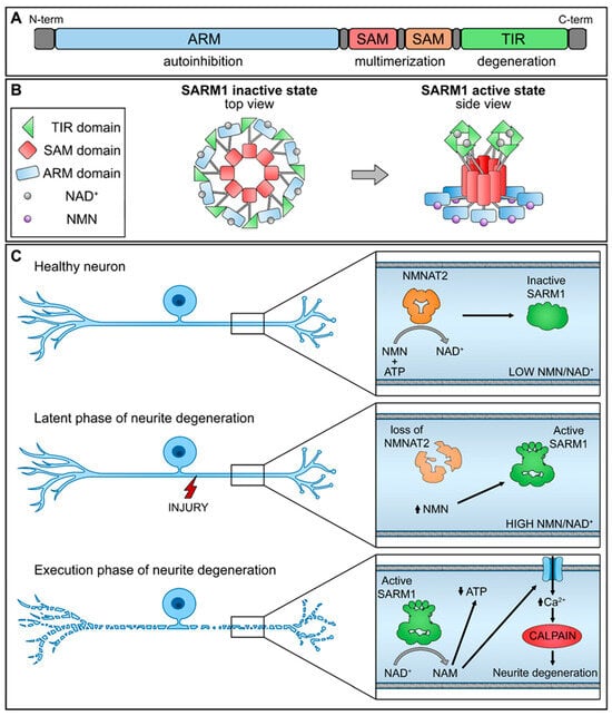
Chemotherapy-induced peripheral neuropathy (CIPN) commonly arises as a side effect of diverse cancer chemotherapy treatments. This condition presents symptoms such as numbness, tingling, and altered sensation in patients, often accompanied by neuropathic pain. Pathologically, CIPN is characterized by an intensive “dying-back” axonopathy, starting at the intra-epidermal sensory innervations and advancing retrogradely. The lack of comprehensive understanding regarding its underlying mechanisms explains the absence of effective treatments for CIPN. Recent investigations into axon degeneration mechanisms have pinpointed nicotinamide mononucleotide adenylyltransferase 2 (NMNAT2) and sterile alpha and TIR motif-containing 1 protein (SARM1) as pivotal mediators of injury-induced axonal degeneration. 

  • NMNAT2
  • SARM1
  • CIPN
  • neuropathy
  • inhibitors

1. Introduction

NMNAT, a highly conserved nicotinamide adenine dinucleotide (NAD+) synthase, plays a crucial role in cell survival by controlling the compartment-specific levels of NAD+ and influencing various cellular processes such as protein balance, cell differentiation, DNA repair, maintenance of neurons, and more. Three NMNAT isoenzymes exist: NMNAT1 which is localized in the nucleus and is widely distributed in tissues; NMNAT2 which is localized in the cytoplasm and Golgi apparatus and mainly exists in the nervous system, heart, and skeletal muscle; NMNAT3 localized in mitochondria and expressed in the spleen and lungs. NMNAT1 stands out as the most prevalent among the isoforms, demonstrating also the highest catalytic efficiency. Additionally, NMNAT1 exhibits about a fourfold greater specificity for nicotinamide mononucleotide (NMN) compared to nicotinic acid mononucleotide (NaMN). NMNAT2 displays the lowest efficiency, utilizing NAMN and NMN with similar effectiveness as NMNAT3, which is also typically found in lower abundance compared to NMNAT1 [1].
In particular, NMNAT2, which predominates in the brain, attracts scientific attention for its possible role in neuroprotection. NMNAT2 has two fundamental biological roles: (1) it acts as a NAD+ synthetase by transferring the adenosyl portion of adenosine triphosphate (ATP) to NMN, resulting in the reversible synthesis of NAD+—this function is crucial for neuroprotection and is strongly linked to the development and outcome of malignant tumors [2][3]; (2) moreover, NMNAT2 acts as a molecular chaperone by activating the distinctive ATP site situated in its C-terminal region. This activation results in the creation of a complex with heat shock protein 90 (HSP90) [4]. The formation of this complex enhances the refoldase activity of NMNAT2, aiding neurons in overcoming protein unfolding and aggregation. Consequently, this process helps alleviate protein-induced stress and preserves the overall health of neurons [5]. NMNAT2 has a notable impact on diverse medical conditions, encompassing neurodegenerative diseases and malignant tumors. Apart from its crucial protective role in neurodegenerative diseases, NMNAT2 exhibits high expression in various solid tumors, playing a significant role in tumor occurrence and progression [6]. NAD+ produced by NMNAT2 acts as a redox coenzyme for numerous dehydrogenases in energy metabolism and as a co-substrate for various enzymes that regulate diverse cellular processes. NAD+ acts as a critical substrate for various enzymes, such as PARPs, CD38/157 ectoenzymes, and sirtuins (histone deacetylases), which are involved in DNA repair, apoptosis, calcium signaling, and transcriptional regulation—processes integral to tumor growth and survival [7][8]. It is well established that cancer cells exhibit a higher demand for NAD+ compared to normal cells. Specifically, NMNAT2 levels increase in colorectal cancer, showing a positive correlation with tumor invasiveness and stage [9]. Elevated levels of NMNAT2 are also observed in ovarian cancer cells. In this context, NMNAT2 was found to enhance the activity of PARP16, which, through mono ADP-ribosylation of ribosomal proteins, maintains proteostasis during accelerated cell proliferation. Deletion of NMNAT2 leads to protein aggregation, consequently reducing the growth of cancer cells [10]. Depending on the tissue-specific NAD(P)ase that NMNAT2 (through NAD+ production) regulates, it can have a distinct functional outcome.
SARM1 serves as a key player in programmed axon death, inducing the destruction of NAD+ when activated. In mammals, SARM1 is abundantly present in neurons, is localized both in cell bodies and axons, and can be found in association with mitochondria [11]. Due to its prominent presence in the nervous system, research on SARM1 primarily focuses on neuron degeneration. However, SARM1 is also observable in other tissues, particularly in macrophages and T cells. SARM1-ko effectively prevents axon degeneration [12][13][14]. However, even a partial reduction provides significant protection [15]. Interestingly, SARM1 also plays a role in innate immunity [16]. Its composition incorporates a Toll/interleukin-1 receptor (TIR) domain, a characteristic frequently identified in Toll-like receptors (TLRs) of the innate immune system.

2. NMNAT2/SARM1 and NAD+ Synthesis

SARM1 is composed of distinct domains, including an N-terminal allosteric regulatory armadillo repeat (ARM) domain, two tandem sterile alpha motif (SAM) domains, and a C-terminal catalytic TIR domain (Figure 1A). The presence of the ARM domain is crucial for exerting an autoinhibitory function, effectively preventing the dimerization of TIR domains required for SARM1 enzymatic activity. Recent structural investigations have revealed the inactivated state of SARM1, wherein it forms an octomeric ring structure (Figure 1B) [17].
Figure 1. Schematic representation of NMNAT2/SARM1 regulation mechanism. (A) SARM1 domains; (B) SARM1 active and inactive state; (C) interplay of NMNAT2/SARM1 during neurite degeneration. (up arrow-increase; down arrow-decrease).
NMNAT2 exerts regulatory control over the activation of SARM1 by competitively binding to the allosteric sites within the N-terminal ARM domain of SARM1, utilizing both NAD+ and its precursor NMN. In functioning nerve cells, an elevated concentration of NAD+ engages and stabilizes the ARM domain, promoting its interaction with the TIR domain and efficiently suppressing the NAD+ hydrolase activity. Nevertheless, a decline in NMNAT2 levels, whether triggered by physical injury or pathological stimuli, compromises the conversion of NMN into NAD+, leading to diminished NAD+ levels within the axons. As a result, the NAD+ bound to the ARM dissociates, diminishing its binding affinity with TIR and reinstating the NAD+ hydrolase activity within the TIR domain.
This sequence of events leads to metabolic disruption and axonal rupture (Figure 1C). Changes in the NMN-NAD+ ratio, achieved through elevating NMN levels or diminishing NAD+ levels, can equally trigger the NADase activity of SARM1. NAD+ and NMN engage in direct competition to control the enzymatic activity of SARM1 [18]. Therefore, the equilibrium between the protective factor NMNAT2 and the destabilizing molecule SARM1 is crucial for preserving axonal integrity.

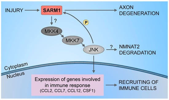
3. NMNAT2/SARM1 Downstream Pathways Regulation

Several studies propose that maintaining NMNAT2/SARM1 balance is crucial for regulating various pathways and highlights the significance of some downstream players involved in neuronal survival and axon degeneration.
The study conducted on various model organisms has elucidated the involvement of SARM1 in controlling mitogen-activated protein kinase (MAPKs) signaling pathways that are relevant for cell life, neuronal plasticity, and innate immunity spanning across both neurons and glial cells [19]. The involvement of MAPKs in axon degeneration has been well established, as inhibiting the MAPK cascade, including dual leucine zipper kinase (DLK) and c-Jun N-terminal kinase (JNK), has shown to provide protection similar to SARM1 knockout following axonal injury [20]. Nevertheless, the recent study suggests that the interplay between MAPK signaling and SARM1 activity may be more complex than initially thought. The role of MAPKs in degeneration downstream of SARM1 has been a matter of discussion, but recent research indicates that they might serve alternative signaling functions. It has been shown that both the NADase activity of SARM and a cascade involving cacophony (Cac)/SARM/MAPK are essential for an initial phase of axon transport blockage after axotomy [21].
However, the relationship between SARM1 activation and MAPK needs to be better examined. Notably, it was found that although MAPK signaling is essential for injury-induced NAD+ depletion and axonal degeneration, it does not play a role in NAD+ depletion or axon degeneration triggered by activated SARM1 [22]. This controversially implies that MAPK signaling might function upstream of SARM1, facilitating its activation in response to injury. Inhibition of MAPK signaling leads to a reduction in the turnover of both NMNAT2 and the microtubule-destabilizing factor SCG10, leading to elevated levels of these survival factors. 
Additionally, it is important to notice, several investigations have indicated primarily the involvement of JNK signaling in the progression of axon degeneration. Stress-related signaling mediated by DLK and downstream JNKs has been recognized as activated in various aspects of the axonal injury response [23][24]. These kinases have been observed to activate in damaged axons shortly after nerve injury [20][25][26]. It is suggested that JNK functions downstream of SARM1, given that genetic disruption of all three JNK isoforms (or both MKK4/7) can counteract the degeneration caused by the ectopic activation of SARM1’s TIR domains [20]. However, this aspect requires further investigation, as one study proposes an upstream pro-degenerative role for JNK, where JNK promotes the degradation of NMNAT2 [19].
On the other hand, different noteworthy papers have established a connection between SARM1 activation and the production of downstream cyclic adenosine diphosphate (ADP)-ribose (cADPR) as an important player in axon degeneration. Recent findings have revealed that cADPR is responsible for inducing axonal calcium flux and degeneration in response to PTX. Genetic or pharmacological inhibitors of cADPR signaling have been shown to prevent axon degeneration and alleviate symptoms of allodynia induced by PTX without compromising the anti-neoplastic efficacy of the drug. These findings highlight cADPR as a calcium-modulating factor that promotes axon degeneration, thereby proposing cADPR signaling as a potential therapeutic target [27]

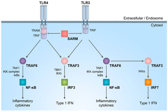
4. NMNAT2/SARM1 Role in Immune System Stimulation

The involvement of glial cells in neuropathic pain is a well-established fact. Increasing evidence suggests that immune cells are not merely passive bystanders in the nervous system but active influencers in the onset and/or development of neuropathies. It is well demonstrated that axon injury leads to the recruitment of macrophages to the site of injury, a process orchestrated by different cell populations. Within 2–3 days post-injury, macrophages are the primary and most abundant cells infiltrating the injury site, attracted by factors released by repair Schwann cells. These macrophages, in turn, produce chemoattractants like CCL2, TNF-α, IL-1α, IL-1β [28], NGF, and nitric oxide (NO) [29] enhancing further macrophage infiltration.
It is important to discuss that SARM1 plays a role in innate immunity, participating in the regulation of TLR signaling, as well as the synthesis of cytokines and chemokines within neurons [30][31] and innate immune cells [32][33]. In complex in vivo models, immune response activated by SARM1 could potentially be linked to axon degeneration. While the enzymatic activity of SARM1 NADase has been intensively studied in neurons, its role in the immune system is not yet fully understood. SARM1 is one of the six adaptor proteins involved in TLR signaling pathways. TLRs serve as indispensable pattern recognition receptors, playing a pivotal role in the innate immune system. Upon stimulation of TLRs, multiple transcription factors, including nuclear factor (NF)-κB, interferon regulatory factor 3 (IRF3), IRF5, and IRF7, are triggered (Figure 2).
Figure 2. SARM1 role in innate immunity activation. Schematic representation of inhibitory effect of SARM1 in TLR signaling pathways.
The distinctive combination of three protein–protein interaction domains in SARM1 implies that, within the TLR adaptor family, SARM1 likely operates in a manner distinct from other adaptor molecules. There is a suggestion that in humans, SARM1 exerts a negative regulation on TRIF-dependent TLR3 and TLR4 signaling, leading to the inactivation of the NF-κB response [34]
Furthermore, it has been demonstrated that SARM1 plays a crucial role in the upregulation of chemokines CCL2, CCL7, and CCL12, along with the cytokine CSF1. However, traumatic axonal injuries did not alter the expression levels of SARM1. Also, increasing the full-length SARM1 protein in cultured neurons did not influence the level of expression of CCL7, CCL2, CCL12, and CSF1. This implies that the neuronal immune response, orchestrated by SARM1, is unlikely to be governed by the levels of SARM1 protein expression. This mechanism is shown to be regulated through the SARM1-JNKs-cJun pathway (Figure 3). 
Figure 3. SARM1/JNK pathway. Schematic representation of SARM1 role in immune cells recruitment to injured neural tissues.

5. NMNAT2/SARM1 in Chemotherapy-Induced Neuropathy

As previously mentioned, an imbalance between NMNAT2 and SARM1 is associated with axon degeneration, particularly in the context of chemotherapy-induced neuropathy). The in vitro study has demonstrated that preserving axonal NAD+ levels and inhibiting SARM1 can effectively prevent axon degeneration. Genetic removal of SARM1 significantly reduces vincristine (VCR) and bortezomib (BTZ)-induced axon degeneration, both in vitro and in vivo [35]. Specifically, exposure to VCR or BTZ leads to a reduction in axonal NMNAT2 levels. The activation of SARM1 occurs through the decrease in NMNAT2, whereas the study demonstrated that preserving a consistent NMNAT isoform in the axon offers strong protection against both VCR and BTZ-induced axon degeneration. SARM1 activation induces a swift and significant reduction in NAD+, leading to subsequent local metabolic collapse and axon degeneration. The prompt decrease in NAD+ within the axon following the administration of VCR and BTZ is facilitated by the activation of SARM1. This illustrates that SARM1 operates via the identical downstream mediator (NAD+) and is controlled by the immediate upstream regulator (NMNAT2).
Additionally, in vivo studies confirm that the absence of SARM1 gene expression counteracts the progression of acute painful neuropathies triggered by chemotherapeutics, such as OXP [36], VCR [14], PTX [35], and CDDP [37]. Nevertheless, in animal models, characterized by a complexity surpassing that of cell cultures, it has been demonstrated that the molecular pathway(s) of axon degeneration involving SARM1 may not universally apply to all instances of axon degeneration. It has been shown that absence of the SARM1 gene elicits exhibited resistance to PTX-induced distal axonal degeneration in CIPN. Partial preservation of thermal sensitivity and epidermal innervation was noted, although no impact was observed on the reduction in tail sensory nerve action potential amplitudes. This result contrasted with the neuroprotection seen in the presumed metabolic neuropathy model, where thermal sensitivity, tail sensory nerve action potential amplitude, and epidermal innervation were all preserved [38]
In another in vivo study, emphasis is placed on the downstream protein calpain in the SARM1 pathway. It is demonstrated that the activation of calpains is crucial for both neurotoxicity and the formation of DNA-platinum adducts in neurons. This is supported by the fact that a calpain inhibitor effectively blocked the formation of DNA-platinum adducts in neuronal cells treated with CDDP. Notably, Sarm1−/− mice treated with CDDP did not exhibit an increase in calpain activity in sciatic nerves. 
A significant discovery is that the overexpression of SARM1 does not trigger axon degeneration in the absence of injury caused by axotomy or vincristine treatment. These findings underscore the pivotal role of SARM1 in the process of axon degeneration, emphasizing that, even when overexpressed, SARM1 necessitates an injury signal to initiate axon degeneration. It is probable that the regulation of SARM1 occurs at the post-translation level. SARM1-dependent axon degeneration is unlikely to involve the transcription of new SARM1 molecules. This suggests two general potential processes: (1) SARM1 is expressed in uninjured neurons at levels sufficient to induce axon degeneration in response to injury without the need for de novo transcription; (2) further, SARM1 is activated after the axon is severed, and it triggers a localized self-destructive pathway within the axon.

6. SARM1 Inhibitors

As mentioned above, there are currently no available treatments specifically addressing disorders linked to axonal degeneration. Inhibition of SARM1 represents an attractive therapeutic target for treating various pathologies of axon degeneration, including peripheral neuropathy, traumatic brain injury, and neurodegenerative disorders.
Using a high throughput screening that utilizes a NAD+ analog that fluoresces upon the release of nicotinamide, five putative SARM1 inhibitor compounds that exhibit competitive or non-competitive modes of inhibition with up to 70% efficiency were identified [39]. Other competitive and non-competitive SARM1 inhibitors were also identified through a modified fluorescence assay that measures the production of cADPR, the hydrolysis product of NAD+. These compounds also showed strong protection against axon degeneration, even if toxicity was manifest for almost all tested compounds [40]
An innovative set of irreversible isothiazole SARM1 inhibitors was studied for their effectiveness in preserving axonal structure and function, both in laboratory settings and, for the first time, in living organisms [41]. A sequence of optimization procedures led to the development of an orally bioavailable compound suitable, thanks to its submicromolar potency, for chronic dosing in vivo.
Dehydronitrosonisodipine (dHNN), a derivative of nisoldipine, was also discovered as an irreversible SARM1 inhibitor [42]. This compound reacts with Cys311 in the ARM domain and blocks its NMN-activation, protecting axons from neurodegeneration. The same cysteine residue was shown to be targeted by a series of tryptoline acrylamides that inhibit the NADase activity of SARM1 and prevent neurite degeneration induced by VCR [43].

This entry is adapted from the peer-reviewed paper 10.3390/biology13010061

References

  1. Fortunato, C.; Mazzola, F.; Raffaelli, N. The key role of the NAD biosynthetic enzyme nicotinamide mononucleotide adenylyltransferase in regulating cell functions. IUBMB Life 2022, 74, 562–572.
  2. Jayaram, H.N.; Kusumanchi, P.; Yalowitz, J.A. NMNAT expression and its relation to NAD metabolism. Curr. Med. Chem. 2011, 18, 1962–1972.
  3. Brunetti, L.; Di Stefano, M.; Ruggieri, S.; Cimadamore, F.; Magni, G. Homology modeling and deletion mutants of human nicotinamide mononucleotide adenylyltransferase isozyme 2: New insights on structure and function relationship. Protein Sci. 2010, 19, 2440–2450.
  4. Berger, F.; Lau, C.; Dahlmann, M.; Ziegler, M. Subcellular compartmentation and differential catalytic properties of the three human nicotinamide mononucleotide adenylyltransferase isoforms. J. Biol. Chem. 2005, 280, 36334–36341.
  5. Ali, Y.O.; Allen, H.M.; Yu, L.; Li-Kroeger, D.; Bakhshizadehmahmoudi, D.; Hatcher, A.; McCabe, C.; Xu, J.; Bjorklund, N.; Taglialatela, G.; et al. NMNAT2:HSP90 Complex Mediates Proteostasis in Proteinopathies. PLoS Biol. 2016, 14, e1002472.
  6. Li, W.; Gao, M.; Hu, C.; Chen, X.; Zhou, Y. NMNAT2: An important metabolic enzyme affecting the disease progression. Biomed. Pharmacother. 2023, 158, 114143.
  7. Amjad, S.; Nisar, S.; Bhat, A.A.; Shah, A.R.; Frenneaux, M.P.; Fakhro, K.; Haris, M.; Reddy, R.; Patay, Z.; Baur, J.; et al. Role of NAD. Mol. Metab. 2021, 49, 101195.
  8. Yaku, K.; Okabe, K.; Hikosaka, K.; Nakagawa, T. NAD Metabolism in Cancer Therapeutics. Front. Oncol. 2018, 8, 622.
  9. Qi, J.; Cui, C.; Deng, Q.; Wang, L.; Chen, R.; Zhai, D.; Xie, L.; Yu, J. Downregulated SIRT6 and upregulated NMNAT2 are associated with the presence, depth and stage of colorectal cancer. Oncol. Lett. 2018, 16, 5829–5837.
  10. Challa, S.; Khulpateea, B.R.; Nandu, T.; Camacho, C.V.; Ryu, K.W.; Chen, H.; Peng, Y.; Lea, J.S.; Kraus, W.L. Ribosome ADP-ribosylation inhibits translation and maintains proteostasis in cancers. Cell 2021, 184, 4531–4546.e26.
  11. Gerdts, J.; Summers, D.W.; Milbrandt, J.; DiAntonio, A. Axon Self-Destruction: New Links among SARM1, MAPKs, and NAD+ Metabolism. Neuron 2016, 89, 449–460.
  12. Bradshaw, D.V.; Knutsen, A.K.; Korotcov, A.; Sullivan, G.M.; Radomski, K.L.; Dardzinski, B.J.; Zi, X.; McDaniel, D.P.; Armstrong, R.C. Genetic inactivation of SARM1 axon degeneration pathway improves outcome trajectory after experimental traumatic brain injury based on pathological, radiological, and functional measures. Acta Neuropathol. Commun. 2021, 9, 89.
  13. Marion, C.M.; McDaniel, D.P.; Armstrong, R.C. Sarm1 deletion reduces axon damage, demyelination, and white matter atrophy after experimental traumatic brain injury. Exp. Neurol. 2019, 321, 113040.
  14. Geisler, S.; Doan, R.A.; Strickland, A.; Huang, X.; Milbrandt, J.; DiAntonio, A. Prevention of vincristine-induced peripheral neuropathy by genetic deletion of SARM1 in mice. Brain 2016, 139, 3092–3108.
  15. Gilley, J.; Orsomando, G.; Nascimento-Ferreira, I.; Coleman, M.P. Absence of SARM1 rescues development and survival of NMNAT2-deficient axons. Cell Rep. 2015, 10, 1974–1981.
  16. Carty, M.; Bowie, A.G. SARM: From immune regulator to cell executioner. Biochem. Pharmacol. 2019, 161, 52–62.
  17. Bratkowski, M.; Xie, T.; Thayer, D.A.; Lad, S.; Mathur, P.; Yang, Y.S.; Danko, G.; Burdett, T.C.; Danao, J.; Cantor, A.; et al. Structural and Mechanistic Regulation of the Pro-degenerative NAD Hydrolase SARM1. Cell Rep. 2020, 32, 107999.
  18. Figley, M.D.; Gu, W.; Nanson, J.D.; Shi, Y.; Sasaki, Y.; Cunnea, K.; Malde, A.K.; Jia, X.; Luo, Z.; Saikot, F.K.; et al. SARM1 is a metabolic sensor activated by an increased NMN/NAD. Neuron 2021, 109, 1118–1136.e11.
  19. Waller, T.J.; Collins, C.A. An NAD+/NMN balancing act by SARM1 and NMNAT2 controls axonal degeneration. Neuron 2021, 109, 1067–1069.
  20. Yang, J.; Wu, Z.; Renier, N.; Simon, D.J.; Uryu, K.; Park, D.S.; Greer, P.A.; Tournier, C.; Davis, R.J.; Tessier-Lavigne, M. Pathological axonal death through a MAPK cascade that triggers a local energy deficit. Cell 2015, 160, 161–176.
  21. Hsu, J.M.; Kang, Y.; Corty, M.M.; Mathieson, D.; Peters, O.M.; Freeman, M.R. Injury-Induced Inhibition of Bystander Neurons Requires dSarm and Signaling from Glia. Neuron 2021, 109, 473–487.e5.
  22. Walker, L.J.; Summers, D.W.; Sasaki, Y.; Brace, E.J.; Milbrandt, J.; DiAntonio, A. MAPK signaling promotes axonal degeneration by speeding the turnover of the axonal maintenance factor NMNAT2. eLife 2017, 6, e22540.
  23. Asghari Adib, E.; Smithson, L.J.; Collins, C.A. An axonal stress response pathway: Degenerative and regenerative signaling by DLK. Curr. Opin. Neurobiol. 2018, 53, 110–119.
  24. Jin, Y.; Zheng, B. Multitasking: Dual Leucine Zipper-Bearing Kinases in Neuronal Development and Stress Management. Annu. Rev. Cell Dev. Biol. 2019, 35, 501–521.
  25. Cavalli, V.; Kujala, P.; Klumperman, J.; Goldstein, L.S. Sunday Driver links axonal transport to damage signaling. J. Cell Biol. 2005, 168, 775–787.
  26. Lindwall, C.; Kanje, M. Retrograde axonal transport of JNK signaling molecules influence injury induced nuclear changes in p-c-Jun and ATF3 in adult rat sensory neurons. Mol. Cell Neurosci. 2005, 29, 269–282.
  27. Li, Y.; Pazyra-Murphy, M.F.; Avizonis, D.; de Sá Tavares Russo, M.; Tang, S.; Chen, C.Y.; Hsueh, Y.P.; Bergholz, J.S.; Jiang, T.; Zhao, J.J.; et al. Sarm1 activation produces cADPR to increase intra-axonal Ca++ and promote axon degeneration in PIPN. J. Cell Biol. 2022, 221, e202106080.
  28. Cattin, A.L.; Burden, J.J.; Van Emmenis, L.; Mackenzie, F.E.; Hoving, J.J.; Garcia Calavia, N.; Guo, Y.; McLaughlin, M.; Rosenberg, L.H.; Quereda, V.; et al. Macrophage-Induced Blood Vessels Guide Schwann Cell-Mediated Regeneration of Peripheral Nerves. Cell 2015, 162, 1127–1139.
  29. Scholz, J.; Woolf, C.J. The neuropathic pain triad: Neurons, immune cells and glia. Nat. Neurosci. 2007, 10, 1361–1368.
  30. Liu, H.Y.; Chen, C.Y.; Hsueh, Y.P. Innate immune responses regulate morphogenesis and degeneration: Roles of Toll-like receptors and Sarm1 in neurons. Neurosci. Bull. 2014, 30, 645–654.
  31. Mukherjee, P.; Winkler, C.W.; Taylor, K.G.; Woods, T.A.; Nair, V.; Khan, B.A.; Peterson, K.E. SARM1, Not MyD88, Mediates TLR7/TLR9-Induced Apoptosis in Neurons. J. Immunol. 2015, 195, 4913–4921.
  32. Loring, H.S.; Thompson, P.R. Emergence of SARM1 as a Potential Therapeutic Target for Wallerian-type Diseases. Cell Chem. Biol. 2020, 27, 1–13.
  33. Tan, R.S.; Ho, B.; Leung, B.P.; Ding, J.L. TLR cross-talk confers specificity to innate immunity. Int. Rev. Immunol. 2014, 33, 443–453.
  34. Carty, M.; Goodbody, R.; Schröder, M.; Stack, J.; Moynagh, P.N.; Bowie, A.G. The human adaptor SARM negatively regulates adaptor protein TRIF-dependent Toll-like receptor signaling. Nat. Immunol. 2006, 7, 1074–1081.
  35. Geisler, S.; Doan, R.A.; Cheng, G.C.; Cetinkaya-Fisgin, A.; Huang, S.X.; Höke, A.; Milbrandt, J.; DiAntonio, A. Vincristine and bortezomib use distinct upstream mechanisms to activate a common SARM1-dependent axon degeneration program. JCI Insight 2019, 4, e129920.
  36. Gould, S.A.; White, M.; Wilbrey, A.L.; Pór, E.; Coleman, M.P.; Adalbert, R. Protection against oxaliplatin-induced mechanical and thermal hypersensitivity in Sarm1. Exp. Neurol. 2021, 338, 113607.
  37. Cetinkaya-Fisgin, A.; Luan, X.; Reed, N.; Jeong, Y.E.; Oh, B.C.; Hoke, A. Cisplatin induced neurotoxicity is mediated by Sarm1 and calpain activation. Sci. Rep. 2020, 10, 21889.
  38. Turkiew, E.; Falconer, D.; Reed, N.; Höke, A. Deletion of Sarm1 gene is neuroprotective in two models of peripheral neuropathy. J. Peripher. Nerv. Syst. 2017, 22, 162–171.
  39. Loring, H.S.; Parelkar, S.S.; Mondal, S.; Thompson, P.R. Identification of the first noncompetitive SARM1 inhibitors. Bioorg. Med. Chem. 2020, 28, 115644.
  40. Khazma, T.; Golan-Vaishenker, Y.; Guez-Haddad, J.; Grossman, A.; Sain, R.; Weitman, M.; Plotnikov, A.; Zalk, R.; Yaron, A.; Hons, M.; et al. A duplex structure of SARM1 octamers stabilized by a new inhibitor. Cell Mol. Life Sci. 2022, 80, 16.
  41. Bosanac, T.; Hughes, R.O.; Engber, T.; Devraj, R.; Brearley, A.; Danker, K.; Young, K.; Kopatz, J.; Hermann, M.; Berthemy, A.; et al. Pharmacological SARM1 inhibition protects axon structure and function in paclitaxel-induced peripheral neuropathy. Brain 2021, 144, 3226–3238.
  42. Li, W.H.; Huang, K.; Cai, Y.; Wang, Q.W.; Zhu, W.J.; Hou, Y.N.; Wang, S.; Cao, S.; Zhao, Z.Y.; Xie, X.J.; et al. Permeant fluorescent probes visualize the activation of SARM1 and uncover an anti-neurodegenerative drug candidate. eLife 2021, 10, e67381.
  43. Feldman, H.C.; Merlini, E.; Guijas, C.; DeMeester, K.E.; Njomen, E.; Kozina, E.M.; Yokoyama, M.; Vinogradova, E.; Reardon, H.T.; Melillo, B.; et al. Selective inhibitors of SARM1 targeting an allosteric cysteine in the autoregulatory ARM domain. Proc. Natl. Acad. Sci. USA 2022, 119, e2208457119.
More
This entry is offline, you can click here to edit this entry!
Video Production Service