Hypothermia is a widespread condition all over the world, with a high risk of mortality in pre-hospital and in-hospital settings when it is not promptly and adequately treated. Hypothermia can occur due to unfavorable environmental conditions as well as internal causes, such as pathological states that result in reduced heat production, increased heat loss or ineffectiveness of the thermal regulation system. The consequences of hypothermia affect several systems in the body—the cardiovascular system, the central and peripheral nervous systems, the respiratory system, the endocrine system and the gastrointestinal system—but also kidney function, electrolyte balance and coagulation.
- hypothermia
- emergency department
- homeostasis
1. Introduction
- -
-
Mild hypothermia: 32 °C < core body temperature < 35 °C.
- -
-
Moderate hypothermia: 28 °C < core body temperature < 32 °C.
- -
-
Severe hypothermia: core body temperature < 28 °C.
- -
-
Primary hypothermia is due to environmental exposure, with no underlying medical condition causing the disruption of temperature regulation [17]. It therefore occurs when a person is exposed to the cold without adequate protection.
- -
-
Secondary hypothermia is a complication of pathological and paraphysiological conditions that determine the hypothermia itself or cause either an alteration of the thermoregulation mechanisms, reduced heat production or increased heat dispersion.
- -
-
Therapeutic hypothermia;
- -
-
Trauma-induced hypothermia [18].
| Classification from Etiopathology | ||
|---|---|---|
| Spontaneous hypothermia | Primary hypothermia | Secondary hypothermia |
| Induced hypothermia | Therapeutic hypothermia | Trauma-induced hypothermia |
2. Physiopathology

-
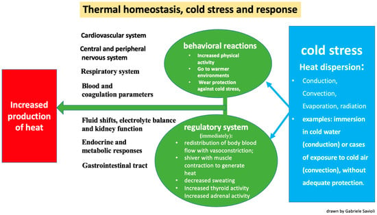
Conduction: the transfer of heat to a cooler object through direct contact (for example, when immersed in cold water).
-
Convection: the transfer of heat at the body surface via air circulation (for example, when exposed to cold air or wind).
-
Evaporation: cooling of the skin surfaces when sweat changes from a liquid to a vapor form.
-
Radiation: occurs through the transmission of electromagnetic waves.
- -
-
The redistribution of blood flow via vasoconstriction and a subsequent reduction in blood volume directed towards the skin and subcutis to reduce heat loss;
- -
-
Shivering, which generates heat via muscle contraction;
- -
-
Decreased sweating;
- -
-
Increased thyroid activity due to hypothalamic stimulus;
- -
-
Increased adrenal activity due to hypothalamic stimulus.
- -
-
Increased physical activity;
- -
-
A shift to warmer environments;
- -
-
Wearing protection against the cold;
- -
-
Taking off wet clothes and replacing them with dry clothes.
3. Etiology and Risk Factors
- -
-
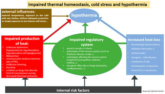
Impaired thermoregulation (Figure 2): This may be paraphysiological in the extreme ages of life (geriatric people or infants). Impaired thermoregulation may also occur following pathological conditions such as mental illnesses, or pathologies of the nervous system (such as Parkinson’s disease, stroke, multiple sclerosis, hypothalamic dysfunction, brain trauma or subarachnoid hemorrhage) or pathologies affecting the peripheral nervous system (neuropathies, diabetes mellitus, trauma to a section of the spinal cord). Impaired thermoregulation may also be due to an iatrogenic effect of drugs such as anxiolytics, antidepressants, phenothiazines, barbiturates, opioids, antipsychotics, oral antihyperglycemics, β-blockers and α-blockers [25][26].
 Figure 2. Mechanisms by which hypothermia develops: disruption of thermal homeostasis and dysregulated response to cold stress.
 - -
-
Increased heat loss (Figure 2): This can be caused by dermatologic diseases such as burns, exfoliative dermatitis or psoriasis [27][28][29]. Heat loss can also be increased in the case of indulgent habits such as alcohol intake [30][31] but it can also have an iatrogenic origin, as in the case of cold infusions, the transfusion of cold hematopoietic components, hasty birth or anesthesia.
- -
-
Decreased heat production (Figure 2): This is secondary to endocrine dysfunction (hypopituitarism, hypothyroidism, hypoadrenalism and hypoglycemia), malnutrition or either conditions or drugs that alter the level of consciousness, causing an impaired shivering mechanism.
4. From Pathophysiology to Clinical Manifestations
4.1. Cardiovascular System
4.2. Central and Peripheral Nervous Systems
4.3. Respiratory System
4.4. Fluid Shifts, Electrolyte Balance and Kidney Function
4.5. Blood and Coagulation Parameters
4.6. Endocrine and Metabolic Responses
4.7. Gastrointestinal Tract
5. Clinical Diagnosis
This entry is adapted from the peer-reviewed paper 10.3390/jpm13121690
References
- Hypothermia: Background, Pathophysiology, Etiology. Available online: https://emedicine.medscape.com/article/770542-overview (accessed on 15 August 2023).
- Petrone, P.; Asensio, J.A.; Marini, C.P. In brief: Hypothermia. Curr. Probl. Surg. 2014, 51, 414–415.
- Epstein, E.; Anna, K. Accidental shypothermia. BMJ 2006, 332, 706–709.
- Spencer, M.R.; Hedegaard, H.B. QuickStats: Death Rates* Attributed to Excessive Cold or Hypothermia† Among Persons Aged ≥15 Years, by Urban-Rural Status§ and Age Group—National Vital Statistics System, United States, 2019. MMWR Morb. Mortal. Wkly. Rep. 2021, 70, 258.
- van Veelen, M.J.; Brodmann Maeder, M. Hypothermia in Trauma. Int. J. Environ. Res. Public Health 2021, 18, 8719.
- Luna, G.K.; Maier, R.V.; Pavlin, E.G.; Anardi, D.; Copass, M.K.; Oreskovich, M.R. Incidence and effect of hypothermia in seriously injured patients. J. Trauma 1987, 27, 1014–1018.
- Forristal, C.; Van Aarsen, K.; Columbus, M.; Wei, J.; Vogt, K.; Mal, S. Predictors of Hypothermia upon Trauma Center Arrival in Severe Trauma Patients Transported to Hospital via EMS. Prehosp. Emerg. Care 2020, 24, 15–22.
- Meiman, J.; Anderson, H.; Tomasallo, C.; Centers for Disease Control and Prevention (CDC). Hypothermia-related deaths—Wisconsin, 2014, and United States, 2003–2013. MMWR Morb. Mortal. Wkly. Rep. 2015, 64, 141–143.
- Cheshire, W.P., Jr. Thermoregulatory disorders and illness related to heat and cold stress. Auton. Neurosci. 2016, 196, 91–104.
- Polderman, K.H. Mechanisms of action, physiological effects, and complications of hypothermia. Crit. Care Med. 2009, 37 (Suppl. 7), S186–S202.
- Cappaert, T.A.; Stone, J.A.; Castellani, J.W.; Krause, B.A.; Smith, D.; Stephens, B.A. National Athletic Trainers’ Association Position Statement: Environmental Cold Injuries. J. Athl. Train. 2008, 43, 640–658.
- Jurkovich, G.J. Environmental Cold-Induced Injury. Surg. Clin. N. Am. 2007, 87, 247–267.
- Ulrich, A.S.; Rathlev, N.K. Hypothermia and localized cold injuries. Emerg. Med. Clin. N. Am. 2004, 22, 281–298.
- Giesbrecht, G.G. Cold stress, near drowning and accidental hypothermia: A review. Aviat. Space Environ. Med. 2000, 71, 733–752.
- Durrer, B.; Brugger, H.; Syme, D.; Elsensohn, F.; Deslarzes, T.; Yersin, B.; Paal, P.; Gordon, L.; Strapazzon, G.; Maeder, M.B.; et al. The Medical On-site Treatment of Hypothermia: ICAR-MEDCOM Recommendation. High Alt. Med. Biol. 2003, 4, 99–103.
- Long, W.B.; Edlich, R.F.; Winters, K.L.; Britt, L.D. Cold injuries. J. Long Term Eff. Med. Implant. 2005, 15, 67–78.
- Søreide, K. Clinical and translational aspects of hypothermia in major trauma patients: From pathophysiology to prevention, prognosis and potential preservation. Injury 2014, 45, 647–654.
- Schumacker, P.T.; Rowland, J.; Saltz, S.; Nelson, D.P.; Wood, L.D. Effects of hyperthermia and hypothermia on oxygen extraction by tissues during hypovolemia. J. Appl. Physiol. 1987, 63, 1246–1252.
- D’Angelo, J. Treating heat-related illness in the elderly. Emerg. Med. Serv. 2004, 33, 111–113.
- Morrison, S.F.; Nakamura, K. Central Mechanisms for Thermoregulation. Annu. Rev. Physiol. 2019, 81, 285–308.
- Romanovsky, A.A. The thermoregulation system and how it works. Handb. Clin. Neurol. 2018, 156, 3–43.
- Osilla, E.V.; Marsidi, J.L.; Sharma, S. Physiology, Temperature Regulation; StatPearls Publishing: Treasure Island, FL, USA, 2023. Available online: http://www.ncbi.nlm.nih.gov/books/NBK507838/ (accessed on 15 August 2023).
- Atha, W.F. Heat-related illness. Emerg. Med. Clin. N. Am. 2013, 31, 1097–1108.
- Severe Recurrent Hypothermia in an Elderly Patient with Refractory Mania Associated with Atypical Antipsychotic, Valproic Acid and Oxcarbazepine Therapy|BMJ Case Reports. Available online: https://casereports.bmj.com/content/2017/bcr-2017-222462 (accessed on 17 August 2023).
- Rumbus, Z.; Garami, A. Fever, hypothermia, and mortality in sepsis. Temperature 2018, 6, 101–103.
- Fox, R.H.; Shuster, S.; Williams, R.; Marks, J.; Goldsmith, R.; Condon, R.E. Cardiovascular, Metabolic, and Ther Moregulatory Disturbances in Patients with Erythrodermic Skin Diseases. Br. Med. J. 1965, 1, 619–622.
- Cucinell, S.A. Hypothermia and Generalized Skin Disease. Arch. Dermatol. 1978, 114, 1244–1245.
- Krook, G. Hypothermia in patients with exfoliative dermatitis. Acta Derm. Venereol. 1960, 40, 142–160.
- Kalant, H.; Lê, A. Effects of ethanol on thermoregulation. Pharmacol. Ther. 1983, 23, 313–364.
- Ristuccia, R.C.; Hernandez, M.; Wilmouth, C.E.; Spear, L.P. Differential Expression of Ethanol-Induced Hypothermia in Adolescent and Adult Rats Induced by Pretest Familiarization to the Handling/Injection Procedure. Alcohol. Clin. Exp. Res. 2007, 31, 575–581.
- Brown, D.J.A.; Brugger, H.; Boyd, J.; Paal, P. Accidental hypothermia. N. Engl. J. Med. 2012, 367, 1930–1938.
- Périard, J.D.; Eijsvogels, T.M.H.; Daanen, H.A.M. Exercise under heat stress: Thermoregulation, hydration, performance implications, and mitigation strategies. Physiol. Rev. 2021, 101, 1873–1979.
- Biem, J.; Koehncke, N.; Classen, D.; Dosman, J. Out of the cold: Management of hypothermia and frostbite. CMAJ 2003, 168, 305–311.
- Hislop, L.J.; Wyatt, J.P.; McNaughton, G.W.; Ireland, A.J.; Rainer, T.H.; Olverman, G.; Laughton, L.M. Urban hypothermia in the west of Scotland. West of Scotland Accident and Emergency Trainees Research Group. BMJ 1995, 311, 725.
- Tsuei, B.J.; Kearney, P.A. Hypothermia in the trauma patient. Injury 2004, 35, 7–15.
- Ireland, S.; Endacott, R.; Cameron, P.; Fitzgerald, M.; Paul, E. The incidence and significance of accidental hypothermia in major trauma—A prospective observational study. Resuscitation 2011, 82, 300–306.
- Danzl, D.F.; Pozos, R.S. Accidental hypothermia. N. Engl. J. Med. 1994, 331, 1756–1760.
- Deussen, A. Hyperthermia and hypothermia. Effects on the cardiovascular system. Anaesthesist 2007, 56, 907–911.
- Brooks, D.P.; Chapman, B.J.; Munday, K.A. The Effect of Hypothermia on the Cardiovascular System and the Pressor Actions of Angiotensin II. J. Therm. Biol. 1984, 9, 243–246.
- Dietrichs, E.S.; McGlynn, K.; Allan, A.; Connolly, A.; Bishop, M.; Burton, F.; Kettlewell, S.; Myles, R.; Tveita, T.; Smith, G.L. Moderate but not severe hypothermia causes pro-arrhythmic changes in cardiac electrophysiology. Cardiovasc. Res. 2020, 116, 2081–2090.
- Prec, O.; Rosenman, R.; Braun, K.; Rodbard, S.; Katz, L.N. The cardiovascular effects of acutely induced hypothermia. J. Clin. Investig. 1949, 28, 293–300.
- Covino, B.G.; D’Amato, H.E. Mechanism of Ventricular Fibrillation in Hypothermia. Circ. Res. 1962, 10, 148–155.
- Bjørnstad, H.; Tande, P.M.; Refsum, H. Cardiac electrophysiology during hypothermia. Implications for medical treatment. Arctic Med. Res. 1991, 50 (Suppl. 6), 71–75.
- Roscher, R.; Arlock, P.; Sjöberg, T.; Steen, S. Effects of dopamine on porcine myocardial action potentials and contractions at 37 degrees C and 32 degrees C. Acta Anaesthesiol. Scand. 2001, 45, 421–426.
- Ehrlich, M.P.; McCullough, J.N.; Zhang, N.; Weisz, D.J.; Juvonen, T.; Bodian, C.A.; Griepp, R.B. Effect of hypothermia on cerebral blood flow and metabolism in the pig. Ann. Thorac. Surg. 2002, 73, 191–197.
- McCullough, J.N.; Zhang, N.; Reich, D.L.; Juvonen, T.S.; Klein, J.J.; Spielvogel, D.; Ergin, M.; Griepp, R.B. Cerebral metabolic suppression during hypothermic circulatory arrest in humans. Ann. Thorac. Surg. 1999, 67, 1895–1899.
- Yager, J.Y.; Asselin, J. Effect of Mild Hypothermia on Cerebral Energy Metabolism During the Evolution of Hypoxic-Ischemic Brain Damage in the Immature Rat. Stroke 1996, 27, 919–926.
- Erecinska, M.; Thoresen, M.; Silver, I.A. Effects of Hypothermia on Energy Metabolism in Mammalian Central Nervous System. J. Cereb. Blood Flow Metab. 2003, 23, 513–530.
- Frappell, P. Experimental Biology 1997 Symposium on Neurobiology of Thermoregulation: Role of Stress: Hypothermia and physiological control: The respiratory system. Clin. Exp. Pharmacol. Physiol. 1998, 25, 159–164.
- Dill, D.B.; Forbes, W.H. Respiratory and metabolic effects of hypothermia. Am. J. Physiol. Leg. Content 1941, 132, 685–697.
- Taiji, S.; Nishino, T.; Jin, H.; Shinozuka, N.; Isono, S.; Nozaki-Taguchi, N. Changes in breathing pattern during severe hypothermia and autoresuscitation from hypothermic respiratory arrest in anesthetized mice. Physiol. Rep. 2021, 9, e15139.
- D’Amato, H.E.; Hegnauer, A.H. Blood Volume in the Hypothermic Dog. Am. J. Physiol. Content 1953, 173, 100–102.
- Moyer, J.H. The effect of hypothermia on renal function and renal damage from ischemia. Ann. N. Y. Acad. Sci. 1959, 80, 424–434.
- Rosenfeld, J.B. Acid-base and electrolyte disturbances in hypothermia. Am. J. Cardiol. 1963, 12, 678–682.
- Sharma, T.; Kunkes, J.; O’Sullivan, D.; Fernandez, A.B. Elevated risk of venous thromboembolism in patients undergoing therapeutic hypothermia after cardiac arrest. Resuscitation 2021, 162, 251–256.
- Polderman, K.H. Hypothermia and coagulation. Crit. Care 2012, 16 (Suppl. 2), A20.
- Li, L.; Chen, X.; Ma, W.; Li, Y. The effects of hypothermia in thrombosis: A systematic review and meta-analysis. Ann. Palliat. Med. 2021, 10, 9564–9571.
- Van Poucke, S.; Stevens, K.; Marcus, A.E.; Lancé, M. Hypothermia: Effects on platelet function and hemostasis. Thromb. J. 2014, 12, 31.
- Paal, P.; Brugger, H.; Boyd, J. Accidental hypothermia. N. Engl. J. Med. 2013, 368, 682.
- Rohrer, M.J.; Natale, A.M. Effect of hypothermia on the coagulation cascade. Crit. Care Med. 1992, 20, 1402–1405.
- Watts, D.D. Hypothermic Coagulopathy in Trauma: Effect of Varying Levels of Hypothermia on Enzyme Speed, Platelet Function and Fibrinolytic Activity. 1997. Available online: https://archive.hshsl.umaryland.edu/handle/10713/1405 (accessed on 15 August 2023).
- Martini, W.Z. Coagulopathy by Hypothermia and Acidosis: Mechanisms of Thrombin Generation and Fibrinogen Availability. J. Trauma Inj. Infect. Crit. Care 2009, 67, 202–209.
- Watts, D.D.; Trask, A.; Soeken, K.; Perdue, P.; Dols, S.; Kaufmann, C. Hypothermic Coagulopathy in Trauma: Effect of Varying Levels of Hypothermia on Enzyme Speed, Platelet Function, and Fibrinolytic Activity. J. Trauma Acute Care Surg. 1998, 44, 846.
- Gale, E.A.M.; Bennett, T.; Green, J.H.; MacDonald, I.A. Hypoglycaemia, Hypothermia and Shivering in Man. Clin. Sci. 1981, 61, 463–469.
- Strapazzon, G.; Nardin, M.; Zanon, P.; Kaufmann, M.; Kritzinger, M.; Brugger, H. Respiratory Failure and Spontaneous Hypoglycemia During Noninvasive Rewarming From 24.7 °C (76.5 °F) Core Body Temperature After Prolonged Avalanche Burial. Ann. Emerg. Med. 2012, 60, 193–196.
- Savides, E.P.; Hoffbrand, B.I. Hypothermia, thrombosis, and acute pancreatitis. Br. Med. J. 1974, 1, 614.
- Castellani, J.W.; Young, A.J.; Ducharme, M.B.; Giesbrecht, G.G.; Glickman, E.; Sallis, R.E.; American College of Sports Medicine. American College of Sports Medicine position stand: Prevention of cold injuries during exercise. Med. Sci. Sports Exerc. 2006, 38, 2012–2029.
- Buchanan, J.T.; Thurman, J. EMS Management of Traumatic and Medical Disorders in a Wilderness Environment; StatPearls Publishing: Treasure Island, FL, USA, 2023. Available online: http://www.ncbi.nlm.nih.gov/books/NBK553188/ (accessed on 15 August 2023).
- Dow, J.; Giesbrecht, G.G.; Danzl, D.F.; Brugger, H.; Sagalyn, E.B.; Walpoth, B.; Auerbach, P.S.; McIntosh, S.E.; Némethy, M.; McDevitt, M.; et al. Wilderness Medical Society Clinical Practice Guidelines for the Out-of-Hospital Evaluation and Treatment of Accidental Hypothermia: 2019 Update. Wilderness Environ. Med. 2019, 30, S47–S69.
- Masè, M.; Micarelli, A.; Falla, M.; Regli, I.B.; Strapazzon, G. Insight into the use of tympanic temperature during target temperature management in emergency and critical care: A scoping review. J. Intensiv. Care 2021, 9, 43.
- Hasper, D.; Nee, J.; Schefold, J.C.; Krueger, A.; Storm, C. Tympanic temperature during therapeutic hypothermia. Emerg. Med. J. 2010, 28, 483–485.
- Childs, C.; Harrison, R.; Hodkinson, C. Tympanic membrane temperature as a measure of core temperature. Arch. Dis. Child. 1999, 80, 262–266.
- Duong, H.; Patel, G. Hypothermia; StatPearls Publishing: Treasure Island, FL, USA, 2023. Available online: http://www.ncbi.nlm.nih.gov/books/NBK545239/ (accessed on 17 August 2023).
- Reed, R.L.; Johnston, T.D.; Hudson, J.D.; Fischer, R.P. The disparity between hypothermic coagulopathy and clotting studies. J. Trauma Inj. Infect. Crit. Care 1992, 33, 465–470.
- Brändström, H.; Johansson, G.; Giesbrecht, G.G.; Ängquist, K.-A.; Haney, M.F. Accidental Cold-Related Injury Leading to Hospitalization in Northern Sweden: An Eight-Year Retrospective Analysis. Scand. J. Trauma Resusc. Emerg. Med. 2014, 22, 6.
