Pathophysiology, Etiology, and Complications of ICP
ICP is a disorder that is potentially harmful to the fetus. A clear relationship between elevated BA levels in maternal serum and fetal disorders has been confirmed in clinical practice, but the underlying mechanisms remain uncertain. One of these mechanisms is undoubtedly the inflammatory mechanism that underlies the pathophysiology of ICP [
11,
12,
13,
14]. Elevated levels of BAs induce the production of pro-inflammatory mediators in hepatocytes, attracting immune cells and initiating inflammation in the liver, eventually leading to cholestatic liver damage [
15]. Furthermore, besides direct cytotoxic liver damage from BAs, oxidative stress and BA-induced mitochondrial damage lead to an inflammatory cascade [
13,
16]. The NLR family pyrin domain containing 3 (NLRP3) inflammasome in hepatic stellate cells and Kupffer cells is activated by BAs, causing inflammation or fibrosis [
16]. The level of intracellular γ-glutamyl-L-cysteinyl-glycine (GSH), which protects cells against oxidative stress, cell proliferation, and division, is significantly influenced by inflammation and oxidative stress related to cholestasis [
17]. Activation of the NF-κB pathway through the G protein-coupled BA receptor 1 (GPBAR1) is induced by BAs, resulting in elevated levels of inflammatory genes in trophoblasts, abnormal leukocyte infiltration, and placental inflammation [
12]. ICP, by altering the metabolism of BAs and the fetal intestinal microflora, may increase the offspring’s susceptibility to inflammation [
11]. Lin et al. [
11] suggested that supplementation with
Lactobacillus rhamnosus LRX01 may improve intestinal immunity in ICP offspring by inhibiting the expression of farnesoid X receptor (FXR) in the ileum.
ICP is a disease that is characteristic of the second half of pregnancy, especially the late second and entire third trimester of pregnancy. Despite this, cases of ICP already developing in the first trimester of pregnancy have been described in the literature [
8,
18,
19,
20,
21,
22]. The reported cases were associated with the hyperstimulation of the ovaries after in vitro fertilization.
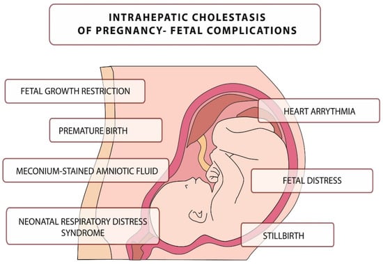
The disease is usually mild in pregnant women, but it can be fatal to the fetus, leading to numerous complications, including intrauterine death [
35]. The main complications related to this disease are presented in
Figure 1. They include, but are not limited to, neonatal respiratory distress syndrome (associated with the presence of BA in the lungs), meconium-stained amniotic fluid, preterm birth, and an increased risk of stillbirth [
36,
37,
38,
39,
40,
41,
42,
43].
Figure 1. Intrahepatic cholestasis of pregnancy–fetal complications.
Fetal death in ICP may be caused by sudden vasoconstriction of the placental surface vessels or the development of arrhythmias induced by elevated BA levels, although pathophysiology has not been confirmed [
38,
39,
40].
Bile Acids
Synthesis and Enterohepatic Circulation of Bile Acids
BAs are synthesized in the liver and constitute the major end product of cholesterol catabolism which proceeds by a multi-enzymatic pathway involving at least 17 different enzymes. In an adult human, approximately 500 mg of cholesterol is converted into BAs per day [
51,
52]. The rate of BA biosynthesis in the initial stage is partially limited by cholesterol 7α-hydroxylase, an enzyme from the cytochrome P450 family. The expression of the CYP7A1 gene encoding cholesterol 7α-hydroxylase, as well as the activity of the enzyme itself, are subject to strict, multifactorial regulation, including such factors as: BAs concentration, hormones (including insulin, glucagon, glucocorticosteroids), cholesterol (oxysterols), cytokines, and the daily cycle [
45,
53,
54,
55]. During the synthesis process, primary BAs, CA, and CDCA are first formed, which in the final stage are coupled with the amino acids: glycine or taurine (in a ratio of approximately 3:1). As a result of the conjugation process, primary BAs lose their ability to cross cell membranes. The process of bile formation is a process that requires the participation of energy from the breakdown of ATP (adenosine-5′-triphosphate) and consists of the transport of bile components through cell membranes, from hepatocytes to the bile ducts, against their concentration gradient. BAs are cleared into the bile ducts by a specific ATP-dependent transporter, bile salt export pump (BSEP), a product of the ABCB11 gene, which is highly specific and only transports conjugated BAs. BAs are the main component of bile, and their concentration in bile can be up to 1000 times higher than in the interior of the hepatocyte [
56]. The second major component of bile is phosphatidylcholine (PC), which is excreted into the biliary tract by the ATP-dependent multidrug resistance protein (MRP) 3 transporter, a product of the ABCB4 gene. Phosphatidylcholine plays an important, protective role in the interstitial space. During bile formation, BAs transported by BSEP form mixed micelles with phosphatidylcholine. These complexes have a protective effect on the epithelium lining the bile ducts against the toxic and detergent effects of bile salts and thus allow their secretion without damaging the surrounding cells. Phosphatidylcholine secretion, in parallel with bile salts, is necessary to maintain adequate bile flow [
57].
Bile produced in the liver is accumulated in the gallbladder until it is released into the gastrointestinal tract under the influence of postprandial cholecystokinin release—a peptide hormone that initiates postprandial contraction of the gallbladder [
59]. In the small intestine, BAs emulsify dietary fats, fat-soluble vitamins, and other lipids. Under the influence of the anaerobic bacterial flora in the intestine, a number of primary BA transformations occur, including deconjugation and dehydroxylation, which lead to the formation of secondary BAs, i.e., DCA and LCA [
60,
61,
62,
63,
64]. As a result of further changes, under the influence of both hepatic and intestinal mechanisms, substances of minor importance are formed, i.e., tertiary BAs: HDCA and UDCA. Through specific protein transport systems in enterocytes, bile salts are reabsorbed and reach the liver via the portal vein and are taken up by hepatocytes within the sinusoidal membrane [
58,
65].
Enterohepatic circulation is extremely efficient; the liver takes up about 95% of BAs and the remaining 5% is excreted in the feces. This loss is replaced by de novo synthesis of BAs in the liver [
51,
66].
The Biological Role of Bile Acids
The basic function of BAs in the human body is above all participation in digestive processes, and additionally participation in other physiological processes occurring in the human body [
66]. These processes include the emulsification and absorption of lipids and lipophilic vitamins contained in food, and the absorption of calcium. In combination with phospholipids, they form complexes that facilitate the dissolution of cholesterol and other lipids in bile. BAs affect the secretion of pancreatic enzymes and cholecystokinins. The osmotic pressure gradient that arises during the secretion of BAs into the bile ducts is one of the most important factors ensuring the proper flow of bile through the liver [
45,
57]. Due to their detergent properties, BAs can damage the cell membranes of the biliary epithelium and, consequently, damage the liver parenchyma [
67,
68,
69,
70].
Originally, four major functions of BAs were identified:
-
constitute the main important mechanism for the elimination of excess cholesterol through their synthesis and subsequent fecal excretion.
-
BAs and phospholipids prevent cholesterol from precipitating in the gallbladder by dissolving cholesterol in the bile.
-
they act as emulsifiers, increasing the availability of fats for pancreatic lipases, facilitating the digestion of triacylglycerols in the diet.
-
they enable the intestinal absorption of fat-soluble vitamins [
56,
67,
69].
After the characterization and isolation of FXR, for which bile acids are physiological ligands, the functions of Bas in the regulation of glucose and lipid homeostasis were confirmed. As indicated above, BAs binding to FXR impair the expression of genes that participate in overall BA homeostasis (e.g., FGF19). However, genes that participate in BA metabolism are not the only ones that are controlled by FXR action as an effect of binding BA [
73,
74,
75].
FXR controls genes that metabolize in the liver lipids (e.g., SREBP-1c), lipoproteins (e.g., apoC-II), glucose (e.g., PEPCK), and that are involved in hepatoprotection (e.g., CYP3A4, which is nifedipine oxidase).
Bile Acids in the Fetus
During intrauterine life, due to the anatomical and functional immaturity of the developing fetus’ liver, there are some functional and structural differences in the transformation and elimination of harmful substances. The main organ responsible for metabolism and elimination of metabolic products during fetal intrauterine life is the placenta [
77,
78,
79,
80]. The processes in the placenta are significantly similar to those in the liver of an adult human.
In utero, hepatic biosynthesis of BAs and bilirubin begins relatively early, and BAs themselves reach relatively high concentrations [
81,
82]. The dominant BA present in fetal serum is CDCA, while CA is present in lower concentrations. It has been observed that already in the 12th week of pregnancy, the CA/CDCA bile acid concentration ratio is 0.85, around the date of delivery (38–40 weeks of pregnancy) it is 1.9, in the neonatal period it is 2.5, and in an adult 1.6 [
83]. In addition, more BAs unbound or bound to glycine than to taurine are observed in fetal serum compared to adult serum. The above differences in the composition and concentration of BAs in the fetus, compared to adults, are caused by the immaturity of the enzyme systems involved in the metabolism of BAs in the fetus and their selective, transplacental exchange.
The process of BA synthesis in the fetus develops before the fetus has developed mechanisms that enable the effective secretion of BAs into bile. As a result, most of the synthesized BAs pass into the fetal blood serum, then a small fraction is excreted via the fetal kidneys into the amniotic fluid, and the remainder, the greater part, is eliminated through the placenta into the mother’s body. Most of the fetal pool of BAs is eliminated from the mother’s body via the gastrointestinal tract [
84,
85]. The identified factors influencing the maintenance of BA balance in the maternal-fetal circulation include: activity of hepatic metabolic pathways involved in the biosynthesis and transformation of BAs in both the mother and the fetus, the rate of BA elimination from the maternal organism, and the transport properties of the placenta [
81,
86].
Bile Acids in Physiological Pregnancy
In physiological pregnancy, the transplacental flow of BAs is supported by a gradient of BAs and bicarbonate concentrations and proceeds from the fetus to the mother [
6,
87]. This process depends on the protein transporters from the organic anion transporting polypeptides (OATP) family and ATP-dependent protein carriers from the ATP—binding cassette (ABC) family [
58,
88]. Simple diffusion, despite the possible free two-way flow of BAs through the placenta, does not appear to be the main mechanism responsible for the transport of BAs.
In physiologically running pregnancy, a slight increase in the total concentration of BAs in the blood serum is observed with the advancement of pregnancy. In studies that assessed the level of individual BAs, it was found that the concentration of secondary BAs did not change significantly, while the level of CDCA doubled around the time of delivery [
89]. The data on CA levels are inconclusive as some studies have shown a significant increase in CA levels in the third trimester compared to the first trimester, while others have not shown changes in CA levels [
89,
90].
Bile Acids in Pregnancy Complicated by ICP
The most important role in the pathomechanism of ICP is played by the increased concentration of BAs and their detergent properties, which accumulate in hepatocytes, causing damage to cell membranes and the release of aminotransferases, bilirubin, γ-glutamyl transpeptidases (GGTP), and alkaline phosphatase into the blood serum.
Most guidelines agree that typical clinical symptoms include pruritus of the skin, which is frequently generalized, but commonly begins and predominates on the palms and soles. The itching sensation strengthens at night, oftentimes involves the right upper quadrant pain and may be accompanied by nausea, poor appetite, sleep deprivation, or steatorrhea. Abnormalities in the biochemical functions of the liver should be identified in the absence of diseases with similar clinical symptoms and laboratory abnormalities [
94]. However, the type and reference values of laboratory markers of liver dysfunction that should be considered diagnostic for the diagnosis of ICP may differ significantly between societies [
94,
95,
96,
97,
98,
99,
100,
101,
102].
Pregnant skin pruritus is accompanied by elevated levels of hepatic function tests or BAs, not caused by other diseases, which normalize after delivery. In addition, the Royal College of Obstetricians and Gynaecologists (RCOG) guidelines emphasize that in the presence of typical clinical symptoms and abnormal levels of hepatic function tests, an increased concentration of BAs is not necessary for the diagnosis of ICP, and the definitive confirmation of the diagnosis is the resolution of clinical symptoms and normalization of laboratory markers of liver function after delivery [
98]. It is important to repeat the laboratory tests every week when the initial level of total aminotransferase and BAs are normal due to the fact that pruritus may precede an increase in serum BAs by several weeks. However, if UDCA becomes empirical, aminotransferases and BAs may never increase. Nevertheless 23% of pregnancies are affected by pruritus, but only a minority are caused by ICP.
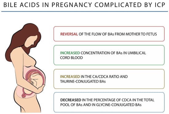
An altered BA profile is observed in women with ICP. CA remains the major BA and its concentration is significantly higher than that of CDCA, resulting in an increase in the CA/CDCA ratio and a decrease in the percentage of CDCA in the total pool of BAs. There is also an increase in the level of secondary BAs, mainly DCA, less than that of the primary BAs, which may indicate an impairment of their enterohepatic circulation [
109]. In addition, ICP shows an increase in taurine-conjugated BAs and a decrease in glycine-conjugated BAs, resulting in a lowered glycine/taurine ratio. Changes in BAs in pregnancy complicated by ICP are shown in
Figure 2.
Figure 2. Bile acids in pregnancy complicated by ICP.