Despite significant advances in biomedical research, osteochondral defects resulting from injury, an autoimmune condition, cancer, or other pathological conditions still represent a significant medical problem. Cell-based therapies and tissue engineering have gradually become promising alternatives. They combine the use of different types of cells and biomaterials to induce regeneration processes or replace damaged osteochondral tissue. One of the main challenges of this approach before clinical translation is the large-scale in vitro expansion of cells without changing their biological properties, while the use of conditioned media which contains various bioactive molecules appears to be very important.
1. Introduction
Diseases of the musculoskeletal system need to be strictly managed daily in clinical medicine. Affecting patients of all age groups, these pathologies significantly impact an individual’s health, as well as their psychical, social, and economic status [
1]. Acute traumas, autoimmune diseases, or tumors heavily affect both bone and cartilage tissues. If treated unproperly, patients may be severely disabled [
2]. The physiological regeneration of cartilage and bone is a complex process that involves tight cooperation between cellular and molecular agents near the affected site [
3]. Continuous remodeling, as a direct response to physiological or pathological stimuli, is typical for osteochondral tissue regeneration [
4]. In the case of bone regeneration, strict coordination between bone resorption provided by osteoclasts and new tissue formation arranged by osteoblasts needs to be maintained to keep the required integrity and functionality of the bone [
5]. Cartilage regeneration is affected by the fact that it is an avascular and aneural tissue. Moreover, resident chondrocytes, which are responsible for the production of important extracellular matrix (ECM) components, have a very low proliferative capacity, and, therefore, regeneration is considerably slowed down and, in many cases, it is not possible to achieve a satisfactory result.
Fractures represent the most common injuries of bone tissue, and the healing process involves inflammatory, proliferative, and remodeling stages [
6]. Effective blood supply to the affected area is crucial for the overall process of new bone formation and is related to the recruitment of adjacent stem cells that can differentiate into blood vessels [
7]. Although the tissue’s propensity for self-repair is high, lower-quality tissue can still be formed and negatively affect patients´ life. Limited vascularization of the cartilage is one of the reasons why its intrinsic self-healing capacity is so restricted and why it is practically unable to repair [
8]. Currently used surgical techniques often lead to the formation of scar tissue [
9]. Novel approaches provided by tissue engineering and regenerative medicine might offer promising alternatives for the effective healing of even large defects affecting bone, as well as cartilage. Due to their differential healing capacity, mesenchymal stromal cells (MSCs) have been intensively studied concerning osteochondral regeneration [
10].
Many in vitro studies have investigated their potential when applied as stem-cell therapy or combined with scaffolds [
11]. Long-running and detailed investigations of MSCs have helped us to elucidate complex processes which are responsible for their biological actions. It is accepted that MSCs operate via the secretion of active molecules such as proteins, lipids, cytokines, mRNAs, and growth factors, which seem to have a direct effect on tissue regeneration [
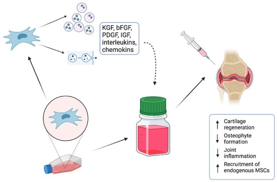
12]. To minimize the risks for patients, attention is currently drawn to the evaluation of the effect of the MSC-conditioned medium (MSC-CM) as a potential candidate for cell-free therapy (
Figure 1). In addition, various studies that focused on clinical MSC application have suggested that their regenerative properties are not related to the proximity of the applied cells to the targeted tissue [
13]. Moreover, it was also described that the applied cells could not maintain their lifespan for a long time, and, therefore, their effect needed to be provided by secreted bioactive molecules [
13]. The MSC secretome presents a package of bioactive agents secreted into extracellular space [
14]. It can be easily obtained with collection of CM and various techniques and protocols have been already applied for its analysis [
15].
Figure 1. MSC-conditioned medium (MSC-CM) containing various proteins, lipids, cytokines, mRNAs, and growth factors is a potential candidate for cell-free therapy of osteochondral defects.
2. Preparation of MSC-Conditioned Medium
MSC-CM is a type of cell culture medium that has been conditioned or modified by the paracrine action of MSCs. This medium contains various growth factors, cytokines, and other molecules secreted by the MSCs and can be used for various purposes, such as promoting cell growth and differentiation, studying cell signaling pathways, and evaluating the therapeutic potential of MSCs [
16]. The most common sources of stem cells which are used for MSC-CM “fabrication” are bone marrow MSCs, adipose tissue MSCs, and dental pulp MSCs [
17,
18].
The first step of MSC-CM production is the isolation and in vitro expansion of MSCs under static or dynamic conditions to obtain a suitable number of cells before starting the conditioning process. Recently, a fully closed, automated, and GMP-compliant cell expansion system (e.g., CliniMACS Prodigy
®) was developed which can be used to allow the large-scale production of MSCs. After a precise characterization of MSCs, culture media are replaced by “starving” serum-free basal media. In this step, MSCs start to secrete various bioactive molecules and extracellular vesicles. In some cases, MSCs are cultured under specific conditions, such as low oxygen tension or the addition of specific growth factors, to encourage the secretion of bioactive molecules into the media. After a certain period of time, typically 24–48 h, the conditioned media are harvested and filtered to remove any cell debris. Afterward, MSC-CM can be concentrated to increase the concentration of bioactive molecules. This can be carried out using various methods, such as centrifugation or ultrafiltration. The final step is the quality control of the fabricated MSC-CM. MSC-CM is usually analyzed for the presence of specific bioactive molecules, such as growth factors and cytokines, using techniques such as ELISA or mass spectrometry [
19,
20].
3. Scaffold Pretreating Using MSC-Conditioned Medium
Within the fields of tissue engineering and regenerative medicine, cells, scaffolds, and growth factors create the main pillars for effective tissue regeneration. Various studies confirmed that the application of scaffolds could enhance bone healing when compared to only SC application [
21]. Moreover, recent studies suggested that pretreatment of the scaffolds with MSC-CM might have provided even better results. Scaffold pretreating using MSC-conditioned medium refers to a technique that involves treating a scaffold material with a conditioned medium derived from MSCs before seeding cells onto the scaffold. MSCs are known to secrete various growth factors and cytokines that can stimulate cellular proliferation, migration, and differentiation. By pretreating the scaffold with MSC-CM, the scaffold can be “primed” with these growth factors and cytokines, providing a more favorable environment for seeded cells to adhere, proliferate, and differentiate [
22].
Garcìa-Ruìz and colleagues applied MSC-CM from bone marrow MSCs and used it to improve the biological properties of a 3D-printed composite scaffold [
23]. The results showed that cells seeded on pretreated scaffolds could attach and proliferate better on the scaffold´s surface. In addition, chondrogenic differentiation was promoted at a higher rate as well, making this approach a promising strategy used for osteochondral regeneration. A similar technique was utilized in the following studies to repair bone tissue under in vivo conditions [
5,
12]. Seeded scaffolds affected by MSC-CM repaired rat calvarial defects more successfully. These outcomes were also supported by in vitro results which revealed a higher expression of osteogenic genes in applied cells. In a more recent study performed by Chang and colleagues, tissue-engineered bones were fabricated with a combination of demineralized bone matrix and MSCs to treat large segmental bone defects. They also used the pretreatment of scaffolds with MSC-CM. Their results demonstrated that MSC-CM containing significant concentrations of various growth factors had a positive effect on MSC migration, proliferation, and osteogenic differentiation [
24].
The improved bone-healing capacity of MCS-CM related to electrical stimuli and a 3D culture system was investigated in a study carried out by Hwang and colleagues [
25]. MSCs were seeded on collagen sponges and constructs were/were not exposed to electric stimuli. Combining the mentioned culture techniques seemed to significantly improve inflammatory-mediated bone loss repair. Ogata and coworkers used atelocollagen in combination with human MSC-CM to treat bone defects. They showed an enhanced migration of endogenous osteoprogenitor cells and accelerated bone regeneration [
26]. More recently, Dilogo and coworkers conducted a large animal study investigating the effect of hydroxyapatite and MSC-CM on the treatment of critical-sized bone defects. They applied MSC-CM alone or with the supplementation of BMP-2. They demonstrated the most significant effect on total callus formation in the group treated with MSC-CM and BMP-2, while the osseous area was found to be highest in the MSC-CM group [
27].
This entry is adapted from the peer-reviewed paper 10.3390/ijms24109054