Mechanisms Governing the Acquisition of Castration-Resistant Prostate Cancer: Comparison
Please note this is a comparison between Version 1 by Mingqiu Hu and Version 2 by Vivi Li.

Despite aggressive treatment and androgen-deprivation therapy, most prostate cancer patients ultimately develop castration-resistant prostate cancer (CRPC), which is associated with high mortality rates. However, the mechanisms governing the development of CRPC are poorly understood, and androgen receptor (AR) signaling has been shown to be important in CRPC through AR gene mutations, gene overexpression, co-regulatory factors, AR shear variants, and androgen resynthesis. A growing number of non-AR pathways have also been shown to influence the CRPC progression, including the Wnt and Hh pathways. Moreover, non-coding RNAs have been identified as important regulators of the CRPC pathogenesis. 

  • destructive resistance prostate cancer
  • androgen receptor
  • Wnt pathway
  • Hh pathway
  • ncRNAs

1. Introduction

Prostate cancer is the second leading cause of cancer-related death among men worldwide [1]. In China, relative to the rates from before 1990, PCA incidence has risen by 98.21% while corresponding mortality rates have fallen by 3.82%. Consistently, PCA incidence continues to rise [2]. At the time of initial diagnosis, most of the PCA patients exhibit progressive or metastatic disease in China, with androgen-deprivation therapy (ADT) being the treatment of choice for most of the patients with metastatic PCA (European Society of Urology; Guidelines for the Treatment of Prostate Cancer, 2020) [3]. While initially highly effective, however, the median time that patients respond well to ADT is just 18–24 months, after which the patient will progress to develop castration-resistant prostate cancer (CRPC), which is generally defined based upon serum testosterone levels (<50 ng/dL or 1.7 nmol/L) and biochemical (prostate-specific antigen (PSA) levels increasing three times in a row within one week, with at least two of these increases being by more than 50% of the lowest levels, PSA > 2 ng/mL) or radiological (such as two or more new bone lesions in bone scans or soft tissue lesions based upon solid tumor response assessment criteria) evidence of progression after castration treatment [4]

2. Androgen Receptor Pathways

The androgen receptor (AR) is a 110 kDa 919 amino acid nuclear receptor (NR) family member encoded on the X chromosome (q11-q12) (Figure 1A). The AR is composed of a central DNA-binding domain (DBD), and C-terminal ligand-binding domain (LBD), an N-terminal structural domain (NTD), and a hinge region linking the LBD and DBD [5][6][7][8][5,6,7,8]. The AR is encoded by eight exons (Figure 1), with exon 1 encoding the NTD, exons 2,3 encoding the DBD, and exons 4–8 encoding the LBD [9][10][11][9,10,11].
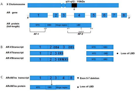
Figure 1. Structural overview of ARs and AR-Vs (AR-V3,7,9 and ARv567es). (A) Structural overview of the AR gene, located on the X chromosome q11-q12, encoding 919 amino acids and consisting of eight exons. The DHT and T ligands to the AR LBD; (B) The mechanisms underlying AR-V3, AR-V7, and AR-V9 production. Exons 4–8 are sheared to produce truncated AR-Vs that lack a LBD and Hinge region; (C) Mechanisms governing the production of ARv567es. Exons 5–7 are missing, resulting in truncated AR proteins and the lack of a LBD. AR = Androgen receptor; AR-Vs = AR variants; NTD  =  N-terminal transcriptional domain; DBD  =  DNA-binding domain; LBD  =  C-terminal ligand-binding domain; CE5  =  cryptic exon 5; CE3  =  cryptic exon 3; PAS  =  polyadenylation site; CE4  =  cryptic exon.
Several different mechanisms link AR to the development of CRPC, including AR upregulation, de novo androgen synthesis, co-regulatory factor activity, AR gene mutations, and altered splicing (Figure 2).
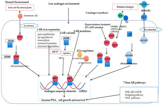
Figure 2. Mechanisms governing the progression of prostate cancer to castration-resistant prostate cancer (CRPC). T: Testosterone; DHT: Dihydrotestosterone; AR: Androgen receptor; AR-Vs: AR variants; DHEA: Dehydroepiandrosterone; A2: androstenedione; 11OXHA4: 11-oxygenated androgens; 11KT: 11-ketotestosterone; 11KDHT: 11-ketodihydrotestosterone.

3. Classic Mechanisms of AR Pathway Enhancement

3.1. AR Overexpression

The AR signaling reactivation most commonly occurs at the level of gene amplification or protein upregulation, with up to 80% of the CRPC patients harboring high AR gene copy numbers, among whom 20–30% of patients exhibit high gene amplification levels [12][13][14][12,13,14]. Such amplification, however, is uncommon among PCA patients that have not undergone hormone therapy. In one fluorescence in situ hybridization (FISH) study, the researchers found AR amplifications to be absent in the benign prostatic hyperplasia (BPH) samples, present in just 2% of the primary PCA tumors, but present in 23.4% of the CRPC tumors [11][15][11,15]. At the genetic level, two-fold increases in the AR mRNA expression levels have been reported in the CRPC tumors [16]. The AR overexpression can additionally occur through the enhanced stabilization of AR mRNA or proteins, or through increased transduction rates mediated by heat-shock proteins (HSPs), with HSP40 and HSP70 having been shown to bind the AR NTD and to then interact with the AR LBD, thereby contributing to its overexpression [17][18][17,18].

3.2. De Novo Androgen Synthesis

The persistent de novo production of androgens within the CRPC tumors can additionally contribute to enhanced AR activation and the progression of hormone-refractory prostate tumors [17]. Even following ADT, dihydrotestosterone (DHT) levels in the prostate tissue remain at ~25% of baseline; these levels are sufficient to drive altered gene expression and tumor progression through tumor epithelial cell signaling [19][20][21][22][23][24][25][19,20,21,22,23,24,25]. Nishiyama et al. further conducted a Gleason score analysis of the CRPC patients in which they found that the low levels of DHT in these patients were sufficient to promote AR receptor activation and tumor progression [19][26][27][19,26,27]. When multiple steroids (HSD3B2AKR1C3CYP17A1, and CYP11A1) are present [28][29][30][31][28,29,30,31], adrenal androgen precursors can be used to promote DHT synthesis within tumors through the 5α-diketone pathway, leading to dehydroepiandrosterone (DHEA) and androstenedione conversion into DHT without any requirement for testosterone [32][33][34][35][32,33,34,35]. Abiraterone is a specific inhibitor of CYP17A1, and it was also the first drug approved to treat CRPC [32][36][32,36]. In recent work, 11-oxygenated androgen (11OHA4) was additionally identified as a critical source of intratumoral androgen production [25][37][38][25,37,38], as it can serve as a precursor for the production of the peripherally active androgens, 11KT and 11KDHT. Functionally, 11KT-mediated AR activation has been shown to be similar to that mediated by testosterone, with 11KT and 11KDHT binding to the AR receptors with an affinity comparable to that for testosterone and DHT, respectively [39][40][41][42][39,40,41,42]. Pretorius et al. consistently found that 1KT and 11KDHT were capable of driving cellular growth through the upregulation of the AR regulatory genes, including KLK3, TMPRSS2, and FKBP5 in the LNCaP and VCaP PCA cell lines [40]. In vitro conversion experiments have also shown that 11KT and 11KDHT can remain present in the LNCaP and VCaP cells for longer than testosterone and DHT [40][43][40,43].

3.3. AR Co-Regulatory Proteins

Over 180 such AR co-regulatory proteins have been identified to date, with both co-repressors and co-activators functioning in a synergistic manner to regulate AR transcription [44]. These proteins can modulate transcription, RNA splicing, and epigenetic regulatory mechanisms, including methylation, acetylation, phosphorylation, and ubiquitination, thereby shaping PCA development and progression [45][46][47][48][49][50][45,46,47,48,49,50]. Notably, these co-regulatory proteins can promote the sustained transcriptional activity of AR even under low levels of androgen availability. The interaction of specific co-activating proteins with AR, including JMJD2C, LSD1, 37CBP/P300, p160/SRC, and SUV39H2, can drive enhanced AR activation and the concomitant upregulation of the AR-dependent genes to augment tumor growth. Consistently, the inhibition of these activators has been linked to reduced AR expression and PCA tumor growth in ex vivo analyses [51][52][53][54][51,52,53,54]. Askew et al. reported that SUV39H2 can function as a co-activator for AR that enhances its androgen-dependent transcriptional regulatory activity through interactions with MAGE-A11 and AR under androgen-deficient conditions [49]. Moreover, the AEEB1 gene encoding the regulatory protein βArr1 was found to be upregulated in the CRPC tumor tissues, with the βArr1 deletion resulting in impaired PCA tumor growth, invasion, and metastatic progression in vitro and in murine model systems [55]. The AR co-repressors, in contrast, function to counteract the activity of the co-activator proteins [56][57][56,57]. Tan et al. reported that the CRPC tumor cells exhibited decreased CKβBP2/CRIF1 expression relative to the levels observed in androgen-dependent PCA tumor cells, with a corresponding increase in the expression of the co-activator STAT3, contributing to synergistic AR signaling enhancement [58]. As such, the downregulation of the co-repressors and the upregulation of the co-activators can spur the CRPC onset and progression [11].
Video Production Service