Your browser does not fully support modern features. Please upgrade for a smoother experience.
Please note this is a comparison between Version 1 by Xiaoding Lou and Version 2 by Camila Xu.
Organelles are important subsystems of cells. The damage and inactivation of organelles are closely related to the occurrence of diseases. Organelles’ functional activity can be observed by fluorescence molecular tools. Nowadays, a series of aggregation-induced emission (AIE) bioprobes with organelles-targeting ability have emerged, showing great potential in visualizing the interactions between probes and different organelles.
- organelles
- peptide
- fluorescence imaging
- aggregation-induced emission
1. Introduction
One of the reasons why eukaryotes are more complicated than prokaryotes is that eukaryotes contain many independent inner membrane systems, which are called organelles or subcellular compartments [1][2][1,2]. Each organelle has unique structural characteristics and plays a different role in various physiological processes. They maintain cell biochemical reactions efficiently, diversely and stably. The organelles of eukaryotes contain a cell membrane, mitochondria, a nucleus, lysosomes, an endoplasmic reticulum, a Golgi apparatus, lipid droplets, ribosomes, etc. [3]. These organelles can not only work independently but also cooperate with each other. On one hand, working independently could guarantee orderly physiological activities. For instance, mitochondria are mostly responsible for producing energy, controlling the apoptosis progress and regulating intracellular calcium and reactive oxygen species (ROS) [4][5][6][4,5,6]. Lysosomes contain many hydrolases that can hydrolyze multiple waste or abnormal proteins [7][8][7,8]. On the other hand, complicated physiological activities require the cooperation of multiple organelles, such as the synthesis of proteins. At first, proteins need to be transcribed in the nucleus and translated in ribosomes. Subsequently, they are modified and packaged by the Golgi apparatus to obtain the active proteins with intended functions. In this case, dysfunction of organelles was able to cause various diseases such as cancer, metabolic disorders, cardiovascular and neurodegenerative diseases and so on [9][10][11][12][13][14][9,10,11,12,13,14]. For example, when mitochondria are damaged, cytochrome c and mitochondria outer membrane proteins are released into the cytoplasm and interact with apoptotic protease activator 1, which leads to the activation and recruitment of the caspase family, ultimately resulting in the apoptosis of cells [15][16][17][15,16,17]. Thus, it is very important to image and detect these organelles. It helps us to study the different physiological and biochemical processes of cells, understand the operation mechanism of the life system and establish the foundation for biomedical research.
Fluorescence imaging technology is widely used in various fields due to its non-invasiveness, high sensitivity and high temporal spatial resolution [18][19][20][18,19,20]. Aggregation-induced emission (AIE) is a phenomenon by which luminogen does not emit fluorescence in a dilute solution but emits strong fluorescence in a high concentration or solid state [21][22][23][24][21,22,23,24]. AIE luminogens (AIEgens) generally have a molecular rotor structure. In the free state, the molecular rotor can rotate freely and activate non-radiative transitions to consume excitation energy. In the aggregated state, molecular rotor movement is restricted, and excitation energy is mainly released through radiative transitions, which results in the bright and stable fluorescence emission [25][26][27][25,26,27]. In recent years, due to their outstanding properties, such as a high quantum yield, a resistance to photobleaching, a large Stokes shift and photosensitivity, AIEgens have been widely used in biomarker detection and imaging [28][29][28,29], drug delivery [30][31][30,31], surgery navigation [32][33][32,33], anti-bacteria processes [34], phototherapy [35][36][37][38][39][40][35,36,37,38,39,40] and a series of biomedical application areas.
AIEgens can be conjugated with biological macromolecules (peptides, polysaccharides, nucleic acids, proteins, etc.) through covalent bonds [22][41][42][22,41,42]. Among them, peptides are widely developed in the biomedical field. When peptides bind to AIEgens, the addition of peptides restricts the rotation of AIEgens, which leads to AIEgens emitting fluorescence [27]. AIEgens have the advantages of photobleaching resistance, photosensitivity and a high quantum yield. At the same time, AIEgens are generally lipid-soluble and more hydrophobic and enter the cell difficultly. The introduction of peptides can improve the hydrophilicity of AIEgens and make them have good biocompatibility, which is conducive to the application of bioprobes in the biomedical field. In addition, the introduction of peptides can also give AIEgens various functions, such as the specific recognition function, toxic function, targeting function, etc. Simultaneously, the immunogenicity of bioprobes can be reduced. Therefore, the design of biomacromolecule-functionalized AIEgens can be used as a practical tool to obtain complex information at the biological level, analyze biomacromolecule interactions and understand disease mechanisms. At the same time, peptides are unstable and easily degraded by bioactive substances. ResWearchers hope that obstacles can be overcome in future research [43][44][45][46][43,44,45,46].
There are various strategies for designing peptide-targeting organelles. Among them, the strategy for the design of cell membrane-targeted peptides is the electrostatic interaction of arginine rich in positive charge with phosphate groups on the cell membrane. In addition, the cell membrane can be targeted by peptides that can bind to cell membrane receptors. The nuclear localization peptide (NLS) peptides could bind to the nucleoprotein. The peptides targeting the mitochondria are usually liposoluble cationic peptides because mitochondria have negative membrane potential and a hydrophobic environment. The lysosomal-targeted peptides are generally reactive under acidic conditions and can bind to lysosomal proteins. ER-targeted peptides could bind to the KDEL receptor (Figure 1). According to different organelles, this researchticle reviews the recent progress of AIEgen-peptide bioprobes in organelles imaging. The structural characteristics and application strategies of AIEgen-peptide bioprobes in different organelles are summarized.
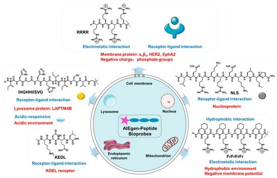
Figure 1. The mechanism of bioprobes-targeting organelles.
2. Cell Membrane-Targeted Bioprobes
The cell membrane is composed of an amphiphilic phospholipid bilayer with a large negative potential. It plays an important role in maintaining homeostasis, controlling substance transport and regulating signal transduction [16][17][47][16,17,47]. At present, cell membrane-targeting fluorescent probes usually consist of a fluorophore and an anchoring element [15]. The cell membrane-anchoring element is generally the substance including: penetrating peptides [48], alkyl chains [49], cholesterol [50], protein ligands [51], antibodies, aptamers [52], etc. Protein ligands can be utilized to target cell membrane receptors; for example, a peptide sequence RGD often acts as a targeted peptide of the integrin receptor in the cell membrane. As shown in Figure 2 compound (1), Liu et al. reported a new bioprobe TPS-2cRGD by integrating an AIEgen (TPS) with cyclic arginine-glycine-aspartic tripeptide (cRGD), a targeting ligand to the cell membrane integrin αvβ3 receptor [53]. The excitation wavelength of the bioprobe is 356 nm, and the maximum emission wavelength is 480 nm. The bioprobe overlapped well with commercial cell membrane dyes and was used to track αvβ3-positive cancer cells. The negative membrane potential and lipid solubility of the phospholipid bilayer lead to the enrichment of lipophilic and cations molecules in the cell membrane. Because of the abundant positive charge, cell-penetrating peptide RRRR was usually used to improve the targeting ability of the cell membrane. At the same time, palmitic acid was usually used to target the cell membrane due to its lipid solubility. Liang et al. reported an AIEgen-peptide bioprobe TR4 [54]. As shown in Figure 2 compound (2), the bioprobe consisted of three elements: palmitic acid, cell-penetrating peptide (RRRR) and tetraphenylethylene (TPE). The excitation wavelength of the bioprobe is 330 nm, and the maximum emission wavelength is 466 nm. When MCF-7 cells were incubated with TR4, the cell membrane was labeled. TR4 showed good photostability and biocompatibility and low toxicity. It opened the door of the TR4 in cell membrane imaging. To further improve cell membrane-targeting ability, ouresearchers' group combined RRRR, RGD and palmitic acid to AIEgen to track the imaging of the cell membrane [55]. As shown in Figure 2 compound (3), RTP (λex: 330 nm, λem: 500 nm) consisted of three elements: a RGD-targeted peptide, a palmitic acid-modified hydrophilic peptide (Pal-RRRR) and AIEgen (T-MY). The palmitic acid had a cell membrane-targeting function and was inserted into the cell membrane through hydrophobic interactions. A RGD-targeted peptide was bound to integrin receptors on the cell membrane. A hydrophilic peptide (RRRR) was bound to the cell membrane through electrostatic interactions. Under the comprehensive combination of RGD and Pal-RRRR, RTP successfully achieved the imaging of the cell membrane precisely and robustly. Simultaneously, RTP showed durable stability and a strong resistance to photobleaching.
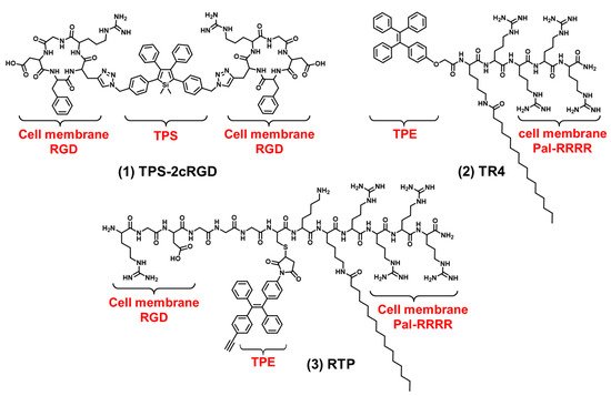
Figure 2. The chemical structures of cell membrane-targeting bioprobes.
In addition to the integrin receptors overexpressed in most cancer cells, some cancer cells also have specific receptors. For example, human epidermal growth factor receptor 2 (HER2) is overexpressed in breast cancers. HER2 is a tyrosine receptor kinase that can induce dimer formation with itself and then lead to downstream signal activation. Wang et al. designed a AIEgen-peptide bioprobe (TPM) that targeted the HER2 receptor on the cell membrane [56]. As shown in Figure 3 compound (4), the bis-pyrene (BP) element with the AIE property for fluorescence reporting, the peptide (YCDGFYACYMDV) bound to the HER2 receptor on the surface of cell membrane and the peptide (KLVFF) was used as a hydrophobic element that could be assembled into nanofibers. When the peptide bound to HER2 on the cancer cells’ surface, TPM (λex: 380 nm, λem: 520 nm), was converted into nanofibers and attached to the cell membrane strongly, this restricted the rotation of AIEgen and the imaged cell membrane. In addition, TPM also destroyed the formation of HER2 dimers, thereby blocking the downstream signaling pathway and leading to tumor cell apoptosis. This is a bioprobe targeting cell surface receptors in order to target the cell membrane which has favorable potential for future clinical applications. In addition, Eph receptor A2 (EphA2) is an adrenaline tyrosine receptor kinase overexpressed in tumor-specific membranes. EphA2 plays an important role in promoting cancer malignancy. Therefore, the specific imaging of EphA2 is of great significance for the diagnosis of tumors. Ding et al. constructed a self-assembling bioprobe (DBT-2FFGYSA) that selectively targeted the EphA2 protein on the cell membrane [57]. As shown in Figure 3 compound (5), it consisted of three elements: the middle element was AIEgen (DBT), the two aromatic phenylalanine (FF) were the self-assembled element and the peptide sequence YSAYPDSVPMMS (YSA) could specifically bind to EphA2. The excitation wavelength of the bioprobe is 490 nm, and the maximum emission wavelength is 642 nm. The bioprobe selectively targeted EphA2 receptors and caused AIEgen to aggregate in the cell membrane. In addition, this bioprobe could effectively transform cold tumors into hot tumors to stimulate immune response and inhibit tumor growth. In addition to the strategy of the bioprobe bound to the cell membrane receptors, 16-carbon alkyl chains can adhere firmly to the cell membrane. Zhang et al. reported a membrane-targeted AIEgen-peptide bioprobe (CTGP) imaging the cell membrane [58]. As shown in Figure 3 compound (6), it was composed of the AIEgen element (TPE), cathepsin B (CB) enzyme-responded peptide element (GFLG) and 16-carbon alkyl chain element (C16), which targeted cell membrane. The excitation wavelength of the bioprobe is 370 nm, and the maximum emission wavelength is 470 nm. The bioprobe was cleaved by CB when the bioprobe was present in tumor cells with CB overexpression. CTGP was transformed from spherical nanoparticles into nanofibers. TPE was encapsulated by nanofibers on the cell membrane, so CTGP can image the cell membrane obviously. In addition, this encapsulation characteristic could prevent the DOX efflux in tumor cells and prevent the drug resistance of tumor cells from inhibiting the drug efflux. This encapsulation of the cell membrane opened a new avenue for tumor imaging and drug resistance research.
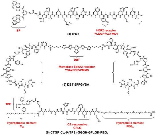
Figure 3. The chemical structures of cell membrane-targeting bioprobes relying on the cell membrane protein.