Gq Signaling in Autophagy Control: Comparison
Please note this is a comparison between Version 3 by Beatrix Zheng and Version 2 by Beatrix Zheng.

All processes in human physiology relies on homeostatic mechanisms which require the activation of specific control circuits to adapt the changes imposed by external stimuli. One of the critical modulators of homeostatic balance is autophagy, a catabolic process that is responsible of the destruction of long-lived proteins and organelles through a lysosome degradative pathway. Identification of the mechanism underlying autophagic flux is considered of great importance as both protective and detrimental functions are linked with deregulated autophagy. At the mechanistic and regulatory levels, autophagy is activated in response to diverse stress conditions (food deprivation, hyperthermia and hypoxia), even a novel perspective highlight the potential role of physical forces in autophagy modulation. To understand the crosstalk between all these controlling mechanisms could give uspeople new clues about the specific contribution of autophagy in a wide range of diseases including vascular disorders, inflammation and cancer.

  • GPCR
  • Gq
  • autophagy
  • oxidative stress
  • mechanotransduction
  • extracellular matrix

1. Introduction

Cells are constantly exposed to a huge number of intra-and extra-cellular signals which requires a proper cell response for adaptation and homeostasis control [1]. Among the different sensors and integrators for such varying signals, the family of G-protein coupled receptors (GPCRs) arise as one of the most important transducers [2][3]. With over 800 GPCRs encoded in the human genome, this family of transmembrane receptors can bind a plethora of stimuli which include hormones, metabolites, and inflammatory mediators, influencing a diverse network of signaling pathways [4].
Apart from the classical chemical inputs, recent studies have begun to unravel the potential of mechanical and architectural properties of the environment as a new and alternative way to dynamically modulate cellular homeostasis [5]. Extracellular Matrix (ECM) is considered as an intricate meshwork of proteins and carbohydrates organized in a specific manner which acts not only as a reservoir of growth factor and bioactive molecules but also as a highly dynamic entity which provide mechanical rigidity and structural support to the cells [6].
All GPCRs contain seven transmembrane domains embedded within the cell membrane with several intracellular domains that trigger guanine nucleotide binding proteins downstream signaling pathways and, interestingly, extracellular domains in GPCRs have been reported as potentially critical elements in the interaction with components of the ECM and as a force sensing mechanism [7][8]. This opens the questions about the potential of GPCRs as linkers in the integration of all these signals (both chemo- and mechanical stimuli) raising the possibility that specific GPCRs and its downstream effectors can mediate the crosstalk between both types of triggers during the control of homeostatic feedback loops. Indeed, the ECM has the potential to significantly impact virtually on every physiological cellular mechanism [9][10]. Excessive deposition and increased stiffness of ECM has been directly linked with the progression of many different pathologies and has the potential to regulate cellular metabolism [11].
An important downstream process in the crossroad between chemo- and mechanosensing regulatory responses is autophagy [12]. The catabolic activity of autophagy is a fundamental cellular process that eliminates molecules and subcellular elements (including proteins, lipids, nucleic acids and even organelles) through a lysosomal-mediated degradative pathway for providing energy sources for ATP production or building blocks for protein synthesis [12]. Activated by different types of stress including those related with DNA damage, hypoxia, oxidative stress, inflammation, and food deprivation and by different challenges arising from mechanical (stretching, shear stress or hypotonic shock), autophagy can have both beneficial and deleterious effects [13][14][15]. Indeed, vascular and heart diseases, infectious diseases, neurodegenerative pathologies, and cancer have all been related to autophagy dysfunctions. Thus, autophagy represents a double-edge sword and for this reason the possibility to regulate autophagy in a time- and local-dependent manner represents a novel and valid therapeutic approach to control most of these disorders.

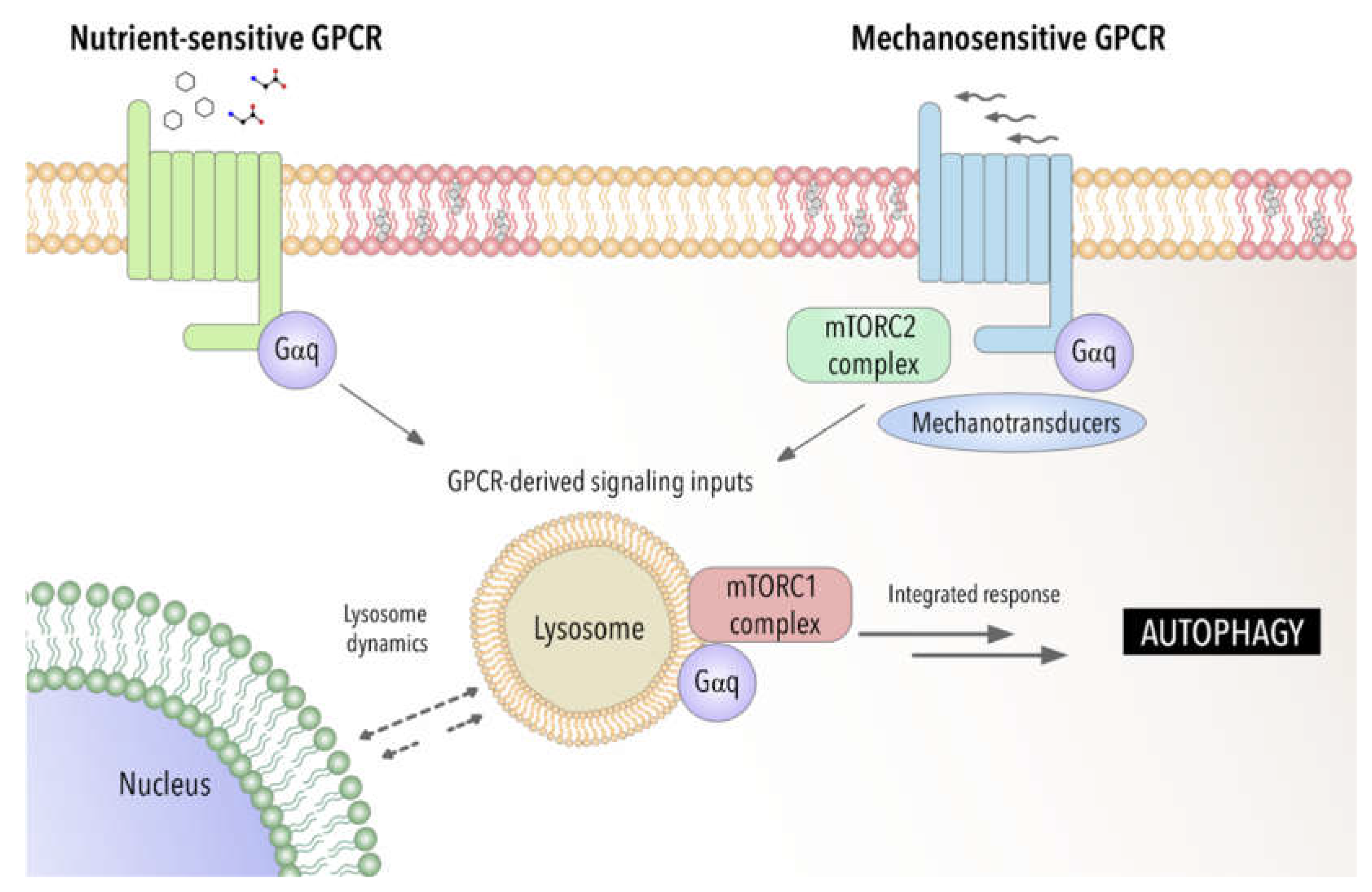
2. Autophagy between Nutrient, Mechanical and Oxidative Stress: An Emerging Role of Gαq

Autophagy is a highly conserved mechanism for cellular degradation in which cytosolic waste, protein aggregates and organelles are sequestered into a double-membrane vesicle (autophagosome) and delivered into lysosomes for breakdown [16]. Autophagy is orchestrated by sequential steps tightly control machinery in which ATG and associated proteins regulate the formation and maturation of autophagosomes into autolysosomes [17]. Under pathophysiological conditions, damaged or excessive accumulation of organelles such as endoplasmic reticulum, ribosomes, mitochondria, lipid droplets and peroxisomes can be degraded through mechanisms mediated by a collection of specific autophagy-related proteins [13][16].
Typically, autophagy stimulation depends on the mTOR system modulation [18]. It is initiated by the ULK1 complex (Unc-51-like-autophagy-activating kinase) which can receive input from cellular energy balance, and the availability of nutrients from mTORC1 (Rapamycin Complex1) and AMPK signaling networks (AMP-activated protein kinase). Canonical initiation of autophagy entails those metabolic stresses (chemical stimuli) such as nutrient deprivation. This causes mTORC1 dissociation from ULK1 which becomes active and binds to ATG13 and FIP200 triggering autophagosome formation [19][20]. In addition, mechanical stresses are also involved in autophagic flux control. Although it is unclear whether mechanical stresses may play a direct role in ULK1 activation, it has been reported that the mechanosensitive mTORC2 complex can indirectly induce ULK1 activation via inactivation of mTORC1 repressor function, through a FAK (focal adhesion kinase)-dependent mechanisms [21][22][23].
The extracellular matrix (ECM) constitutes a dynamic and plastic network of biophysical and biochemical factors that maintains tissue homeostasis. Changes in ECM composition, elasticity, and structure have also been reported to impact on autophagic flux raising the potential of matrix biology modulation as a critical controller of this process [9]. Apart from the interaction with physicochemical environmental imposed by the ECM, cells are also subject, and they have to respond to a great variety of mechanical forces due to other external forces, such shear stresses of fluid pressure (e.g., blood vessels), lateral stretches and compression (such in the case of muscles). Overall, this plethora of short- and large- scale forces elicit an adaptive cellular response in which autophagy seems to be a critical player.
In this adaptive response, cells can sense extracellular mechanical cues in different ways. This includes cellular adhesion complexes with ECM and/or cells, mechanosensors such as proteoglycans localized at the cell surface or mechanically activated ion channels (e.g., Piezo) [24][25] even plasma membrane-associated structures such cilium, caveolae, and clathrin-coated pits [26][27][28]. Moreover, multiple intracellular organelles, including autophagosomes, can also sense mechanical forces [29][30][31].
Mechanical cues imposed by forces or microenvironmental cues may affect the autophagic process through specific crosstalk with autophagy regulatory proteins (such as mTORC system, or AMPK pathway) or via mechanical regulation of cytoskeletal elements or phospholipid membranes, which can be crucial in the autophagic process [32][33][34]. Interestingly, a direct link between cell attachment to ECM and autophagy has also been reported [35]. Loss of cell attachment with the ECM usually results in programmed cell death via anoikis. In some cases, ECM-cell detachment can rapidly activate autophagy allowing for survival and re-attachment to the substrate [36]. However, it remains elusive the mechanism controlling this process with integrin-mediated adhesions emerging as a critical element [37]. Furthermore, ECM and integrin-mediated adhesion may trigger autophagy via FAK and ILK (integrin linked kinase) being relevant in different processes such as for immunosurveillance [38].
Additionally, matrix constituents have been shown to regulate autophagy in both directions, promoting or inhibiting this process, depending on matrix stiffness but also on its specific composition [39]. Indeed, recent studies have demonstrated an active and dynamic signaling role of specific extracellular matrix components on autophagic regulation which can act in both positive (activators) or negative (inhibitors) ways. Among them, decorin, collagen VI, kringle 5, endorepellin and endostatin function as activators and pro-autophagic matrix constituents that engage a diverse array of cell surface receptor for autophagic initiation [40][41][42]. In contrast, laminin α2 acts as an inhibitor. Thus, absence of laminin α2 permits excessive autophagy [43]. Interestingly, the action of all these components seems to be independent of the predominant nutrient concentrations. This unique class of matrix molecules can function as an alternative mechanism to the classical nutrient deprivation mechanism to safeguard cellular homeostatic balance through autophagic control and providing a new mechanism through which GPCRs could also be participating in the regulation of autophagy.
GPCRs are directly linked with mechano-transduction mechanisms. Supporting this idea, it has been demonstrated that mechanical perturbations can modulate GPCR conformational transitions [44]. Moreover, the response to shear stress can be directly modulated through the Gαq-coupled GPCR, GPR68 [45][46]. Interestingly, PKCζ, a protein that wthe researchers have described as new interactor of Gαq [47], can be regulated by shear stress and activated by disturbed flow in atheroprone areas [48]. Further evidence on this mechanical stimulation comes from studies on adhesion GPCRs which display a long extracellular N-terminus with adhesive properties to the extracellular matrix or N-glycans modifications. These glycan chains have been reported to be able to be activated in the context of mechanical traction forces [49][50]. Further investigations are required to really address how these forces can structurally activate GPCRs in different contexts.
As a sequential process involving membrane remodeling events, autophagy is mechanically linked to cytoskeletal dynamics that lead to mechanical deformation and transport. Actin filaments and fibers and microtubule network can act as critical modulators in the control of organelle dynamics and autophagy control [51].
Although the most classical autophagy process relies on the delivery of cytoplasmic material to lysosomes via the double-membraned autophagosome, another form of autophagy, known as chaperone-mediated autophagy (CMA), occurs in the lysosome directly [52]. Thus, lysosomes can be considered critical hubs in the modulation of autophagic control. Lysosomes constitute a highly dynamic organelle which display important changes, including acidification and enhanced enzymatic activity. Furthermore, this organelle can move from the perinuclear localization to the cell periphery with important implications in cell metabolic control [53].
An emerging aspect to be considered is how lysosomal position can directly modulate its function. Lysosomes are transported bidirectionally through the microtubule network by dynein and kinesin motors, with microtubule motors such as dynein modulating the movement of lysosomes from the periphery towards a perinuclear location, while kinesins promote the scattering of lysosomes through the cytoplasm [54][55]. Recent evidence suggest that the distribution of lysosomes can be controlled by stimulation with different inputs. Under cell starvation, autophagosomes and lysosomes move toward the center of the cell facilitating the fusion of both compartments and the degradative capacity [56].
Several protein complexes have been implicated in the regulation of lysosomal positioning. One important regulator is the Rab7, a small GTPase involved in the coordination of appropriate coordination and homeostatic control of late endosomes and lysosomes [57][58]. Recently, WDR91 a Rab7 effector has been reported to be essential for lysosomal function [59]. In addition, the transcription factors TFEB and TFE3 are essential to promote the expression of multiple lysosomal genes. They play critical roles in the modulation of lysosomal biogenesis and distribution through the control of the lysosomal transmembrane protein TMEM55B (transmembrane protein 55B) expression [60]. TFEB acts as a link between autophagic process and lysosome biology [61], interacting with mTORC1 complex but not with mTORC2 controlling the lysosomal localization and function of mTORC1 [62].
OuThe researchers' recent studies strongly suggest that Gαq is a critical autophagy regulator raising the potential to control the shift between mTORC1-mTORC2 switch through lysosome control. We hThe researchers have recently reported a higher number of autolysosomes in cell lacking Gαq/11 compared to the wild type of counterpart which is directly linked with the involvement of this protein in the modulation of autophagy [63]. Immunofluorescence with LAMP1, a lysosomal-endolysosomal compartment marker, revealed that Gαq KO cells showed a predominant perinuclear lysosomal distribution in basal conditions, a phenotype that was mimicked by upon starving conditions. These results are consistent with a not-yet described role of Gαq in the modulation of lysosome dynamic regulation.
Furthermore, although many components of the autophagic machinery and autophagy receptors which are involved in the regulation of the process are being subjected to lysosomal degradation, in the case of Gαq, its presence in lysosomes does not alter its protein expression levels neither in basal nor in nutrient stress conditions which reinforce a critical role of this protein in the modulation of the autophagic control process.
Recent studies have demonstrated by using a biosensor the presence of GTP-loaded Gαq/11 at endosomes [64]. This provides a powerful tool to be applied to other cellular organelles such lysosomal compartment to fully address the specific function of GPCR- Gαq signaling at these organelles. Furthermore, this raises the possibility that GPCR-Gαq signaling may act as a modulator of autophagy by acting as a switch between chemical and mechanical cellular responses (see Figure 1).
Figure 1. Gαq as a potential integrator of chemical and mechanical signals modulating autophagic process. Involvement of Gαq interactome-autophagy control in pathophysiological settings.

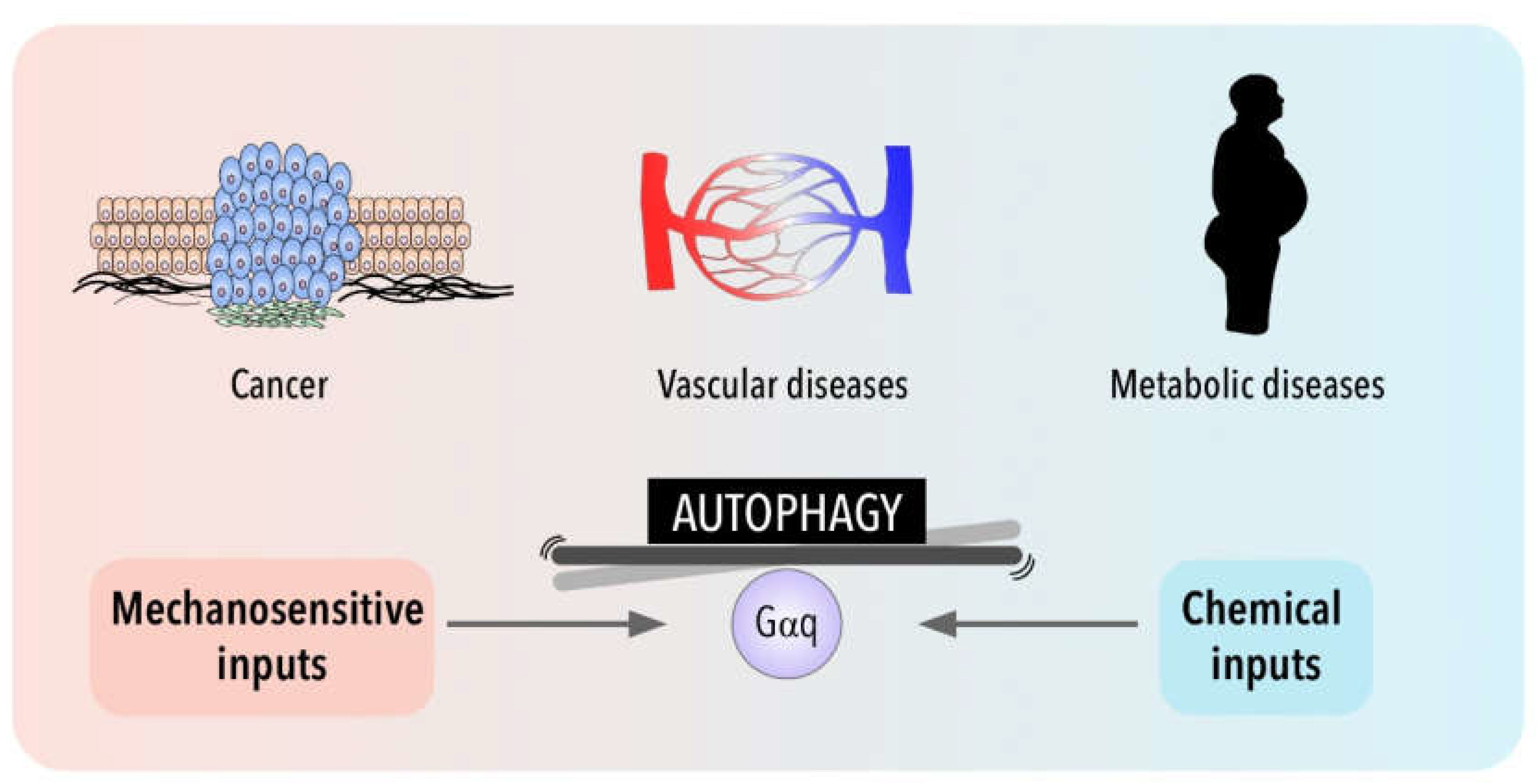
3. Autophagy in Disease for Good and for Bad: Gαq Involvement

Organisms have to constantly adapt to external stimuli and changes in their intracellular environment. Organs, tissues and cells have to face both chemical (e.g., Ca2+, amino acids, cytokines, chemokines and hormones) as well as physical challenges. Among the cytoplasmic responses to mechanical forces, recent studies have uncovered the role of autophagy in the translation of mechanical forces into biological responses [22][23][65][66]. Given the importance of autophagy regulation and dynamics of lysosomal system to ensure cellular fitness, it is not surprising that autophagy disruption can contribute to the development of several diseases such as metabolic disorders, cardiovascular or cancer diseases. Although the involvement of autophagy in these major diseases has been well studied (reviewed in [67], over the years autophagy regulation has grown in complexity and their consequences are less predictable. The importance of Gαq and Gαq-coupled GPCRs in all these contexts, together with its recently described importance in autophagy, strongly suggest that alteration in Gαq modulation signaling pathways can contribute to all these pathological situations. In this part of the revisew, wearch, the researchers will focus on how autophagy may be involved in different pathologies, emphasizing as far as possible the influence of mechanical inputs.
Various metabolic disorders have shown functional defects in autophagy [68][69]. Since the lysosomal disposal of intracellular macromolecules leads to their breakdown into important metabolic intermediates, including amino acids, glucose, nucleotides, and free fatty acids (FAs), autophagy plays an important role in the response to energetic stresses, at both the tissue-specific and systemic levels [70]. Many studies have emphasized the importance of autophagy in conditions such as obesity, insulin resistance and diabetes that are characterized by metabolic alterations and intracellular stresses that have in common the accumulation of damaged cellular components. Silencing of ATG system promotes obesity and induces metabolic alterations [71]. Interestingly autophagy genes are differentially expressed and activated in a tissue and stage-specific manner. In general, nutrient limitation and different stress situations favor autophagy as a mechanism of cytoprotecting, reducing cellular death and limiting inflammatory response. Upon autophagy inhibition alteration of adipocyte differentiation, lipid metabolism and storing of lipids is drastically altered [72][73][74] For example, in obesity, autophagy is suppressed due to an increased in mTOR activity. Moreover, in patients with diabetes, changes in oxidative stress and autophagy have been reported [75]. Therefore, the enhancement of autophagy activity has been suggested as a novel therapeutic approach against organ failure associated to metabolic disorders.
In general, GPCRs regulate virtually all metabolic processes including glucose and energy homeostasis, particularly diabetes and obesity-related diseases. Several endogenous ligands such as free fatty acids and their receptors have been extensively studied in insulin secretion regulation, and glucose metabolism [76]. A growing number of GPCR are being identified as sensors of circulating of local concentrators of energy substrates or metabolic intermediates. Examples of these receptors include the amino-acid responsive receptors GPRC6A taste receptors type 1 members 1 and 3 (T1R1/T1R3), the calcium sensing receptor (CaSR) long chain fatty acid receptors GPR120 and GPR40, short fatty acid receptors GPR41 and GPR43 or hydroxy carboxylic acid receptors [77][78][79]. These GPCR nutrient receptors act via different G proteins including Gαq/11 and might be able to modulate the canonical metabolic regulators AMPK and mTORC1 [80]. In this sense, Gαq-coupled T1R1/T1R3 act as a direct sensor of the fed state and amino acids availability, leading to the activation of mTORC1 [81]. Nutrient and homeostasis fluctuations may also indicate the release of classical hormones and neurotransmitters that activated GPCRs, along a systemic regulation of autophagy. In this sense, β-adrenergic receptors activation has been related with autophagic flux favoring lipolysis [82], and hyperglycemia induces autophagy in pancreatic β cells through P2Y purinergic receptors [83]. In addition, drugs that target metabolic tissues have emerged as attractive diabetes therapeutic targets as well. The p62-mTORC1-autophagy axis has been described to regulate adipogenesis and energy control in a complex manner [84]. The potential and reported connections of Gαq signaling that wthe researchers have described with this axis [63] may provide new insights in the mechanisms underlying these metabolic alterations. Recent studies have further confirmed the relevance of Gαq signaling for driving metabolic reprogramming in uveal melanoma [85] and in the regulation of glucose and lipid homeostasis [86] reinforcing a critical role of GPCR-Gαq system in metabolic diseases. Further investigation will be required to define the mechanisms involved.
Interestingly, cell metabolism is sensitive also to the physical cell microenvironment [87]. Although cell metabolism has recently emerged as one of the processes regulated by mechanical cues, the link between cell mechanics and metabolism is still poorly understood when compared with other pathologies such as cardiovascular diseases or cancer (Figure 2). Thus, in addition to metabolic intermediates, autophagy can influence metabolic reprogramming in epithelial cells through the involvement of mechanical forces such as shear stress [88][89] and, mechanical stretching/tension exerted by exercise has been shown to induce also autophagy in peripheral tissues (liver, pancreas and adipose tissue) [90].
Figure 2. Gαq signaling and autophagy at the crossroads of a balance between mechanical and chemical cues and their impact in cancer, metabolic and cardiovascular pathologies.
The involvement of shear stress and mechanical forces in endothelial function has been well established and although, there is an increasing interest in the role of autophagic flux in vessel wall biology, the mechanosensors upstream of autophagy induction in endothelial cells are not well known [91]. Emerging evidence links alterations in autophagic flux with disease processes that include atherosclerosis, pulmonary hypertension and cardiovascular diseases [92]. Interestingly, a very recent study proposes a protective role of CMA (Chaperon Mediated Autophagy) against atherosclerosis [93]. Loss of autophagy may be a central mechanism through which risk factors elicit endothelial dysfunction. The role of autophagy in vascular development and in sprouting has been associated with a defective autophagy in mice lacking endothelial specific-TFEB factor [94]. The autophagic state of endothelial cells is also critical for vascular permeability [95]. Additionally, endothelial cells require autophagy to regulate tight junction proteins and maintain endothelial barrier integrity during inflammation [96]. Moreover, it has been reported that autophagy may be involved in the regulation of nitric oxide bioavailability, a crucial molecule that maintains vascular homeostasis in endothelial cells. Concomitant with a reduction in NO, loss of autophagy promotes and increase in endothelial ROS and inflammatory cytokine production [97][98]. Shear-stress-dependent autophagy is also important for NO production [98][99]. Indeed, in zones of low shear stress that are prone to develop atherosclerotic plaques, the impairment of autophagic flux induces endothelial NO synthase (eNOS) uncoupling, resulting in the production of superoxide instead of NO. Restoration of the autophagic flux favors the production of NO by endothelial NO synthase [100]. Interestingly, Gαq has been described as an important sensor of shear stress in endothelium [101][102]. Recent studies have demonstrated that changes in the type of flow can activate the same initial mechanosensing pathway involving Piezo1- and Gαq/11-mediated signaling with different atheroprotective response depending on the activation of α5 integrin, which is activated only by disturbed flow, but not by sustained laminar flow [103].
Regarding cardiovascular context, autophagy preserves cardiac structure and function under baseline conditions and is activated during stress, contributing to limit damage and preserve cardiac functionality during ischemia [104]. Cardiac cells are also subjected to tension. Several pathophysiological conditions, lead to an increase in cardiac workload and mechanical forces that are usually associated with pathological cardiac hypertrophy [105]. Mechanical forces can induce autophagy in cardiac cells being protective or detrimental depending on the context [91]. Indeed, during ischemia, autophagy has a protective effect on cardiomyocytes [106][107][108], while inhibition of autophagy improves cardiac function after reperfusion in an ischemia–reperfusion mouse model [107]. Much evidence places Gαq at the center of hypertrophic pathways in the heart (see [109], for more details). Indeed, Gαq signaling is both necessary and sufficient for the development of cardiac hypertrophy. The development of a cardiac hypertrophy phenotype has been correlated with a higher risk of heart failure [110]. Interestingly, it has become increasingly clear that both events are tied to the activation/presence of Gαq that can promote cardiomyocyte apoptosis and heart failure [111] by affecting vascular permeability and hypertension. Indeed, Gαq inhibition using specific drugs have been proposed to have anti-hypertensive role [112]. WThe researchers have also described, in previous studies, a role of Gαq/PKCζ signaling axis in the development of cardiac hypertrophy in response to angiotensin II through a novel binding region on Gαq. Given the involvement of Gαq in cardiovascular function and in the process of autophagy, the potential participation of novel mechanisms downstream Gαq directly linked with autophagic flux in cardiovascular system is a relevant open question.
Autophagy is also recognized as a critical player in a context-dependent manner in cancer. Although it is well accepted that autophagy is important in many diseases, as described above, practically all clinical studies that involve autophagy manipulation are focused on cancer therapy. Autophagy networks are related to multiple aspects of cancer and may play a dual role with tumor-suppressive and tumor promoting functions depending on tumor cell type and stage [113]. Both inhibition of autophagy and its overstimulation are strategies tested in cancer, with the use of different drugs such as hydroxychloroquine, 3-methyl-adenina and everolimus as currently new strategies to be employed in clinics in combination with other chemotherapeutic treatments [114]. However, the high toxicity and adverse effects of these treatments urge a further understanding of the specific mechanisms by which autophagy modulates the different tumor progression steps.
Deficiency of autophagic genes has been found in various cancers. Impaired autophagy can promote tumorigenic environment through ROS dysregulation and inflammation processes [115][116][117]. On the other hand, at advanced cancer stages, increased autophagy can sustain tumor cell growth in nutrient-deficient, hypoxic tumor microenvironment and resistance to anoikis [118]. Upregulation confers chemoresistance and promotes the maintenance and survival of stem cell cancer status. Furthermore, autophagy inhibition can favor tumor cell invasiveness through the induction of de-differentiation mechanism. Thus, it seems that in premalignant lesions, enhanced autophagy might be beneficial preventing cancer, but in advance cancers most therapeutic strategies are focused on inhibiting autophagy [119]. Adding another layer of complexity, the novel Gαq role in modulating autophagy suggests that the balance between these processes characteristic of tumor growth might be altered in different cancer settings.
Furthermore, evidence identifies tumor microenvironment as a central driver of tumorigenesis in cancer [120]. Interestingly, cancer cells can also experience shear stress that can induce autophagy in different tumor cell lines [121][122][123][124]. Interstitial flow can promote the distribution of tumor-derived cells in primary tumor, while circulating tumor cells are also subjected to the shear stress from body fluids (blood, lymph and interstitial fluid) during metastasis [125][126]. It has been suggested that shear stress-induced autophagy can play an important role in controlling important cell responses from the regulation of cell size and metabolism to inflammation and cell death [91]. Moreover, the activation of tumor stromal fibroblasts to a state commonly known as cancer associated fibroblasts (CAFs) is critical. CAFs impact tumor progression by the modulation of multiple secretion functions of different factors (growth factors and inflammatory signals), by remodeling the extracellular matrix and even reprogramming their metabolism to provide nutrients and survival factors [127]. Moreover, autophagy can play a key role in CAFs activation [128]. Recent studies demonstrate that normal fibroblasts can differentiate into CAFs as protective responses to stresses under tumor microenvironment via the p62-Nrf2-pathway [129]. Furthermore, a molecular mechanism for CAFs activation has shown that tumor secreted lactate downregulates p62 in the stroma blocking AP-1-mediated p62 transcription [130]. Interestingly, wthe researchers have described changes in the expression of Gαq affecting some partners such as p62 promoting its downregulation and favoring autophagic flux [63].
Altered GPCR pathways have increasingly been reported in cancer context and activating mutations in Gαq has been identified in approximately 80% of uveal melanomas. Considering Gαq as a component of the nutrient-sensing machinery, able to link nutrient availability with the activation of mTORC1 through its interaction with p62 [63] strongly reinforce its potential contribution in the modulation of both the tumor and its microenvironment, during tumor progression.

References

  1. Gudipaty, S.A.; Conner, C.M.; Rosenblatt, J.; Montell, D.J. Unconventional Ways to Live and Die: Cell Death and Survival in Development, Homeostasis, and Disease. Annu. Rev. Cell Dev. Biol. 2018, 34, 311–332.
  2. Heldin, C.-H.; Lu, B.; Evans, R.; Gutkind, J.S. Signals and Receptors. Cold Spring Harb. Perspect. Biol. 2016, 8, a005900.
  3. Hilger, D.; Masureel, M.; Kobilka, B.K. Structure and Dynamics of GPCR Signaling Complexes. Nat. Struct. Mol. Biol. 2018, 25, 4–12.
  4. Marinissen, M.J.; Gutkind, J.S. G-Protein-Coupled Receptors and Signaling Networks: Emerging Paradigms. Trends Pharmacol. Sci. 2001, 22, 368–376.
  5. Humphrey, J.D.; Dufresne, E.R.; Schwartz, M.A. Mechanotransduction and Extracellular Matrix Homeostasis. Nat. Rev. Mol. Cell Biol. 2014, 15, 802–812.
  6. Cox, T.R.; Erler, J.T. Remodeling and Homeostasis of the Extracellular Matrix: Implications for Fibrotic Diseases and Cancer. Dis. Model. Mech. 2011, 4, 165–178.
  7. Mahoney, J.P.; Sunahara, R.K. Mechanistic Insights into GPCR–G Protein Interactions. Curr. Opin. Struct. Biol. 2016, 41, 247–254.
  8. Pierce, K.L.; Premont, R.T.; Lefkowitz, R.J. Seven-Transmembrane Receptors. Nat. Rev. Mol. Cell Biol. 2002, 3, 639–650.
  9. Chen, C.G.; Iozzo, R.V. Extracellular Matrix Guidance of Autophagy: A Mechanism Regulating Cancer Growth. Open Biol. 2022, 12, 210304.
  10. Hupfer, A.; Brichkina, A.; Koeniger, A.; Keber, C.; Denkert, C.; Pfefferle, P.; Helmprobst, F.; Pagenstecher, A.; Visekruna, A.; Lauth, M. Matrix Stiffness Drives Stromal Autophagy and Promotes Formation of a Protumorigenic Niche. Proc. Natl. Acad. Sci. USA 2021, 118, e2105367118.
  11. Ge, H.; Tian, M.; Pei, Q.; Tan, F.; Pei, H. Extracellular Matrix Stiffness: New Areas Affecting Cell Metabolism. Front. Oncol. 2021, 11, 631991.
  12. Parzych, K.R.; Klionsky, D.J. An Overview of Autophagy: Morphology, Mechanism, and Regulation. Antioxid. Redox Signal. 2014, 20, 460–473.
  13. Mizushima, N.; Levine, B.; Cuervo, A.M.; Klionsky, D.J. Autophagy Fights Disease through Cellular Self-Digestion. Nature 2008, 451, 1069–1075.
  14. Shintani, T.; Klionsky, D.J. Autophagy in Health and Disease: A Double-Edged Sword. Science 2004, 306, 990–995.
  15. Bellot, G.; Garcia-Medina, R.; Gounon, P.; Chiche, J.; Roux, D.; Pouysségur, J.; Mazure, N.M. Hypoxia-Induced Autophagy Is Mediated through Hypoxia-Inducible Factor Induction of BNIP3 and BNIP3L via Their BH3 Domains. Mol. Cell. Biol. 2009, 29, 2570–2581.
  16. Galluzzi, L.; Baehrecke, E.H.; Ballabio, A.; Boya, P.; Bravo-San Pedro, J.M.; Cecconi, F.; Choi, A.M.; Chu, C.T.; Codogno, P.; Colombo, M.I.; et al. Molecular Definitions of Autophagy and Related Processes. EMBO J. 2017, 36, 1811–1836.
  17. Mizushima, N.; Yoshimori, T.; Ohsumi, Y. The Role of Atg Proteins in Autophagosome Formation. Annu. Rev. Cell Dev. Biol. 2011, 27, 107–132.
  18. Dunlop, E.A.; Tee, A.R. MTOR and Autophagy: A Dynamic Relationship Governed by Nutrients and Energy. Semin. Cell Dev. Biol. 2014, 36, 121–129.
  19. Park, J.-M.; Jung, C.H.; Seo, M.; Otto, N.M.; Grunwald, D.; Kim, K.H.; Moriarity, B.; Kim, Y.-M.; Starker, C.; Nho, R.S.; et al. The ULK1 Complex Mediates MTORC1 Signaling to the Autophagy Initiation Machinery via Binding and Phosphorylating ATG14. Autophagy 2016, 12, 547–564.
  20. Hosokawa, N.; Hara, T.; Kaizuka, T.; Kishi, C.; Takamura, A.; Miura, Y.; Iemura, S.; Natsume, T.; Takehana, K.; Yamada, N.; et al. Nutrient-Dependent MTORC1 Association with the ULK1–Atg13–FIP200 Complex Required for Autophagy. Mol. Biol. Cell 2009, 20, 1981–1991.
  21. Case, N.; Thomas, J.; Sen, B.; Styner, M.; Xie, Z.; Galior, K.; Rubin, J. Mechanical Regulation of Glycogen Synthase Kinase 3β (GSK3β) in Mesenchymal Stem Cells Is Dependent on Akt Protein Serine 473 Phosphorylation via MTORC2 Protein. J. Biol. Chem. 2011, 286, 39450–39456.
  22. Hernández-Cáceres, M.P.; Munoz, L.; Pradenas, J.M.; Pena, F.; Lagos, P.; Aceiton, P.; Owen, G.I.; Morselli, E.; Criollo, A.; Ravasio, A.; et al. Mechanobiology of Autophagy: The Unexplored Side of Cancer. Front. Oncol. 2021, 11, 632956.
  23. Dupont, N.; Codogno, P. Autophagy Transduces Physical Constraints into Biological Responses. Int. J. Biochem. Cell Biol. 2016, 79, 419–426.
  24. Martinac, B.; Poole, K. Mechanically Activated Ion Channels. Int. J. Biochem. Cell Biol. 2018, 97, 104–107.
  25. Saotome, K.; Murthy, S.E.; Kefauver, J.M.; Whitwam, T.; Patapoutian, A.; Ward, A.B. Structure of the Mechanically Activated Ion Channel Piezo1. Nature 2018, 554, 481–486.
  26. Sinha, B.; Köster, D.; Ruez, R.; Gonnord, P.; Bastiani, M.; Abankwa, D.; Stan, R.V.; Butler-Browne, G.; Vedie, B.; Johannes, L.; et al. Cells Respond to Mechanical Stress by Rapid Disassembly of Caveolae. Cell 2011, 144, 402–413.
  27. Praetorius, H.A. The Primary Cilium as Sensor of Fluid Flow: New Building Blocks to the Model. A Review in the Theme: Cell Signaling: Proteins, Pathways and Mechanisms. Am. J. Physiol. -Cell Physiol. 2015, 308, C198–C208.
  28. Baschieri, F.; Dayot, S.; Elkhatib, N.; Ly, N.; Capmany, A.; Schauer, K.; Betz, T.; Vignjevic, D.M.; Poincloux, R.; Montagnac, G. Frustrated Endocytosis Controls Contractility-Independent Mechanotransduction at Clathrin-Coated Structures. Nat. Commun. 2018, 9, 3825.
  29. Kirby, T.J.; Lammerding, J. Emerging Views of the Nucleus as a Cellular Mechanosensor. Nat. Cell Biol. 2018, 20, 373–381.
  30. Helle, S.C.J.; Feng, Q.; Aebersold, M.J.; Hirt, L.; Grüter, R.R.; Vahid, A.; Sirianni, A.; Mostowy, S.; Snedeker, J.G.; Šarić, A.; et al. Mechanical Force Induces Mitochondrial Fission. eLife 2017, 6, e30292.
  31. Guet, D.; Mandal, K.; Pinot, M.; Hoffmann, J.; Abidine, Y.; Sigaut, W.; Bardin, S.; Schauer, K.; Goud, B.; Manneville, J.-B. Mechanical Role of Actin Dynamics in the Rheology of the Golgi Complex and in Golgi-Associated Trafficking Events. Curr. Biol. 2014, 24, 1700–1711.
  32. Aguilera, M.O.; Berón, W.; Colombo, M.I. The Actin Cytoskeleton Participates in the Early Events of Autophagosome Formation upon Starvation Induced Autophagy. Autophagy 2012, 8, 1590–1603.
  33. Kast, D.J.; Dominguez, R. The Cytoskeleton–Autophagy Connection. Curr. Biol. 2017, 27, R318–R326.
  34. Hsu, P.; Shi, Y. Regulation of Autophagy by Mitochondrial Phospholipids in Health and Diseases. Biochim. Et Biophys. Acta (BBA)—Mol. Cell Biol. Lipids 2017, 1862, 114–129.
  35. Avivar-Valderas, A.; Bobrovnikova-Marjon, E.; Alan Diehl, J.; Bardeesy, N.; Debnath, J.; Aguirre-Ghiso, J.A. Regulation of Autophagy during ECM Detachment Is Linked to a Selective Inhibition of MTORC1 by PERK. Oncogene 2013, 32, 4932–4940.
  36. Cai, Z.; Zhao, D.; Sun, Y.; Gao, D.; Li, X.; Yang, J.; Ma, Z. Detachment-Based Equilibrium of Anoikic Cell Death and Autophagic Cell Survival through Adaptor Protein P66Shc. Anat. Rec. 2016, 299, 325–333.
  37. Vlahakis, A.; Debnath, J. The Interconnections between Autophagy and Integrin-Mediated Cell Adhesion. J. Mol. Biol. 2017, 429, 515–530.
  38. Anlaş, A.A.; Nelson, C.M. Soft Microenvironments Induce Chemoresistance by Increasing Autophagy Downstream of Integrin-Linked Kinase. Cancer Res. 2020, 80, 4103–4113.
  39. Neill, T.; Schaefer, L.; Iozzo, R.V. Instructive Roles of Extracellular Matrix on Autophagy. Am. J. Pathol. 2014, 184, 2146–2153.
  40. Buraschi, S.; Neill, T.; Goyal, A.; Poluzzi, C.; Smythies, J.; Owens, R.T.; Schaefer, L.; Torres, A.; Iozzo, R.V. Decorin Causes Autophagy in Endothelial Cells via Peg3. Proc. Natl. Acad. Sci. USA 2013, 110, E2582–E2591.
  41. Nguyen, T.M.B.; Subramanian, I.V.; Xiao, X.; Ghosh, G.; Nguyen, P.; Kelekar, A.; Ramakrishnan, S. Endostatin Induces Autophagy in Endothelial Cells by Modulating Beclin 1 and β-Catenin Levels. J. Cell. Mol. Med. 2009, 13, 3687–3698.
  42. Castagnaro, S.; Gambarotto, L.; Cescon, M.; Bonaldo, P. Autophagy in the Mesh of Collagen VI. Matrix Biol. 2021, 100–101, 162–172.
  43. Carmignac, V.; Svensson, M.; Körner, Z.; Elowsson, L.; Matsumura, C.; Gawlik, K.I.; Allamand, V.; Durbeej, M. Autophagy Is Increased in Laminin A2 Chain-Deficient Muscle and Its Inhibition Improves Muscle Morphology in a Mouse Model of MDC1A. Hum. Mol. Genet. 2011, 20, 4891–4902.
  44. Marullo, S.; Doly, S.; Saha, K.; Enslen, H.; Scott, M.G.H.; Coureuil, M. Mechanical GPCR Activation by Traction Forces Exerted on Receptor N-Glycans. ACS Pharmacol. Transl. Sci. 2020, 3, 171–178.
  45. Iliff, A.J.; Xu, X.Z.S. A Mechanosensitive GPCR That Detects the Bloody Force. Cell 2018, 173, 542–544.
  46. Ozkan, A.D.; Gettas, T.; Sogata, A.; Phaychanpheng, W.; Zhou, M.; Lacroix, J.J. Mechanical and Chemical Activation of GPR68 Probed with a Genetically Encoded Fluorescent Reporter. J. Cell Sci. 2021, 134, jcs255455.
  47. García-Hoz, C.; Sánchez-Fernández, G.; Díaz-Meco, M.T.; Moscat, J.; Mayor, F.; Ribas, C. Gαq Acts as an Adaptor Protein in Protein Kinase Cζ (PKCζ)-Mediated ERK5 Activation by G Protein-Coupled Receptors (GPCR). J. Biol. Chem. 2010, 285, 13480–13489.
  48. Nigro, P.; Abe, J.; Berk, B.C. Flow Shear Stress and Atherosclerosis: A Matter of Site Specificity. Antioxid. Redox Signal. 2011, 15, 1405–1414.
  49. Folts, C.J.; Giera, S.; Li, T.; Piao, X. Adhesion G Protein-Coupled Receptors as Drug Targets for Neurological Diseases. Trends Pharmacol. Sci. 2019, 40, 278–293.
  50. Scholz, N. Cancer Cell Mechanics: Adhesion G Protein-Coupled Receptors in Action? Front. Oncol. 2018, 8, 59.
  51. Aplin, A.; Jasionowski, T.; Tuttle, D.L.; Lenk, S.E.; Dunn, W.A. Cytoskeletal Elements Are Required for the Formation and Maturation of Autophagic Vacuoles. J. Cell. Physiol. 1992, 152, 458–466.
  52. Kaushik, S.; Cuervo, A.M. The Coming of Age of Chaperone-Mediated Autophagy. Nat. Rev. Mol. Cell Biol. 2018, 19, 365–381.
  53. Lim, C.-Y.; Zoncu, R. The Lysosome as a Command-and-Control Center for Cellular Metabolism. J. Cell Biol. 2016, 214, 653–664.
  54. Cabukusta, B.; Neefjes, J. Mechanisms of Lysosomal Positioning and Movement. Traffic 2018, 19, 761–769.
  55. Kesisova, I.A.; Robinson, B.P.; Spiliotis, E.T. A Septin GTPase Scaffold of Dynein–Dynactin Motors Triggers Retrograde Lysosome Transport. J. Cell Biol. 2021, 220, e202005219.
  56. Korolchuk, V.I.; Saiki, S.; Lichtenberg, M.; Siddiqi, F.H.; Roberts, E.A.; Imarisio, S.; Jahreiss, L.; Sarkar, S.; Futter, M.; Menzies, F.M.; et al. Lysosomal Positioning Coordinates Cellular Nutrient Responses. Nat. Cell Biol. 2011, 13, 453–460.
  57. Jordens, I.; Fernandez-Borja, M.; Marsman, M.; Dusseljee, S.; Janssen, L.; Calafat, J.; Janssen, H.; Wubbolts, R.; Neefjes, J. The Rab7 Effector Protein RILP Controls Lysosomal Transport by Inducing the Recruitment of Dynein-Dynactin Motors. Curr. Biol. 2001, 11, 1680–1685.
  58. Bucci, C.; Thomsen, P.; Nicoziani, P.; McCarthy, J.; van Deurs, B. Rab7: A Key to Lysosome Biogenesis. Mol. Biol. Cell 2000, 11, 467–480.
  59. Xing, R.; Zhou, H.; Jian, Y.; Li, L.; Wang, M.; Liu, N.; Yin, Q.; Liang, Z.; Guo, W.; Yang, C. The Rab7 Effector WDR91 Promotes Autophagy-Lysosome Degradation in Neurons by Regulating Lysosome Fusion. J. Cell Biol. 2021, 220, e202007061.
  60. Willett, R.; Martina, J.A.; Zewe, J.P.; Wills, R.; Hammond, G.R.V.; Puertollano, R. TFEB Regulates Lysosomal Positioning by Modulating TMEM55B Expression and JIP4 Recruitment to Lysosomes. Nat. Commun. 2017, 8, 1580.
  61. Settembre, C.; di Malta, C.; Polito, V.A.; Arencibia, M.G.; Vetrini, F.; Erdin, S.; Erdin, S.U.; Huynh, T.; Medina, D.; Colella, P.; et al. TFEB Links Autophagy to Lysosomal Biogenesis. Science 2011, 332, 1429–1433.
  62. Settembre, C.; Zoncu, R.; Medina, D.L.; Vetrini, F.; Erdin, S.; Erdin, S.; Huynh, T.; Ferron, M.; Karsenty, G.; Vellard, M.C.; et al. A Lysosome-to-Nucleus Signalling Mechanism Senses and Regulates the Lysosome via MTOR and TFEB. EMBO J. 2012, 31, 1095–1108.
  63. Cabezudo, S.; Sanz-Flores, M.; Caballero, A.; Tasset, I.; Rebollo, E.; Diaz, A.; Aragay, A.M.; Cuervo, A.M.; Mayor, F.; Ribas, C. Gαq Activation Modulates Autophagy by Promoting MTORC1 Signaling. Nat. Commun. 2021, 12, 4540.
  64. Wright, S.C.; Lukasheva, V.; le Gouill, C.; Kobayashi, H.; Breton, B.; Mailhot-Larouche, S.; Blondel-Tepaz, É.; Antunes Vieira, N.; Costa-Neto, C.; Héroux, M.; et al. BRET-Based Effector Membrane Translocation Assay Monitors GPCR-Promoted and Endocytosis-Mediated Gq Activation at Early Endosomes. Proc. Natl. Acad. Sci. USA 2021, 118, e2025846118.
  65. Kenific, C.M.; Wittmann, T.; Debnath, J. Autophagy in Adhesion and Migration. J. Cell Sci. 2016, 129, 3685–3693.
  66. King, J.S. Mechanical Stress Meets Autophagy: Potential Implications for Physiology and Pathology. Trends Mol. Med. 2012, 18, 583–588.
  67. Klionsky, D.J.; Petroni, G.; Amaravadi, R.K.; Baehrecke, E.H.; Ballabio, A.; Boya, P.; Bravo-San Pedro, J.M.; Cadwell, K.; Cecconi, F.; Choi, A.M.K.; et al. Autophagy in Major Human Diseases. EMBO J. 2021, 40, e108863.
  68. Ichimura, Y.; Komatsu, M. Pathophysiological Role of Autophagy: Lesson from Autophagy-Deficient Mouse Models. Exp. Anim. 2011, 60, 329–345.
  69. Kitada, M.; Koya, D. Autophagy in Metabolic Disease and Ageing. Nat. Rev. Endocrinol. 2021, 17, 647–661.
  70. Rabinowitz, J.D.; White, E. Autophagy and Metabolism. Science 2010, 330, 1344–1348.
  71. Van Beek, N.; Klionsky, D.J.; Reggiori, F. Genetic Aberrations in Macroautophagy Genes Leading to Diseases. Biochim. Et Biophys. Acta (BBA)—Mol. Cell Res. 2018, 1865, 803–816.
  72. Zhang, X.; Wu, D.; Wang, C.; Luo, Y.; Ding, X.; Yang, X.; Silva, F.; Arenas, S.; Weaver, J.M.; Mandell, M.; et al. Sustained Activation of Autophagy Suppresses Adipocyte Maturation via a Lipolysis-Dependent Mechanism. Autophagy 2020, 16, 1668–1682.
  73. Codogno, P.; Meijer, A.J. Autophagy: A Potential Link between Obesity and Insulin Resistance. Cell Metab. 2010, 11, 449–451.
  74. Madrigal-Matute, J.; Cuervo, A.M. Regulation of Liver Metabolism by Autophagy. Gastroenterology 2016, 150, 328–339.
  75. Park, E.-Y.; Park, J.-B. High Glucose-Induced Oxidative Stress Promotes Autophagy through Mitochondrial Damage in Rat Notochordal Cells. Int. Orthop. 2013, 37, 2507–2514.
  76. Oliveira de Souza, C.; Sun, X.; Oh, D. Metabolic Functions of G Protein-Coupled Receptors and β-Arrestin-Mediated Signaling Pathways in the Pathophysiology of Type 2 Diabetes and Obesity. Front. Endocrinol. 2021, 12, 1005.
  77. Blad, C.C.; Tang, C.; Offermanns, S. G Protein-Coupled Receptors for Energy Metabolites as New Therapeutic Targets. Nat. Rev. Drug Discov. 2012, 11, 603–619.
  78. Offermanns, S. Free Fatty Acid (FFA) and Hydroxy Carboxylic Acid (HCA) Receptors. Annu. Rev. Pharmacol. Toxicol. 2014, 54, 407–434.
  79. Wauson, E.M.; Zaganjor, E.; Cobb, M.H. Amino Acid Regulation of Autophagy through the GPCR TAS1R1-TAS1R3. Autophagy 2013, 9, 418–419.
  80. Wauson, E.M.; Zaganjor, E.; Lee, A.-Y.; Guerra, M.L.; Ghosh, A.B.; Bookout, A.L.; Chambers, C.P.; Jivan, A.; McGlynn, K.; Hutchison, M.R.; et al. The G Protein-Coupled Taste Receptor T1R1/T1R3 Regulates MTORC1 and Autophagy. Mol. Cell 2012, 47, 851–862.
  81. Wauson, E.M.; Lorente-Rodríguez, A.; Cobb, M.H. Minireview: Nutrient Sensing by G Protein-Coupled Receptors. Mol. Endocrinol. 2013, 27, 1188–1197.
  82. Lizaso, A.; Tan, K.-T.; Lee, Y.-H. β-Adrenergic Receptor-Stimulated Lipolysis Requires the RAB7-Mediated Autolysosomal Lipid Degradation. Autophagy 2013, 9, 1228–1243.
  83. Jung, H.S.; Chung, K.W.; Won Kim, J.; Kim, J.; Komatsu, M.; Tanaka, K.; Nguyen, Y.H.; Kang, T.M.; Yoon, K.-H.; Kim, J.-W.; et al. Loss of Autophagy Diminishes Pancreatic β Cell Mass and Function with Resultant Hyperglycemia. Cell Metab. 2008, 8, 318–324.
  84. Moscat, J.; Diaz-Meco, M.T. Feedback on Fat: P62-MTORC1-Autophagy Connections. Cell 2011, 147, 724–727.
  85. Onken, M.D.; Noda, S.E.; Kaltenbronn, K.M.; Frankfater, C.; Makepeace, C.M.; Fettig, N.; Piggott, K.D.; Custer, P.L.; Ippolito, J.E.; Blumer, K.J. Oncogenic Gq/11 Signaling Acutely Drives and Chronically Sustains Metabolic Reprogramming in Uveal Melanoma. J. Biol. Chem. 2022, 298, 101495.
  86. Kimura, T.; Pydi, S.P.; Wang, L.; Haspula, D.; Cui, Y.; Lu, H.; König, G.M.; Kostenis, E.; Steinberg, G.R.; Gavrilova, O.; et al. Adipocyte Gq Signaling Is a Regulator of Glucose and Lipid Homeostasis in Mice. Nat. Commun. 2022, 13, 1652.
  87. Romani, P.; Valcarcel-Jimenez, L.; Frezza, C.; Dupont, S. Crosstalk between Mechanotransduction and Metabolism. Nat. Rev. Mol. Cell Biol. 2021, 22, 22–38.
  88. Kim, S.W.; Ehrman, J.; Ahn, M.-R.; Kondo, J.; Lopez, A.A.M.; Oh, Y.S.; Kim, X.H.; Crawley, S.W.; Goldenring, J.R.; Tyska, M.J.; et al. Shear Stress Induces Noncanonical Autophagy in Intestinal Epithelial Monolayers. Mol. Biol. Cell 2017, 28, 3043–3056.
  89. Miceli, C.; Roccio, F.; Penalva-Mousset, L.; Burtin, M.; Leroy, C.; Nemazanyy, I.; Kuperwasser, N.; Pontoglio, M.; Friedlander, G.; Morel, E.; et al. The Primary Cilium and Lipophagy Translate Mechanical Forces to Direct Metabolic Adaptation of Kidney Epithelial Cells. Nat. Cell Biol. 2020, 22, 1091–1102.
  90. He, C.; Sumpter, R., Jr.; Levine, B. Exercise Induces Autophagy in Peripheral Tissues and in the Brain. Autophagy 2012, 8, 1548–1551.
  91. Claude-Taupin, A.; Codogno, P.; Dupont, N. Links between Autophagy and Tissue Mechanics. J. Cell Sci. 2021, 134, jcs258589.
  92. Mameli, E.; Martello, A.; Caporali, A. Autophagy at the Interface of Endothelial Cell Homeostasis and Vascular Disease. FEBS J. 2021, 289, 2976–2991.
  93. Madrigal-Matute, J.; de Bruijn, J.; van Kuijk, K.; Riascos-Bernal, D.F.; Diaz, A.; Tasset, I.; Martín-Segura, A.; Gijbels, M.J.J.; Sander, B.; Kaushik, S.; et al. Protective Role of Chaperone-Mediated Autophagy against Atherosclerosis. Proc. Natl. Acad. Sci. USA 2022, 119, e2121133119.
  94. Doronzo, G.; Astanina, E.; Corà, D.; Chiabotto, G.; Comunanza, V.; Noghero, A.; Neri, F.; Puliafito, A.; Primo, L.; Spampanato, C.; et al. TFEB Controls Vascular Development by Regulating the Proliferation of Endothelial Cells. EMBO J. 2019, 38, e98250.
  95. Shadab, M.; Millar, M.W.; Slavin, S.A.; Leonard, A.; Fazal, F.; Rahman, A. Autophagy Protein ATG7 Is a Critical Regulator of Endothelial Cell Inflammation and Permeability. Sci. Rep. 2020, 10, 13708.
  96. Reglero-Real, N.; Pérez-Gutiérrez, L.; Yoshimura, A.; Rolas, L.; Garrido-Mesa, J.; Barkaway, A.; Pickworth, C.; Saleeb, R.S.; Gonzalez-Nuñez, M.; Austin-Williams, S.N.; et al. Autophagy Modulates Endothelial Junctions to Restrain Neutrophil Diapedesis during Inflammation. Immunity 2021, 54, 1989–2004.e9.
  97. Deretic, V.; Saitoh, T.; Akira, S. Autophagy in Infection, Inflammation and Immunity. Nat. Rev. Immunol. 2013, 13, 722–737.
  98. Bharath, L.P.; Cho, J.M.; Park, S.-K.; Ruan, T.; Li, Y.; Mueller, R.; Bean, T.; Reese, V.; Richardson, R.S.; Cai, J.; et al. Endothelial Cell Autophagy Maintains Shear Stress–Induced Nitric Oxide Generation via Glycolysis-Dependent Purinergic Signaling to Endothelial Nitric Oxide Synthase. Arterioscler. Thromb. Vasc. Biol. 2017, 37, 1646–1656.
  99. Bharath, L.P.; Mueller, R.; Li, Y.; Ruan, T.; Kunz, D.; Goodrich, R.; Mills, T.; Deeter, L.; Sargsyan, A.; Anandh Babu, P.V.; et al. Impairment of Autophagy in Endothelial Cells Prevents Shear-Stress-Induced Increases in Nitric Oxide Bioavailability. Can. J. Physiol. Pharmacol. 2014, 92, 605–612.
  100. Zhang, J.-X.; Qu, X.-L.; Chu, P.; Xie, D.-J.; Zhu, L.-L.; Chao, Y.-L.; Li, L.; Zhang, J.-J.; Chen, S.-L. Low Shear Stress Induces Vascular ENOS Uncoupling via Autophagy-Mediated ENOS Phosphorylation. Biochim. Et Biophys. Acta (BBA)—Mol. Cell Res. 2018, 1865, 709–720.
  101. Wang, S.; Iring, A.; Strilic, B.; Albarrán Juárez, J.; Kaur, H.; Troidl, K.; Tonack, S.; Burbiel, J.C.; Müller, C.E.; Fleming, I.; et al. P2Y2 and Gq/G11 Control Blood Pressure by Mediating Endothelial Mechanotransduction. J. Clin. Investig. 2015, 125, 3077–3086.
  102. Dela Paz, N.G.; Melchior, B.; Frangos, J.A. Shear Stress Induces Gαq/11 Activation Independently of G Protein-Coupled Receptor Activation in Endothelial Cells. Am. J. Physiol. -Cell Physiol. 2017, 312, C428–C437.
  103. Albarrán-Juárez, J.; Iring, A.; Wang, S.; Joseph, S.; Grimm, M.; Strilic, B.; Wettschureck, N.; Althoff, T.F.; Offermanns, S. Piezo1 and Gq/G11 Promote Endothelial Inflammation Depending on Flow Pattern and Integrin Activation. J. Exp. Med. 2018, 215, 2655–2672.
  104. Sciarretta, S.; Maejima, Y.; Zablocki, D.; Sadoshima, J. The Role of Autophagy in the Heart. Annu. Rev. Physiol. 2018, 80, 1–26.
  105. Nakamura, M.; Sadoshima, J. Mechanisms of Physiological and Pathological Cardiac Hypertrophy. Nat. Rev. Cardiol. 2018, 15, 387–407.
  106. Kanamori, H.; Takemura, G.; Goto, K.; Maruyama, R.; Tsujimoto, A.; Ogino, A.; Takeyama, T.; Kawaguchi, T.; Watanabe, T.; Fujiwara, T.; et al. The Role of Autophagy Emerging in Postinfarction Cardiac Remodelling. Cardiovasc. Res. 2011, 91, 330–339.
  107. Matsui, Y.; Takagi, H.; Qu, X.; Abdellatif, M.; Sakoda, H.; Asano, T.; Levine, B.; Sadoshima, J. Distinct Roles of Autophagy in the Heart During Ischemia and Reperfusion. Circ. Res. 2007, 100, 914–922.
  108. Yan, L.; Vatner, D.E.; Kim, S.-J.; Ge, H.; Masurekar, M.; Massover, W.H.; Yang, G.; Matsui, Y.; Sadoshima, J.; Vatner, S.F. Autophagy in Chronically Ischemic Myocardium. Proc. Natl. Acad. Sci. USA 2005, 102, 13807–13812.
  109. García-Hoz, C.; Sánchez-Fernández, G.; García-Escudero, R.; Fernández-Velasco, M.; Palacios-García, J.; Ruiz-Meana, M.; Díaz-Meco, M.T.; Leitges, M.; Moscat, J.; García-Dorado, D.; et al. Protein Kinase C (PKC) ζ-Mediated Gαq Stimulation of ERK5 Protein Pathway in Cardiomyocytes and Cardiac Fibroblasts. J. Biol. Chem. 2012, 287, 7792–7802.
  110. Bartolomé, A.; García-Aguilar, A.; Asahara, S.-I.; Kido, Y.; Guillén, C.; Pajvani, U.B.; Benito, M. MTORC1 Regulates Both General Autophagy and Mitophagy Induction after Oxidative Phosphorylation Uncoupling. Mol. Cell. Biol. 2017, 37, e00441-17.
  111. Adams, J.W.; Pagel, A.L.; Means, C.K.; Oksenberg, D.; Armstrong, R.C.; Brown, J.H. Cardiomyocyte Apoptosis Induced by Gαq Signaling Is Mediated by Permeability Transition Pore Formation and Activation of the Mitochondrial Death Pathway. Circ. Res. 2000, 87, 1180–1187.
  112. Meleka, M.M.; Edwards, A.J.; Xia, J.; Dahlen, S.A.; Mohanty, I.; Medcalf, M.; Aggarwal, S.; Moeller, K.D.; Mortensen, O.V.; Osei-Owusu, P. Anti-Hypertensive Mechanisms of Cyclic Depsipeptide Inhibitor Ligands for Gq/11 Class G Proteins. Pharmacol. Res. 2019, 141, 264–275.
  113. Yun, C.W.; Jeon, J.; Go, G.; Lee, J.H.; Lee, S.H. The Dual Role of Autophagy in Cancer Development and a Therapeutic Strategy for Cancer by Targeting Autophagy. Int. J. Mol. Sci. 2020, 22, 179.
  114. Chude, C.; Amaravadi, R. Targeting Autophagy in Cancer: Update on Clinical Trials and Novel Inhibitors. Int. J. Mol. Sci. 2017, 18, 1279.
  115. Filomeni, G.; de Zio, D.; Cecconi, F. Oxidative Stress and Autophagy: The Clash between Damage and Metabolic Needs. Cell Death Differ. 2015, 22, 377–388.
  116. Qu, X.; Yu, J.; Bhagat, G.; Furuya, N.; Hibshoosh, H.; Troxel, A.; Rosen, J.; Eskelinen, E.-L.; Mizushima, N.; Ohsumi, Y.; et al. Promotion of Tumorigenesis by Heterozygous Disruption of the Beclin 1 Autophagy Gene. J. Clin. Investig. 2003, 112, 1809–1820.
  117. Shen, Y.; Li, D.-D.; Wang, L.-L.; Deng, R.; Zhu, X.-F. Decreased Expression of Autophagy-Related Proteins in Malignant Epithelial Ovarian Cancer. Autophagy 2008, 4, 1067–1068.
  118. Peng, Y.-F.; Shi, Y.-H.; Ding, Z.-B.; Ke, A.-W.; Gu, C.-Y.; Hui, B.; Zhou, J.; Qiu, S.-J.; Dai, Z.; Fan, J. Autophagy Inhibition Suppresses Pulmonary Metastasis of HCC in Mice via Impairing Anoikis Resistance and Colonization of HCC Cells. Autophagy 2013, 9, 2056–2068.
  119. Chmurska, A.; Matczak, K.; Marczak, A. Two Faces of Autophagy in the Struggle against Cancer. Int. J. Mol. Sci. 2021, 22, 2981.
  120. Tsai, M.-J.; Chang, W.-A.; Huang, M.-S.; Kuo, P.-L. Tumor Microenvironment: A New Treatment Target for Cancer. ISRN Biochem. 2014, 2014, 351959.
  121. Das, J.; Maji, S.; Agarwal, T.; Chakraborty, S.; Maiti, T.K. Hemodynamic Shear Stress Induces Protective Autophagy in HeLa Cells through Lipid Raft-Mediated Mechanotransduction. Clin. Exp. Metastasis 2018, 35, 135–148.
  122. Lien, S.-C.; Chang, S.-F.; Lee, P.-L.; Wei, S.-Y.; Chang, M.D.-T.; Chang, J.-Y.; Chiu, J.-J. Mechanical Regulation of Cancer Cell Apoptosis and Autophagy: Roles of Bone Morphogenetic Protein Receptor, Smad1/5, and P38 MAPK. Biochim. Et Biophys. Acta (BBA)—Mol. Cell Res. 2013, 1833, 3124–3133.
  123. Wang, K.; Wei, Y.; Liu, W.; Liu, L.; Guo, Z.; Fan, C.; Wang, L.; Hu, J.; Li, B. Mechanical Stress-Dependent Autophagy Component Release via Extracellular Nanovesicles in Tumor Cells. ACS Nano 2019, 13, 4589–4602.
  124. Yan, Z.; Su, G.; Gao, W.; He, J.; Shen, Y.; Zeng, Y.; Liu, X. Fluid Shear Stress Induces Cell Migration and Invasion via Activating Autophagy in HepG2 Cells. Cell Adhes. Migr. 2019, 13, 152–163.
  125. Follain, G.; Herrmann, D.; Harlepp, S.; Hyenne, V.; Osmani, N.; Warren, S.C.; Timpson, P.; Goetz, J.G. Fluids and Their Mechanics in Tumour Transit: Shaping Metastasis. Nat. Rev. Cancer 2020, 20, 107–124.
  126. Swartz, M.A.; Lund, A.W. Lymphatic and Interstitial Flow in the Tumour Microenvironment: Linking Mechanobiology with Immunity. Nat. Rev. Cancer 2012, 12, 210–219.
  127. Kalluri, R.; Zeisberg, M. Fibroblasts in Cancer. Nat. Rev. Cancer 2006, 6, 392–401.
  128. Goruppi, S.; Clocchiatti, A.; Dotto, G.P. A Role for Stromal Autophagy in Cancer-Associated Fibroblast Activation. Autophagy 2019, 15, 738–739.
  129. Kang, J.I.; Kim, D.H.; Sung, K.W.; Shim, S.M.; Cha-Molstad, H.; Soung, N.K.; Lee, K.H.; Hwang, J.; Lee, H.G.; Kwon, Y.T.; et al. P62-Induced Cancer-Associated Fibroblast Activation via the Nrf2-ATF6 Pathway Promotes Lung Tumorigenesis. Cancers 2021, 13, 864.
  130. Linares, J.F.; Cid-Diaz, T.; Duran, A.; Osrodek, M.; Martinez-Ordoñez, A.; Reina-Campos, M.; Kuo, H.-H.; Elemento, O.; Martin, M.L.; Cordes, T.; et al. The Lactate-NAD+ Axis Activates Cancer-Associated Fibroblasts by Downregulating P62. Cell Rep. 2022, 39, 110792.
More
Video Production Service