Your browser does not fully support modern features. Please upgrade for a smoother experience.
Please note this is a comparison between Version 1 by Giovanni Ferrandino and Version 2 by Nora Tang.
Acute coronary syndrome (ACS) refers to a group of diseases in which blood flow to the heart is decreased. It is a manifestation of CHD (coronary heart disease) which usually results in plaque disruption in the coronary arteries (atherosclerosis) and the formation of thrombus. In some cases, ACS may be due to vasospasm with or without underlying atherosclerosis, but in both cases it determines a reduction in blood flow to a part of the heart, resulting first in ischemia and then infarction.
- cardiac radiology
- cardiac-CT
- cardiac MRI
- CT angiography
- acute coronary syndrome
1. Introduction
ACS includes several conditions: ST-segment elevation myocardial infarction (STEMI), non-ST-segment elevation myocardial infarction (NSTEMI), and unstable angina, and it is responsible for one third of total deaths in people older than 35 [1][49].
In patients with suspected ACS, it will first be necessary to classify the patient in one of the two major sub-categories (STEMI and NSTEMI) and subsequently to distinguish the N-STEMI subtype (N-STEMI and unstable angina). STEMI is representative of an acute myocardial infarction with evidence of myocardial necrosis. Most of the patients presenting with ischemic symptoms and persistent ST-segment elevation on the electrocardiogram (ECG) > 20 min will show a typical rise in biomarkers of myocardial necrosis and progress to Q-wave myocardial infarction [2][50]. Patients with acute chest pain but no persistent ST-segment elevation may have a variety of ECG abnormalities such as transient ST-segment elevation, persistent or transient ST-segment depression, inversion of the T wave, flat T waves, or pseudo-normalization of the T waves up to a normal ECG. Cardiomyocyte necrosis [myocardial infarction without ST-segment elevation (NSTEMI)] or, less frequently, myocardial ischemia without cell damage (unstable angina) may be observed in the myocardium [3][10].
2. Suspected Acute Coronary Syndrome
In patients with a high risk of ACS, a prompt invasive coronary angiography and/or revascularization should be performed to salvage the viable myocardium. In such a scenario, radiology techniques (CT or MRI) should not be employed, they should only be used in the post-treatment phase for assessing the extent of myocardial damage. In contrast, in patients with a low to moderate risk of ACS alongside the non-invasive clinical instrumental standard evaluation, radiological techniques and in particular CT can play a key role in making a faster diagnosis, compared to standard of care [4][5][6][51,52,53]. Standard protocol is associated with a long stay in the emergency room or coronary care unit waiting for any changes in the ECG or troponin to occur or waiting for stress tests, the results of which are emergency room overcrowding and an increase in cost. State-of-the-art CT scans (≥64 slices) can acquire images in moments, thereby reducing the possibility of artifacts and allowing the quick and reliable exclusion of ACS without ST-segment elevation by displaying the main coronary vessels and their main branches. The ROMICAT II and ACRIN studies have shown that CTA can lead to rapid and safe discharge from ED (47% vs. 12%, p < 0.001 and 49.6% vs. 22.7%, p < 0.001, respectively), reducing hospitalization times (8.6 h vs. 26.7 h, p < 0.001 and 18.0 h vs. 24.8 h, p < 0.001, respectively) and showing a better cost–benefit ratio than standard-of-care, thanks to the high negative predictive value of the CTA (excluding coronary stenosis > 50%) [7][8][54,55]. The main advantage of coronary CT is the possibility to evaluate the vessel wall with consequent plaque characterization. CT can also exclude or detect any serious complications of acute myocardial infarction (pulmonary edema and acute mitral regurgitation, due to the ischemic involvement of the papillary muscles, ventricular septal defect, free wall rupture, etc.) [9][10][11][56,57,58]. A type 1 acute myocardial infarction (AMI) is triggered by fissuring and the subsequent rupture of the vulnerable plaque, resulting in obstructive coronary stenosis or occlusion. Thus, a culprit lesion is often characterized at CT evaluation by a ≥70% stenosis with mixed or mainly noncalcified plaque [12][59]. A Type 2 AMI is caused by a mismatch of blood supply and demand. Type 2 AMI is often referred to as myocardial infarction with nonobstructive coronary arteries (MINOCA). In this context, cardiac-CT allows the evaluation of the presence of non-obstructive plaques (<50%) with vulnerable characteristics, as well as the presence of myocardial damage in the possible distribution area of the suspected lesion [5][52] (thanks to the acquisition of LIE-Late Iodine Enhancement). There are still major limitations related to available instruments, radiation dosage, and low positive predictive value in prognostic terms, especially in patients with calcifications, obesity and suboptimal heart rate. Coronary CT can identify coronary stenosis that is not functionally significant and decrease the number of coronary angiography procedures not followed by revascularization [13][60]. Theoretically, cardiac MRI is the ideal method for studying ACS. It allows not only the evaluation of myocardial perfusion and parietal motility abnormalities but the distinction between scar tissue and recent infarction. CMR, combining examination at rest and with stress can be used in acute chest pain that is suspected ACS. In clinical practice, in the emergency setting it carries very little application in the diagnostic phase [14][61] (Figure 15).
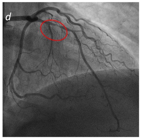
Figure 1. Acute Coronary Syndrome. A patient presenting to the emergency department with acute chest pain. No significant cardiac enzyme elevation and ECG alteration. Cardiac CTA shows absence of significant stenoses in the right coronary artery (a) and the left ording to recircumflex (b); a significant stenosis in the proximal anterior descending artery (c) is present, confirmed by subsequent coronliterary angiogram (d).
According to recent literature the integration of derived fractional flow reserve in CT (FFR-CT) studies using computational fluid dynamics gives the chance to enhance CT diagnostic accuracy. Moreover, in the emergency setting the use of FFR-CT allows a safer patient management thanks to the opportunity to detect, in the case of negative study, cardiac revascularization in chest pain patients. At the same time, it allows a significant reduction in medical cost [15][62].