ncRNA Subtypes, Biogenesis, and Turnover: Comparison
Please note this is a comparison between Version 2 by Conner Chen and Version 1 by Karla Rubio.

Epigenetics investigates modifications in the expression of genes that do not depend on the underlying DNA sequence. Some studies have confirmed that environmental factors, such as toxicants, may promote a phenotype or a disease in an individual or even in the subsequent progeny through epigenetic alterations. Several epigenetic mechanisms, including modifications in DNA (e.g., methylation), histones, and non-protein coding RNAs (ncRNAs) can change genome expression under the exogenous influenc. Notably, the role of long noncoding RNAs (lncRNAs) in epigenetic processes has recently been highlighted in more detail. The latent interest in epigenetics has resulted in breakthroughs in seminal concepts in diseases ranging from autoimmune conditions to cancer, congenital diseases, mental retardation, endocrine diseases, pediatric diseases, neuropsychiatric disorders, and many others.

  • ncRNA
  • epigenetic reprogramming
  • environment-related toxicants
  • tumorigenesis
  • biomarkers
  • exposome

1. Introduction

The central dogma in genetics is that information in our cells flows only in one direction, from DNA to RNA, then to proteins. It was an absolute dogma that has now been essentially debunked due to the role of the environment in the modulation of gene expression [1]. Environmental factors, including pollutants and lifestyle, constitute a significant role in severe, chronic pathologies with social and economic consequences [2].
The measurement of all environmental exposures and assessing their correlation with effects on individual health is defined as the exposome [3]. An individual’s exposome begins before birth and includes insults from environmental and occupational sources. In fact, the exposome interacts with our unique characteristics such as genetics, physiology, and epigenetics. The “environmental exposure” is a complex construct that encompasses exposures either physical, chemical, biological, or societal [4]. Historically, Christopher Paul Wild defined the exposome in 2005 as the totality of an individual’s exposure experience from conception until death and its impact on chronic diseases [5]. As a concept, it was put forward to stress the necessity of appropriate tools development for exposure assessment when applied to the study of human disease’s etiology [6].
The adaptation of cells to environmental factors causing stress relies on a wide range of tightly controlled regulatory mechanisms. The epigenetic landscape represents the platform where multiple environmental factors interact with the complex genetic milieu, resulting in alterations in the expression gene that shape many aspects of health and disease [7]. Changes in the organization and the structure of chromatin structure are associated with the transcriptional response to stress caused by the environment, and in some cases, can impart the memory of stress exposure to subsequent generations through mechanisms of epigenetic inheritance [8].
Epigenetics investigates modifications in the expression of genes that do not depend on the underlying DNA sequence. Some studies have confirmed that environmental factors, such as toxicants, may promote a phenotype or a disease in an individual or even in the subsequent progeny through epigenetic alterations [9,10][9][10]. Several epigenetic mechanisms, including modifications in DNA (e.g., methylation), histones, and non-protein coding RNAs (ncRNAs) can change genome expression under the exogenous influence [11]. Notably, the role of long noncoding RNAs (lncRNAs) in epigenetic processes has recently been highlighted in more detail [12]. The latent interest in epigenetics has resulted in breakthroughs in seminal concepts in diseases ranging from autoimmune conditions to cancer, congenital diseases, mental retardation, endocrine diseases, pediatric diseases, neuropsychiatric disorders, and many others [1,13][1][13].
Discrepancies in the homeostatic functions of the epigenetic machinery cause a range of different disorders since these mechanisms (“epigenome”) are more sensitive to the environmental status (lack of nutrient consumption, physicochemical exposures, and psychological stress) than the genome, especially during early development due to the inherently dynamic nature of the epigenetic landscape [14]. Besides the factors above, exposure to pollutants such as arsenic, nickel, cadmium, mercury, benzene, dioxin, bisphenol A, and diethylstilbestrol can alter the epigenetic regulation of the genome. These changes can cause abnormal gene expression, leading to various cancer types and other diseases [15]. Here, wthe review current evidence indicating that epigenetic alterations mediate those detrimental effects caused by exposure to environmental toxicants, focusing mainly on a direct regulation by a diversity of ncRNAs subtypes (
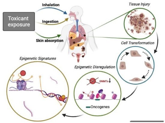
Figure 1).
Figure 1. Epigenetic toxicants lead to the phenotypic transformation of normal cells. People exposed (by inhalation, food or water ingestion, and skin contact) to certain pollutants suffer potential tissue injury in the lung, mammary gland, liver, pancreas, skin, colon, ovary, and hematological tissue among other target organs. It is well known that acute or chronic exposures to toxicants are associated with malignant cell transformation and, thus, pollution-related diseases, including cancer. Aberrant genetic modifications (DNA methylation, histone modifications, and ncRNAs can transform cells and disturb the expression of genes involved in homeostasis maintenance. Recently much attention has been given to ncRNAs with their role in pathophysiological conditions and signaling pathways such as oncogenesis, cell survival, altered apoptosis, and cell adhesion. Thus, ncRNAs (miRNA, lncRNA) are vital mediator molecules. Moreover, other important regulators, such as ncRNA-associated proteins forming multi-component complexes on specific loci at specific time points that conform complex epigenetic signatures, are also crucial during the cell transformation process. Studying those epigenetic signatures may improve the understanding of the biology of different pollution-related cancer types.

2. ncRNA Subtypes, Biogenesis, and Turnover

2.1. miRNAs

MicroRNAs (miRNAs) are the smallest ncRNAs, 20–25 nucleotides long, and do not encode proteins [16]. They bind in a complementary manner in the 3′ untranslated region (3′ UTR) of messenger RNAs (mRNAs) and mostly target them for degradation and blocking of their translation [17]. A miRNA can bind numerous mRNAs to inhibit their translation. Therefore, miRNAs are considered crucial post-transcriptional regulators of gene expression [16]. However, under certain cellular contexts (for instance, cell cycle arrest) they are recruited with AGO2 and FXR1 to specific loci with AU-rich elements (AREs) to ultimately activate translation [18,19,20][18][19][20]. Interestingly, recent evidence has called into question that the function of miRNAs is exclusively in the cytoplasm as a post-transcriptional mechanism targeting only mRNAs. In contrast, nuclear miRNAs potentially act during transcriptional silencing of bi-directionally expressed genes involving the formation of miRNA-ncRNA duplexes and nucleolus organization [21,22][21][22].
Genes coding for miRNAs are transcribed by RNA polymerase II (POLII) in the nucleus, where the primary miRNAs (pri-miRNAs) are capped, spliced, and polyadenylated. One miRNA, or clusters of two or more miRNAs, are produced from a primary transcript [16]. The double-stranded RNAse III DROSHA, inside a nuclear protein complex called Microprocessor, exerts the important role of cleaving the immature long pri-miRNAs. One essential cofactor that supports the activity of this protein complex is the double-stranded RNA (dsRNA)-binding protein DiGeorge syndrome critical region 8, DGCR8. Two RNase III domains inside DROSHA are necessary for the cleavage of one strand of the dsRNA to release a hairpin-shaped precursor miRNAs (pre-miRNAs) of approximately 60–70-nucleotides long [17]. Next, the export of the pre-miRNAs to the cytoplasm is mediated by exportin 5 (XPO5), and these are then cleaved by DICER1, an RNase III enzyme, originating from dsRNA to produce the mature miRNA duplex with 2-nucleotide 3′ overhangs of 22 nucleotides. DICER1 associates with transactivation-responsive RNA-binding protein (TRBP), which attaches dsRNA. TRBP physically bridges DICER1 with the Argonaute proteins (AGO1, AGO2, AGO3, or AGO4) and with members of the GW182 protein family during the assembly of the miRNA-induced silencing complex (miRISC). The silencing domain within GW proteins interacts with the deadenylase complex, which shortens the miRNA poly(A) tail. The mRNA-decapping enzyme 1 (DCP1)–DCP2 complex removes the 7-methylguanylate (m7G) cap. Consequently, the unprotected 5′ end is degraded by 5′–3′ exoribonuclease 1 (XRN1). In this manner, a specific mRNA is still stable at early stages, but its translation is inhibited, whereas, at later stages, mRNAs with short poly(A) tails are degraded [17]. Recent evidence showed that the increased production of miRNAs is proportional to the concentration of target mRNAs. Increased processivity of AGO2-associated DICER in the presence of target mRNA also contributes to the higher biogenesis of mature miRNAs [23].
Several classes of non-canonical, Drosha-independent or Dicer-independent miRNAs have been identified. One example of miRNAs that are Microprocessor-independent is related to “mirtrons”, which originate from spliced introns that function as pre-miRNAs and can be immediately exported to the cytoplasm for further processing by DICER [16]. Potential miRNAs can also originate from small nucleolar RNAs (snoRNAs) and transfer RNA (tRNA) fragments, and some of them can even be loaded into the RISC complex. Another Microprocessor-independent miRNA type is originated from the 5′-end of POLII-transcribed genes. Early transcription termination liberates the hairpins and serves as a DICER substrate. These miRNA precursors have a 5′7-methylguanylate (m7G) cap, which is not removed by the Microprocessor and facilitates its nuclear export by the cap-binding complex–exportin 1 (EXP1) pathway [16]. In particular, the existence of DICER-independent miRNAs arose from the detailed analysis of AGO2 activity, an RNase H-like endonuclease in AGO2-knockout mice [24] and zebrafish [25]. In other studies, focused on the precursor of the erythrocyte-specific miR-451, it was demonstrated that sequences that are too short to be processed by DICER produce abundant miRNA loads. Thus, miR-451 is bound by AGO2 and cleaved within its stem for mature and functional miRNA formation. This crucial process is conserved during erythrocyte maturation [24,25][24][25].

2.2. lncRNAs

Long noncoding RNAs (lncRNAs) lack protein-coding properties and extend more than 200 nucleotides. Although the proportion of functional lncRNAs is still undetermined and investigation is required to support evidence of functionality of the majority of lncRNAs, it is well documented that a growing number of lncRNAs have determinant roles in different mechanisms of gene regulation [26]. These large molecules of RNA can be spliced and polyadenylated, although some of them bind to ribosomes [27]. LncRNAs were originally described to have equivalent chromatin features to protein-coding genes [28]. However, recent work has highlighted the differences in the abundance of precise histone marks and splicing efficiency between lncRNAs and coding RNAs [29], as well as additional specifications among lncRNA subsets that differ in their chromatin landscapes [30].
LncRNAs are fundamental to different mechanisms such as gene imprinting, cells, and tissue differentiation, antiviral response, and the development of cancers. Among the broad mechanisms of action of transcriptional regulation by lncRNAs, those considered most relevant and universal is the interaction with chromatin-modifying complexes, obstruction of the transcriptional machinery, maintenance of the structure of nuclear speckles, regulation of splicing, regulation of mRNA degradation, modulation of protein translation and stability, and acting as molecular sponges for miRNAs [31]. However, while lncRNAs can be dysregulated in various human diseases known to include environmental factors as etiology, it is unclear how specific the interactions between lncRNA and mediators of the cellular response to environmental exposures could be. Most of the available data are derived from cell studies, and no data generated from population-based studies have been published [32]. In addition, as highly specific as lncRNAs are, depending on the target tissue, they represent potential diagnostic markers and, in consequence, therapeutic targets in cancer biology. Developing RNA-targeting therapeutics means a tremendous opportunity for modulating lncRNAs endogenous activity. Several preclinical approaches have been developed in this context, and others are currently under development by several pharmaceutical companies [33].

2.3. Other ncRNA Biotypes (siRNA, piRNAs, snoRNAs)

Although miRNAs are by far the most studied class of small-ncRNA biotypes in many fields, including cancer, environmental toxicology, and risk assessment, research of other RNA biotypes such as small nucleolar RNAs (snoRNAs), short interfering RNAs (siRNAs), and PIWI-interacting RNAs (piRNAs) may lead to similar breakthroughs based on their distinct cellular functions. First, siRNAs are 21- and 22-nucleotides RNAs generated by ribonuclease III (RNase III) and cleaved from longer double-stranded RNAs (dsRNAs) involved in post-transcriptional gene silencing in mammals and plants. Silencing is initiated by dsRNA that is homologous in sequence to the silenced gene [12]. Similar to miRNAs, siRNAs depend on DICER enzymes to excise them from their precursors [34] and AGO proteins to support their silencing-effector functions [35]. Generally, siRNA assembles into functional siRISC; one strand is separated and degraded. Then, siRNA-containing siRISC binds to AGO2 protein. This complex finally recognizes its targets by base pairing, and silencing occurs through one of several molecular mechanisms [36]. PIWI-interacting RNAs (piRNAs) are 26–32 nucleotides in length and bind to the PIWI protein family; they are abundant in spermatogenic cells, responsible for stem cell self-renewal, and involved in transposon silencing. piRNAs have been found to act in somatic cells and are crucial in guiding epigenetic regulation [37]. The biogenesis of piRNAs, initially described in Drosophila spp. and in C. elegans, begins with the transcription of a long, single-stranded precursor derived from a coding- gene transcript, transposons, tRNA, ribosomal RNA (rRNA), or intergenic loci by RNA POLII. Furthermore, a DICER/DROSHA-independent manner makes their processing. The precursor is exported from the nucleus into yeast bodies and further processed into smaller segments in yeast. piRNAs then form a complex with PIWI proteins assisted by shutdown (SHU) and heat shock protein 90 (HSP90). Their mature form is completed when the enzyme HENMT1 adds a methyl group to the 2′ carbon of the ribose on the 3′ end of the transcript. It can reenter the nucleus to induce transposon silencing and epigenetic regulation [38]. The abnormal expression of piRNA is associated with various cancers such as gastric, breast, renal, colorectal, and lung cancer [39].
Small nucleolar RNAs (snoRNAs) are short (60–300 nucleotides long) crucial biomolecules that participate importantly in ribosome biogenesis. They also have a role in the chemical modifications of some RNA biotypes, such as rRNAs, tRNAs, and small nuclear RNAs. There are two classes according to their conserved sequence elements: the C/D box snoRNAs (hairpins containing a sizeable internal loop, bounded by the C/C′ box and the Box D/D’ motifs), and the H/ACA box snoRNAs (two stem-loop structures separated by a conserved single-stranded motif with a consensus sequence of ANANNA) that are predicted to direct site-specific pseudo-uridylation and 2′-O-methylation of rRNA, to guide pre-rRNA processing and to act as molecular chaperones [40]. snoRNAs are derived from either mono- or polycistronic transcription units. In contrast, most of them are encoded within introns of pre-mRNAs and are transcribed mainly by RNA POLII. However, RNA POLIII and their final maturation require exonucleolytic trimming via two pathways: a splicing-dependent and splicing-independent pathway [41]. Splicing of the pre-mRNA results in a debranched precursor further processed by exonucleases to develop a mature snoRNA [42]. Because of their essential roles in biological processes, dysregulations in their expression can actively contribute to the promotion of carcinogenesis in the lung [43] and breast tissue [44].
Video Production Service