Cytokine Receptors Involved in Antimycobacterial Immune Response: Comparison
Please note this is a comparison between Version 1 by Magdalena Druszczyńska and Version 2 by Nora Tang.

Cytokine receptors are membrane-bound or soluble glycoproteins that serve as cytokine docking sites and inductors of a signaling cascade inside the cells. They are involved in the initiation of intracellular signaling that regulates a diverse range of biological functions including metabolism control, neural stem cell activation, inflammatory responses as well as blood cell and immune cell development and growth. The classification of cytokine receptor families is based on the structural homology of the extracellular cytokine binding domains and common intracellular signaling mechanisms. The main families include type I cytokine receptors, type II cytokine receptors, chemokine receptors, the tumor necrosis factor (TNF) receptor family, the transforming growth factor (TGF)-β receptor family, the immunoglobulin (Ig) superfamily, and the interleukin (IL)-17 receptor family.

  • cytokine
  • cytokine receptors
  • immune response
  • mycobacteria

1. Type I Cytokine Receptors

The type I cytokine receptors family includes cell surface expressed transmembrane receptors that recognize four-helix bundle cytokines such as IL-2, IL-4, IL-6, IL-12, IL-23, the granulocyte colony-stimulating factor (G-CSF), and the granulocyte-macrophage colony-stimulating factor (GM-CSF). The unifying feature of these receptors is the lack of intrinsic protein tyrosine kinase activity and transduction of the signaling pathway through the involvement of the non-receptor Janus kinases (JAKs) as well as the signal transducer and activator of transcription (STATs) factors. They are composed of several amino acid chains with conserved intracellular and extracellular features (Figure 12). The extracellular domains contain a region known as the haemopoietin domain or the cytokine receptor homology region (CHR) formed by a pair of Fibronectin type III (FnIII) modules, at the junction of which the primary cytokine binding site is located. CHRs contain four conserved cysteine residues within the first FnIII domain and a tryptophan-serine-X-tryptophan-serine (WSXWS) motif in the second FnIII domain [1][43]. Cysteines are critical to the maintenance of the structural and functional integrity of the receptors, and the WSXWS sequence serves as a recognition site for protein-protein interactions [2][44]. Many type I cytokine receptors also contain additional extracellular domains such as immunoglobulin (Ig) domains, extra FnIII domains or even a second CHR [1][43]. The cytoplasmic receptor domains, containing the Box 1/Box 2 motifs, provide specific docking sites for JAKs and STATs. In between these two motifs, additional binding sites for negative regulators such as the suppressor of cytokine signaling (SOCS) proteins can be found.
Figure 12. Structure of type I and type II cytokine receptors. The cytokine receptor homology region (CHR) contains four conserved cysteine moieties in the first FnIII domain and a tryptophan-serine-X-tryptophan-serine (WSXWS) motif in the second FnIII domain. Many type I cytokine receptors also contain immunoglobulin (Ig) domains. Cytoplasmic receptor domains contain sites for JAK and STAT binding. Type II cytokine receptors: CHRs have conserved cysteine residues arranged differently and do not contain the WSXWS motif compared to type I cytokine receptors. The cytoplasmic receptor domains contain sites for JAK and STAT binding.
Type I cytokine receptors can function as homodimeric or heterodimeric complexes. They usually consist of a ligand-binding chain and one or more signal-transducing chains (common gamma (γc) chain (CD132), common beta (βc) chain (CD131), or glycoprotein 130 (gp130)), which may be shared between various receptors within this receptor family [3][45]. Binding of type I cytokines to their receptors initiates activation of signal transduction pathways including the JAK-STAT pathway as well as additional signaling systems such as the Ras-mitogen-activated protein kinase (MAPK) pathway, and the phosphatidylinositol 3-kinase (PI3K)/protein kinase B (AKT) pathway.

1.1. IL-2 Receptor (IL-2R)

IL-2, known as a major T-lymphocyte growth factor, promotes proliferation and maturation of activated T cells as well as controls B cell proliferation and natural killer (NK) cell cytolytic activity [4][46]. IL-2 is the first short-chain type I cytokine, for which the receptor structure has been discovered [5][47]. The functional IL-2 receptor (IL-2R) has three forms, consisting of different combinations of three different chains, IL-2Rα (CD25), IL-2Rβ (CD122), and γc (CD132) (Figure 23). The IL-2Rβ and γc subunits belong to the type I cytokine receptor family and are responsible for signal transduction. The chains are expressed separately and differently on various cell types, and can assemble in different combinations, binding IL-2 with low, intermediate, or high-affinity. The IL-2Rα subunit binds IL-2 with low affinity, the combination of the IL-2Rβ and γc forms an intermediate-affinity IL-2R, and the three-chain complex binds the cytokine with high affinity [5][47]. IL-2 is first bound by IL2Rα, which leads to a conformational change and increases its affinity for the IL-2Rβ and γc subunits. IL-2 stimulation induces the activation of the Janus family of tyrosine kinases JAK1 and JAK3, associated with IL-2Rβ and γc, respectively, which phosphorylate IL-2Rβ and induce tyrosine phosphorylation of STATs and various other downstream targets. The downstream signaling pathways activated by IL-2 occur via three major signal transduction systems, the JAK-STAT pathway, the PI3K/AKT pathway, and the MAPK pathway, leading to the transcription of target genes that contribute to IL-2-dependent biological activity [5][47].
Figure 23. IL-2R structure and signaling. The IL-2 receptor (IL-2R) has three forms, consisting of different combinations of three different chains, IL-2Rα (CD25), IL-2Rβ (CD122), and γc (CD132). The interaction of IL-2 with IL-2R causes phosphorylation of JAK1 and JAK3, which causes downstream signaling through STAT5. The PI3/AKT and MAPK pathways are activated, which stimulates gene transcription in the cell nucleus, leading to cytokine production.
Using the soluble form of the IL-2Rα (sIL-2Rα) as a surrogate marker of IL-2-mediated T cell activation, it was found that HIV infection is associated with low serum levels of sIL-2Rα in patients with TB, even when CD4+ lymphocyte counts are relatively well preserved, and that impaired IL-2 signaling could contribute to the profound impact that HIV has had on both the incidence and clinicopathological manifestations of TB [6][48]. It is suggested that sIL-2R together with IFN-γ and neopterin may serve as parameters to monitor the prognosis of TB, particularly in patients with severe pulmonary TB [7][49]. IL-2R gene polymorphism analysis including variants of IL-2RAIL-2RB, and IL-2RG revealed that only IL-2RA gene polymorphisms showed a statistically significant association with susceptibility to TB [8][50]. In the study involving 235 participants with TB and latent Mtb infection, serum levels of IL-2Ra and chemokine CCL1 were higher in TB compared to latently infected individuals; therefore, they can be used as a diagnostic tool to discriminate between these groups [9][51]. IL-2R appears to have the potential to be used in the therapy of both TB and melanoma by binding fusion toxin composed of the catalytic and transmembrane domains of diphtheria toxin fused to human IL-2, leading to selective depletion of cells expressing the high affinity IL-2 receptor, including regulatory T cells (Tregs). Short-term depletion of IL-2R+ cells has been reported to be beneficial during TB infection, as it results in a decrease in the bacterial burden of the lung and spleen both as monotherapy and as adjunctive therapy administered with standard antibiotic treatment for TB [10][52].

1.2. IL-4 Receptor (IL-4R)

IL-4 is a pleiotropic type I cytokine that plays a critical role in the regulation of immune response. IL-4 induces the differentiation of naïve CD4 T cells into the type 2 helper (Th2) phenotype, the immunoglobulin (Ig) class switch to IgG1 and IgE in B cells, and alternative macrophage activation [11][53]. The cytokine exerts its biological activities through interaction with two surface receptor complexes, the type I IL-4R and the type II IL-4R (Figure 34) [12][54]. Both receptor types contain a common IL-4Rα (CD124) chain, which is a functional receptor subunit. The type I IL-4R is formed by the interaction of the IL-4Rα subunit with the γc subunit (CD132), while the type II IL-4R is formed by the interaction of the IL-4Rα subunit with the IL-13 binding chain, IL-13Rα1 (CD213a1) [13][55]. The IL-4Rα chain is also a subunit of the IL-13 receptor (IL-13R), which explains the similarity of the biological effects of IL-4 and IL-13. Binding of IL-4 to the IL-4Rα extracellular domain causes a conformational change in the intracellular receptor domains, activating the receptor-associated Janus kinases, which leads to the recruitment of STAT6 and its phosphorylation. Activated STAT6 forms homodimers that translocate to the nucleus and promote transcription of genes responsive to IL-4. Other phosphorylated tyrosine residues bind to proteins with phosphotyrosine binding (PTB) domains including insulin receptor substrate (IRS) proteins. Phosphorylated IRS proteins can subsequently activate the PI3K/AKT signaling pathway or the MAPK cascade [13][55].
Figure 34. IL-4R structure and signaling. IL-4 receptors consist of three different chains, α, γ and α1. There are two types of IL-4 receptors: type I IL-4R containing α and γ chains, type II IL-4R containing α and α1 (IL-13R) chains. The interaction of IL-4 and IL-13 with IL-4R activates the tyrosine kinases JAK1/JAK3 and TYK2. The PI3/AKT, MAPK and JAK-STAT pathways are activated, which stimulates gene transcription in the cell nucleus, leading to cytokine production.
The role of T helper (Th)2 cell-mediated immunity manifested by IL-4 and IL-13 production in the susceptibility and pathogenesis of TB remains a subject of scientific inquiry. Studies carried out in the Ghanaian cohort, in which genotype frequencies of variants of the genes IL-4IL-13IL-4RIL-13RA1 and IL-13RA2 were assigned to the size and number of cavities in patients with TB, showed that some variants of IL-4RA and IL-13RA2 are associated with greater risk of cavity development or progression, pointing to a role for both IL-4Rα and the IL-13Rα2 in the pathogenesis and progression of TB [14][56]. Experiments with the use of mice with IL-13 overexpression (IL-13tg) and with the absence of IL-4Rα (IL-4Rα−/−) revealed that deletion of IL-4Rα abrogates the increased susceptibility of Mtb-infected IL-13tg mice and the mandatory role for IL-4Rα in mediating the progression dependent on IL-13 of experimental TB [9][51]. IL-13 overexpression was found to result in recrudescence of Mtb growth accompanied by centrally necrotizing granulomas. It is postulated that the mechanisms driven by IL-13/IL-4Rα are directly related to the development of central granuloma necrosis [15][57]. Using BALB/c mice deficient in IL-4Rα specifically on B cells (mb1creIL-4Rα−/lox) it was shown that they had decreased mycobacterial burdens and lung pathology during chronic TB infection. It should be stressed that intranasal transfer of IL-4Rα-sufficient B cells isolated from the spleen of wild-type donor mice abolished the protective effect in mb1creIL-4Rα−/lox mice. Furthermore, adoptive transfer of wild-type B cells restored IFN-β production, but not IL-6, IL-12p40, and IL-10 in the lungs of mb1creIL-4Rα−/lox mice. Furthermore, the absence of IL-4Rα on B cells increased the macrophage inflammatory response ex vivo [16][58].

1.3. IL-6 Receptor (IL-6R)

IL-6, originally discovered as a B-cell differentiation factor, is a multifunctional cytokine with extensive immunomodulatory activity. It plays an important role in the regulation of acute phase response, inflammation, immune response, and haemopoiesis [17][18][59,60]. The cytokine influences various cell types through its unique receptor system. The IL-6-binding receptor complex consists of an IL-6 receptor subunit (IL-6R), existing in both membrane-bound (mIL-6R) and soluble (sIL-6R) forms, and IL-6 signal transducing chain glycoprotein 130 (gp130) (Figure 45). Although cells that do not express IL-6R do not respond to IL-6 alone, they can be stimulated by the complex formed by IL-6 and sIL-6R [19][61]. Upon binding of IL-6 to mIL-6R, homodimerization of gp130 is induced to form a high affinity IL-6/IL-6R/gp130 complex, activating a variety of biological signals through two pathways—the JAK/STAT pathway and the MAPK pathway.
Figure 45. IL-6R structure and signaling. The IL-6-binding receptor complex consists of an IL-6 receptor subunit (IL-6R), existing in both membrane-bound (mIL-6R) and soluble (sIL-6R) forms, and IL-6 signal-transducing chain glycoprotein 130 (gp130). Interaction of IL-6 with IL-6R activates the JAK1/JAK3 tyrosine kinases. The JAK-STAT and MAPK pathways are activated, which stimulates gene transcription in the cell nucleus, leading to cytokine production.
Produced primarily by monocyte-derived and recruited macrophages, IL-6 manages (together with IL-1 and TNF-α) the development of the acute phase response in TB. However, Mtb can affect the production of this cytokine and, by reducing it, leads to disease progression [20][62]. Furthermore, Mtb can dysregulate IL-6 production through the family of cytoplasmic proteins called suppressors of cytokine signaling (SOCS), leading to excessive IL-6 production in the epithelium, which inhibits STAT signaling [21][63]. Susceptibility and severity of TB have been reported to be associated with genetic variants in IL-6/IL-6R [22][64]. Studies conducted by Ritter et al. with the use of IL-6- and T-cell-specific gp130-deficient mice showed that the absence of IL-6 or gp130 in T cells has only a minor effect on the development of Th1 and Th17 antigen-specific cells after aerosol infection with Mtb [23][65]. Delgobo et al. provided interesting data that point to the improvement of IL-6R-mediated myeloid differentiation by human CD34+ cells in vitro by live Mtb [24][66]. The use of advanced tools, including ingenuity pathway analysis (IPA), gene set enrichment analysis (GSEA), STRING network analysis of protein-protein interactions, for comprehensive analysis of large transcriptomic and proteomic data sets allowed us to suggest that  Mtb infection activates a gene module shared by both type I IFN and IL-6, linking downstream “interferon-stimulated genes” (ISG) and lineage-specific regulators of myeloid differentiation—CCAAT/enhancer binding proteins (CEBP). Furthermore, the IL-6/IL-6R/CEBP gene module was found to be a central component correlated with monocyte expansion during Mtb infection in vivo, which is amplified in severe pulmonary and systemic disease. The transcriptional induction of CEBPB and CEBPD controlled by IL-6- and type I IFN signaling appears to be a relatively recent event in mammalian and primate evolution [24][66].

1.4. IL-12 Receptor (IL-12R)

IL-12 is a key immunoregulatory cytokine consisting of two covalently-linked subunits, IL-12p35 (35 kDa) and IL-12p40 (40 kDa), each expressed on different chromosomes [25][27]. The cytokine is involved in the induction of interferon (IFN)-gamma (γ) production and differentiation of CD4+ T cells into the type 1 T helper (Th1) phenotype, which is important for protective cell-mediated immune responses against a variety of intracellular pathogens [26][67]. The biological activities of IL-12 are mediated via binding to the membrane IL-12 receptor (IL-12R) complex, which is composed of two chains: IL-12Rβ1 (CD212) and IL-12Rβ2 (Figure 56) [25][27]. Binding of IL-12p40 and IL-12p35 subunits to IL-12Rβ1 and IL-12Rβ2 is followed by the activation of JAK kinases (Tyk-2 and Jak-2). Phosphorylated IL-12Rβ2 serves as a docking site for STAT4 proteins, which are phosphorylated by the JAK kinases on their tyrosine residues. To regulate IL-12-related gene transcription, phosphorylated STAT4 proteins are homodimerized and translocated to the nucleus.
Figure 56. IL-12R and IL-23R structures and signaling. The IL-12 receptor consists of IL-12/23p40 and IL-12p35 subunits, and IL-23 receptor is formed by IL-23p19 and IL-12/23p40 subunits. The interaction of IL-12/23 with IL-12R/23R activates the tyrosine kinases JAK2 /TYK2. The JAK-STAT pathway is activated, which stimulates the transcription of genes in the cell nucleus, leading to the production of cytokines.
IL-12R expression was found to be important for Th1 IFN-γ-producing cells maturation [27][68]. Zhang et al. found that the percentage of T cells expressing IL-12Rβ1 and IL-12Rβ2 as well as the levels of IL-12Rβ2 mRNA in peripheral blood mononuclear cells stimulated with Mtb, were significantly lower in patients with TB compared to controls [28][69]. On the contrary, the IL-12Rβ2 mRNA expression in patients with active TB was increased in the pleural fluid and lymph nodes. The use of anti-IL-10 and anti-TGF-β antibodies enhanced IL-12Rβ1/IL-12Rβ2 expression and IFN-γ production by Mtb-stimulated peripheral blood T cells from patients with TB suggesting that increased TGF-β production could reduce IL-12Rβ1 and IL-12Rβ2 expression in active TB. It also provides evidence that the expression of IL-12Rβ1 and IL-12Rβ2 plays a key role in mediating a protective Th1 response against mycobacteria.

1.5. IL-23 Receptor (IL-23R)

IL-23 is a heterodimeric cytokine, that is structurally related to IL-12. Both cytokines share a common IL-12p40 subunit, which, in IL-23, forms a biologically active complex with the IL-23p19 subunit [25][29][27,70]. The cytokine induces signals via its specific IL-23 receptor complex formed by two chains: the IL-12Rβ1 subunit, which is also utilized by IL-12, and the unique IL-23R (Figure 56) [29][70]. Despite the shared subunits, IL-23 and IL-12 have different biological functions. The main function of IL-23 is its ability to stimulate Th17 cells to produce IL-17 and induce proliferation of memory T cells [30][71]. Like other type I cytokine receptors, the IL-23 receptor complex lacks intrinsic enzymatic activity and is associated with the JAK family members, Janus kinase 2 (Jak2) and tyrosine kinase 2 (Tyk2). Binding of IL-23 to IL-23R promotes JAK kinases activation and phosphorylation of both the kinases themselves and the cytoplasmic tail IL-23R, creating docking sites for STAT3 monomers. Active Jak2/Tyk2 kinases phosphorylate STAT3 monomers, leading to dimerization, nuclear translocation, and DNA binding to target gene promoters [31][29].
IL-23 can influence both innate and acquired immunity by targeting cells that express IL-23R [32][72]. TCR γδ T cells are one of the populations highly responsive to IL-23, which, along with IL-17, appears to be a key regulator of immune response in all phases of Mtb infection [33][73]. Shen et al. found that Mtb infection in macaques increased the ability of IL-23 to enhance the proliferation of activated Vγ2Vδ2 T cells and production of such cytokines as IL-17, IL-22, IL-2, and IFN-γ [34][74]. Data from human studies using peripheral blood mononuclear cells (PBMCs) of TB patients showed a dysregulation of the IL-23/IL-17 axis by overexposure to stimulation of Mtb antigens [35][75]Mtb-stimulated CD4+ T cells from active TB patients expressed less IL-23R and pSTAT3 than those from latently infected individuals, despite similar levels of IL-23p19 mRNA in Mtb-stimulated monocytes. It is therefore suggested that chronic Mtb infection disrupts the STAT3 signal transducing pathway in T cells, reducing the signaling effect of IL-23 [36][76].

2. Type II Cytokine Receptors

Type II cytokine receptors are transmembrane proteins that bind interferons and members of the IL-10 family. They are structurally related to type I cytokine receptors, but their CHRs have differently arranged conserved cysteine residues and do not contain the WSXWS motif (Figure 12) [37][77]. Type II cytokine receptors are heterodimers or multimers containing components with high and low ligand affinity. The intracellular domain of the receptors is related to a member of the JAK family, which is activated upon ligand binding [2][44]. This results in the phosphorylation of the receptors and the formation of docking sites for STAT proteins. Upon phosphorylation by JAKs, STATs form dimers via their SRC homology (SH2) domain and translocate to the nucleus to activate the transcription of target genes. STAT3 and STAT1 factors are activated by all type II cytokine receptors, and some of them also activate STAT2 and STAT5. In addition to activating the JAK/STAT signal transduction system, some receptors also trigger the MAPK signaling pathway [37][77].

2.1. Interferon (IFN)-Gamma (γ) Receptor (IFN-γR)

IFN-γ, known as type II interferon, exerts a wide range of immunoregulatory activities and is critical for innate and adaptive immunity against infections [38][78]. The cytokine, secreted primarily by activated T cells and NK cells, acts as a factor promoting macrophage activation, regulating Th1/Th2 balance, and controlling cell proliferation and apoptosis [39][79]. The heterodimeric IFN-γ receptor (IFN-γR) is composed of two subunits: an α chain (IFN-γR1; CD119), and a β chain (IFN-γR2) (Figure 67). IFN-γR1 binds IFN-γ with high affinity, however it is not capable of mediating biological responses by itself. Activation of the IFN-γR2 subunit is required for the induction of the intracellular signaling pathway [33][73]. Binding of IFN-γ to IFN-γR1 induces its oligomerization, recruitment of the two IFN-γR2 chains, and activation of the Janus kinases 1 and 2 (Jak1 and Jak2). The activated JAKs phosphorylate a specific C-terminal tyrosine residue in the IFN-γR1 chain that serves as a docking site for STAT1 protein, which undergoes dimerization, migrates to the nucleus and regulates gene expression by binding to gamma-activated sequence (GAS) elements in the promoters of IFN-γ-regulated genes.
Figure 67. IFN-γR structure and signaling. IFN-γR consists of two β chains (IFN-γR2) and two α chains (IFN-γR1). The interaction of IFN-γ with IFN-γR activates the JAK1/JAK2 tyrosine kinases. The JAK-STAT pathway is activated, which stimulates the transcription of genes in the cell nucleus, leading to the production of cytokines.
Since IFN-γ plays a critical role in the host protective response against mycobacteria, blocking IFN-γR mediated signaling is believed to be an important Mtb evasion strategy [40][80]. IFN-γR gene knockout mice or mice having genetic defects in IFN-γR were occurred to be extremely susceptible to Mtb infection [41][81]. In humans, the surface expression of IFN-γR was found to be downregulated in macrophages and peripheral blood mononuclear cells of patients with active TB, which was related to lower IFN-γR mRNA transcription [42][82]. This was associated with altered expression of the transcription factor Sp1, which is required for IFN-γR gene transcription. The resulting phenotype made macrophages resistant to the protective effect of IFN-γ, despite its presence at optimal levels [43][83].

2.2. IL-10 Receptor (IL-10R)

IL-10 is a potent cytokine with multiple pleiotropic effects on immunoregulation [44][84]. It is produced predominantly by leukocytes including T and B lymphocytes, monocytes, macrophages, and DC, as well as by some epithelial cells. The anti-inflammatory activity of IL-10 includes, inter alia (i.a)., downregulating the expression of MHC class II molecules and co-stimulatory molecules on monocytes and macrophages, as well as reducing the production of pro-inflammatory cytokines and chemokines [45][85]. Dysregulation of IL-10 production is associated with increased immunopathology in response to infection as well as an increased risk of developing various autoimmune disorders [45][85]. The IL-10 receptor (IL-10R) complex is composed of two subunits, IL-10Rα (IL-10R1), a ligand-binding subunit, and IL-10Rβ (IL-10R2), a signalling subunit (Figure 78). The IL-10Rα chain is specific to IL-10, however the IL-10Rβ subunit is shared by the receptors for other type II cytokines such as IL-22, IL-26, and IFN-lambda (IFN-λ) [46][86]. Binding of IL-10 to the extracellular domain of IL-10Rα activates phosphorylation of the JAK family kinases, Jak1 and Tyk2 (tyrosine kinase-2). These kinases phosphorylate specific tyrosine residues in the intracellular domain of the IL-10Rα chain, which serve as temporary docking sites for the signal transducer and activator of transcription 3 (STAT3) factor. The Jak1 and Tyk2 phosphorylate STAT3, leading to its homodimerization and subsequent translocation to the nucleus, where it binds to STAT3-binding elements (SBE) in promoters of various IL-10-responsive genes and drives the expression of anti-inflammatory mediators [46][47][86,87].
Figure 78. IL-10R structure and signaling. IL-10R consists of two β chains (IL-10R2) and two α chains (IL-10R1). The interaction of IL-10 with IL-10R activates the JAK1/TYK2 tyrosine kinases. The JAK-STAT pathway is activated, which stimulates the transcription of genes in the cell nucleus, leading to the production of cytokines.
Many studies have shown that IL-10 negatively regulates the immune response during Mtb infection [48][49][50][51][88,89,90,91]. Beamer et al. demonstrated in a mouse model that blockade of IL-10 activity with anti-IL-10R1 antibodies (αIL-10R1) during the first 21 days of Mtb infection resulted in increased recruitment of Th1 lymphocytes to the lungs and improved control of bacterial load [50][90]. This effect was found to be associated with the formation of mature fibrotic granulomas [51][91]. Furthermore, Pitt et al. revealed that, for 3 weeks following vaccination with M. bovis BCG, the IL-10R1 signal blockade enhanced antigen-specific Th1, Th17, and innate lymphoid IFN-γ and IL-17 responses in the lungs that subsequently increased protection against Mtb for up to 16 weeks after infection [52][92]. The results by Dwivedi et al. confirmed that a single dose of αIL-10R1 delivered simultaneously with the BCG vaccine was capable of maintaining long-term control of Mtb infection by reducing the pro-inflammatory cytokine profile in the lungs and increasing the production of antigen-specific IFN-γ and IL-17 [53][93]. These studies indicate the key role of IL-10R1 blockade in the establishment of long-term antigen-specific memory immunity.

2.3. IL-22 Receptor (IL-22R)

IL-22, a member of the IL-10 family, is a potent mediator of cellular inflammatory response [54][94]. The cytokine is produced by many types of immune cells including activated T cells, innate lymphoid cells (ILCs) and NK T cells [55][56][95,96]. IL-22 acts via a heterodimeric receptor complex composed of the IL-22α1 and IL-10β2 subunits [57][97]. The cytokine binding to the IL-22 receptor complex activates the Janus kinases, Jak1 and Tyk2, followed by phosphorylation and activation of STAT3, STAT1 and STAT5 proteins that migrate to the nucleus, induce expression of specific genes, and trigger the biological activity of IL-22 (Figure 89). IL-22 bioactivity may be negatively regulated by a soluble IL-22Rα2 receptor, known as the IL-22 binding protein (IL-22BP), which prevents binding of the cytokine to the functional cell receptor complex and neutralizes its activity.
Figure 89. IL-22R structure and signaling. IL-22R consists of 2 subunits (IL-22α1 and IL-10b2). Interaction of IL-22 with IL-22R activates JAK1/TYK2 tyrosine kinases. The JAK-STAT pathway is then activated, which stimulates the transcription of genes in the cell nucleus, leading to the production of cytokines.
Although the IL-22 receptor complex is predominantly expressed on epithelial cells at mucosal sites, recent studies have found that Mtb-infected macrophages also express IL-22R [58][59][60][61][98,99,100,101]. Treerat et al. demonstrated that IL-22R was expressed on macrophages accumulated in tuberculous granulomas in the lungs and that IL-22 could directly induce TNF-α production and macrophage activation to control the infection [62][102]. Reduced circulating IL-22 levels and a lower percentage of Mtb-specific IL-22-producing T cells in TB patients suggest an important role for IL-22 in TB immunology. However, it is still unclear whether the role of IL-22 in antimycobacterial immunity is protective or pathological.

3. Tumor Necrosis Factor (TNF) Receptor (TNFR) Superfamily

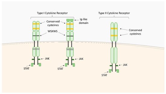
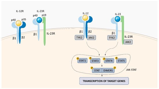
Within the TNFSF/TNFRSF superfamily consisting of 19 ligands and 29 receptors, TNF-α and its two main receptors: TNFR1 (TNFRp55/CD120a) and TNFR2 (TNFRp75/CD120b) are among the best characterized members. Soluble TNF-α (sTNF-α) preferentially binds to TNFR1, while membrane bound TNF-α (mTNF-α) is attached to TNFR2 [63][64][65][66][67][103,104,105,106,107]. Both receptors are single transmembrane glycoproteins [68][108]. Similarly to TNF-α, both TNFR1 and TNFR2, are synthesized in a membrane-bound form, which can be cleaved by metalloproteases to release soluble receptors (sTNFR) [69][109]. TNFR1 is expressed on nearly all nucleated cells [70][110]. As a death domain (DD) is present in the structure of TNFR1, this receptor belongs to cell sensors termed death receptors (DR) [71][111]. Two types of the DR signaling complex have been distinguished: the first group comprises the death-inducing signaling complexes (DISCs) and the second one, including TNFR1, transduces both apoptotic and survival signals [72][112]. TNFR1 is constitutively expressed on most cell types and undergoes activation not only by sTNF-α but also by mTNF-α [71][111]. TNFR1 stimulation is associated with the formation of two signaling complexes: Complex I, which leads to cytokine signaling and cell survival via activation of NF-kB, JNK, and p38 pathways, Complex II (in different variants) leading to apoptotic or necrotic cell death [71][73][111,113] (
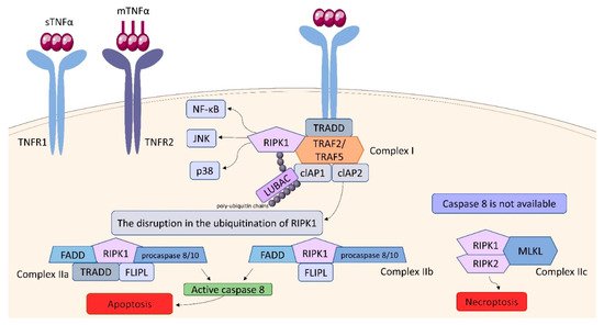
Figure 910). TNF binding toTNFR1 entails trimerization of the receptor and formation of a core signaling complex within the cytoplasmic tail of TNFR1. Trimerization enables the recruitment of TRADD (TNFR1-associated death domain) through DD. Acting as a scaffold, TRADD recruits the next elements of the forming complex: receptor-interacting serine/threonine-protein kinase 1 (RIPK1) and TNF receptor-associated factor (TRAF) 2, or TRAF5, whereby TRAF2 is responsible for recruiting other elements: cellular inhibitor of apoptosis protein (cIAP) 1 and cIAP2. Thus, the core signaling complex formed on the membrane consists of TRADD, TRAF2 (or TRAF5), RIPK1, and cIAP1/2. There are two factors determining whether it will be a platform for developing Complex I or Complex II: ubiquitination of RIPK1 and the availability of caspase molecules. As a result of the activity of TRAF and cIAP1/2, the ubiquitin chains are added to RIPK1 acting as scaffolds for other signaling factors, including K63, K11, K48 poly-ubiquitin chains and the linear ubiquitin chain assembly complex (LUBAC). The final stage in the formation of Complex I is the attachment of an M1 polyubiquitin chain to RIPK1 by LUBAC. Fully assembled Complex I activates NF-kB, JNK, and p38 pathways via recruitment of the inhibitor of the IkB kinase (IKK) complex and the TGFβ-activated kinase 1 (TAK1)-dependent mechanism, as it was described in detail by Gough and Myles [71][111]. The disruption in the ubiquitination of RIPK1 resulting in its release into cytosol leads to the formation of Complex II, which can be assembled in two variants: IIa and IIb. The attachment of TRADD, Fas-associated death domain (FADD), FLICE-like inhibitory protein (FLIPL) and procaspase 8 or 10 to RIPK1 forms Complex IIa. Variant IIb includes the same components as Complex IIa, except it lacks TRADD. The assembly of these proteins allow converting procaspase 8/10 to the active form that triggers the cell death by apoptosis. If caspase 8 is not available, Complex IIc is formed. It involves RIPK1 and RIPK3 assembling in an amyloid-like structure to form the necrosome, which leads to cell lysis (necroptosis) via mixed-lineage kinase domain-like protein (MLKL) and phosphoinositides binding [71][111]. It is suggested, that the effectiveness of complex II formation, caspase-8 activation, and the availability of FLIP in the cell cytosol, which prevents procaspase-8 activation at Complex II, determine whether a cell stays alive/active or dies. This model seems to be an attractive framework for making life-or-death decisions, however it requires more experimental proof [64][104].
Figure 910. TNFR structure and signaling. Binding of TNF-α to cell surface receptors engages multiple signal transduction pathways, including the IKK/NF-κB, JNK/AP-1 and p38 MAP signaling cascades. TNF also can trigger apoptosis via caspase-8 or necroptosis by activating intracellular RIPK kinases.
Video Production Service