Translational Stroke: Comparison
Please note this is a comparison between Version 2 by Camila Xu and Version 1 by benedetta piccardi.

The approach to reperfusion therapies in stroke patients is rapidly evolving, but there is still no explanation why a substantial proportion of patients have a poor clinical prognosis despite successful flow restoration. This issue of futile recanalization is explained here by three clinical cases which, despite complete recanalization, have very different outcomes. Preclinical research is particularly suited to characterize the highly dynamic changes in acute ischemic stroke and to identify potential treatment targets useful for clinical translation. This entreviewy surveys the efforts taken so far to achieve mouse models capable to investigate of investigating the neurovascular underpinnings of futile recanalization. We highlight the translational potential of targeting tissue reperfusion in fully recanalized mouse models and of investigating the underlying pathophysiological mechanisms from subcellular to tissue scale. We suggest that stroke preclinical research should increasingly drive forward a continuous and circular dialogue with clinical research. When the preclinical and the clinical stroke research are consistent, translational success will follow.


  • brain
  • ischemic stroke
  • reperfusion
  • futile recanalization

1. Introduction

Intravenous thrombolysis (IVT) and mechanical thrombectomy (MT) are established treatments proven to reduce disability after acute ischemic stroke by salvaging the brain. Even in the case of complete vessel recanalization, some patients remain functional dependent, which is thus called “futile recanalization” (FR) [1,2,3][1][2][3]. Moreover, some patients develop early complications, among which hemorrhagic transformation (HT) and cerebral edema (CE) are the most feared.
HT encompasses a broad spectrum of severity grades ranging from small areas of petechial hemorrhage to massive space-occupying hematomas. From the clinical point of view, HT has been divided into symptomatic and asymptomatic, a distinction that is important while evaluating the overall risk-to-benefit ratio of revascularization treatments.
CE is a severe complication of acute ischemic stroke and is the cause of death in 5% of all patients with cerebral infarction [4,5][4][5]. Edema causes tissue shifts and increased intracranial pressure that can cause death, usually between the second and the fifth day after stroke onset [6,7][6][7]. A large and potentially life-threatening infarct in the territory of the middle cerebral artery is often called a malignant middle cerebral artery infarct [4]. If not treated with reperfusion therapies, ≈50% to 80% of patients with this condition die, despite basic life support strategies. Surgical treatment by early decompressive hemicraniectomy decreases mortality, and decompressive hemicraniectomy is recommended by leading practice guidelines in selected patients [8]. Swelling and infarct growth each contribute to total stroke lesion growth in the days after stroke and should be considered a predictor of poor outcome even in patients with moderately sized stroke [9].
A critical role in the molecular mechanisms determining HT and CE is the disruption of the neurovascular unit (NVU), a dynamic terminal structure configuring an elaborate vascular network. The NVU is composed of an arteriole and its endothelial cells, basal lamina matrix, astrocyte end-feet, pericytes, astrocytes, neurons and their axons, and supporting cells (microglia and oligodendroglia) [10[10][11],11], and allows neurons to regulate micro vessels to support the metabolic needs of the tissue. The specialization and cellular composition of the NVU varies spatially along the arteriole–capillary–venule axis in order to allow local neurovascular coupling [12]. Indeed, more recently, a new concept of NVU has been proposed, identifying this complex interaction of segmentally diverse functional modules aimed to coordinate the entire brain vascular system, reacting to central and peripheral signals to maintain homeostasis of the brain, in health and disease [13].
During the ischemic insult, NVU participates in the reperfusion battleground occurring between the ischemic core and the surrounding salvageable tissue. Endothelial basal lamina dissolution starts as soon as 2 h after the onset of ischemia and is rapidly followed by an increase in Blood–Brain Barrier (BBB) permeability [14]. The early phase of BBB leakage occurs at 6 h from symptom onset, while there is a delayed secondary opening that occurs during the neuroinflammatory response (24–72 h after the ischemic insult). The prevailing view attributes the biphasic increase in BBB permeability to the disintegration and redistribution of tight junctions (TJs). However, recent studies suggest that increased endothelial transcytosis precedes and is independent of TJs disintegration [15]. BBB disruption following ischemic stroke contributes to HT, CE, secondary injury, and mortality. Clinical studies show no apparent increase in the risk of CE in ischemic stroke patients receiving IVT. However, there is experimental evidence that IVT could impair the BBB and contribute to reperfusion injury [16].
Age, stroke severity [17[17][18],18], and procedure delay [19], are the main predictors of FR.
Notably, the problem of selection for reperfusion therapies also persists after the introduction of multiparametric imaging techniques, such as multimodal Computed Tomography (CT) protocol and magnetic resonance (MR) imaging. These widely used techniques include: 1—non-contrast CT (NCCT) to detect intracerebral hemorrhage and early ischemic changes, quantified by a semiquantitative method known as ASPECTS (Alberta Stroke Program Early CT Score); 2—CT Angiography (CTA) to identify the occlusion site and to assess collateral circulation with a single-phase (sCTA) or, better, multiphase (mCTA) techniques; 3—CT perfusion (CTP) to define the size of infarct core and ischemic penumbra and consequently their mismatch using its capability to discriminate the different functional components of the ischemic area [20,21,22,23,24][20][21][22][23][24]. Likewise, MR findings can improve the ability to select patients for novel treatment options properly by using experimental approaches measuring selected biochemical parameters in the brain, in addition to the most common sequences (i.e., perfusion and diffusion) [25,26][25][26]. Nonetheless, MR application in the clinical field, despite being feasible, is constrained by the limited availability of this exam, which is hard to reconcile in the context of a time-dependent disease [27]. Aside from clinical and radiological parameters, blood biomarkers may also serve as a practical tool to represent the pathophysiology status before clinical deterioration. A recent observational study showed that the increased levels of some circulating biomarkers (particularly metalloproteinases and inflammatory biomarkers, such as C-Reactive Protein) were independent predictors of FR in acute ischemic stroke patients after recanalization by endovascular treatments [28]. Precision medicine, the initiative to replace a one-size-fits-all approach designed for the average patient with treatments tailored to account for unique differences among individuals, shifts paradigms across many fields of medicine. The treatment of ischemic stroke due to large vessel occlusion (LVO) offers a compelling illustration. Individual intrinsic differences in the flow capacity of collateral vessels, degree of chronic ischemic disease, ischemic preconditioning, oxidative stress tolerance, microvascular blood flow regulation, and other factors influence each patient’s response to treatment [29]. Preclinical research may help understand the neuronal and vascular underpinnings of FR, identify potential treatment targets, and lead to clinical translation.

2. From Bedside

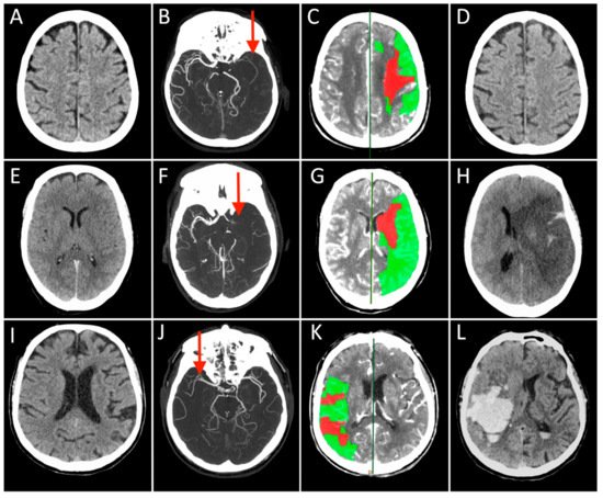
Case 1: A 73-year-old man with a history of hypertension, dyslipidemia, and prior myocardial infarction presented to the emergency department after the sudden onset of right-sided weakness and difficulty speaking. NCCT showed early ischemic changes in the left insular territory with an ASPECT score of 9 (Figure 1A). The mCTA demonstrated occlusion of the distal left M1 segment of the middle cerebral artery (MCA) with good collateral circulation (Figure 1B). A large penumbra in the left MCA territory with CBV lesion volume ≤ 50% of MTT lesion size (Figure 1C). After IVT, started 115 min after symptom onset, he underwent MT with complete recanalization (onset to recanalization time 280 min). In the 24 h follow-up CT scan, no ischemic lesion was visible (Figure 1D), and the patient experienced a great clinical improvement over the subsequent 3 days. At the 3-months follow-up, the patient was functionally independent.
Figure 1. Each row represents a single patient’s images derived in different modalities and timepoints (Case 1 presented in (AD), Case 2 in (EH), and Case 3 in (IL)). The first three columns on the left show the Non-Contrast-CT (NCCT), multiphase CT Angiography (CTA), and CT Perfusion (CTP) performed at hospital arrival, while the last column on the right displays the NCCT acquired at 24 h after stroke. On presenting NCCT (A,E,I) no early ischemic changes can be seen in the brain tissue in Case 2 and 3 (ASPECT score = 10), while a tissue swelling was detected in the right insular lobe in Case 1 (ASPECT score = 9, not shown). The mCTA images identified proximal (M1 segment) Middle Cerebral Artery (MCA) occlusion in the left hemisphere in (B,F), and contralateral side in (J) (red arrows). CTP showed in all cases a small infarct core corresponding to CBV lesion (red) and a large ischemic penumbra consisting of the difference between MTT and CBV lesions (green), representing the expected “salvageable” tissue after recanalization. In these patients, CBV lesion volume ≤ 50% of MTT lesion size. All patients were treated with combined IVT and MT, obtaining a complete recanalization, but the 24 h NCCT showed three different conditions: no ischemic lesion visible (D), complete MCA territory infarct associated with a massive cerebral oedema (H), and a vast hemorrhagic transformation of the ischemic lesion.
Case 2: A 71-year-old woman presented to a local emergency department 1 h after the sudden onset of speech difficulties and hemiparesis involving the right face, arm, and leg. She had a history of poorly controlled cardiovascular risk factors. NCCT showed a left MCA hyperdense sign (Figure 1E). The mCTA demonstrated a proximal left MCA occlusion (Figure 1F). CTP revealed a large penumbra in the left MCA territory with CBV lesion volume ≤ 50% of MTT lesion size (Figure 1G). She had no contraindications for IVT that was started 160 min after symptom onset. The patient then underwent endovascular treatment with complete recanalization that was not followed by clinical recovery (onset to recanalization time 300 min). Serial NCCT scans demonstrated progressive edema with mass effect in the left MCA distribution and 12 mm of midline shift (Figure 1H). She required endotracheal intubation and was admitted to the neurocritical care unit. The patient died the next day.
Case 3: A 70-year-old man had a sudden onset of left hemiplegia and forced eye deviation to the right. His medical comorbidities included hypertension, dyslipidemia, type 2 diabetes mellitus, and coronary artery disease. On neurological examination, he was awake, unable to communicate or follow commands. He had forced right gaze deviation, left hemianopsia, moderate left arm and leg weakness. NCCT identified hyperdense right MCA sign. The ASPECT score was 10 (Figure 1I). The mCTA showed an occlusion of the right proximal M2 segment of the MCA with a good collateral flow (Figure 1J). CTP demonstrated a large penumbra in the right MCA territory with CBV lesion volume ≤ 50% of MTT lesion size (Figure 1K). He received IVT after 90 min from clinical onset and underwent MT with complete recanalization (onset to recanalization time 215 min). The patient showed initial mild clinical improvement that was followed by both a rapid deterioration of consciousness and worsening neurological conditions with extensor posturing. Follow-up NCCT demonstrated an evolving infarct of the right MCA territory with hemorrhagic transformation and 8-mm midline shift (Figure 1L). He underwent surgical hematoma drainage with partial improvement. The patient was unable to walk and dependent on daily activities at the 3-month follow-up.
The three cases presented here show rather different clinical–functional outcomes despite similar neurological severity, neuroimaging at onset, and recanalization degree. Different treatment delays and clinical parameters might only partially account for differences in outcomes. The potential causes underlying this phenomenon are probably multifactorial but poorly understood. Ischemic changes at the tissue level appear to play a critical role and include reperfusion injury and ischemia-related microcirculatory dysfunction. It is of paramount importance to understand in depth what is there inside the hypoperfused tissue in order to find new predictors of clinical deterioration/FR and, consequently, specific prevention strategies.

References

  1. Nie, X.; Pu, Y.; Zhang, Z.; Liu, X.; Duan, W.; Liu, L. Futile Recanalization after Endovascular Therapy in Acute Ischemic Stroke. Biomed. Res. Int. 2018, 2018, 5879548.
  2. Stoll, G.; Pham, M. Beyond recanalization—A call for action in acute stroke. Nat. Rev. Neurol. 2020, 16, 591–592.
  3. Ter Schiphorst, A.; Charron, S.; Hassen, W.B.; Provost, C.; Naggara, O.; Benzakoun, J.; Seners, P.; Turc, G.; Baron, J.C.; Oppenheim, C. Tissue no-reflow despite full recanalization following thrombectomy for anterior circulation stroke with proximal occlusion: A clinical study. J. Cereb. Blood Flow Metab. 2021, 41, 253–266.
  4. Hacke, W.; Schwab, S.; Horn, M.; Spranger, M.; De Georgia, M.; von Kummer, R. ‘Malignant’ middle cerebral artery territory infarction: Clinical course and prognostic signs. Arch. Neurol. 1996, 53, 309–315.
  5. Brogan, M.E.; Manno, E.M. Treatment of malignant brain edema and increased intracranial pressure after stroke. Curr. Treat. Opt. Neurol. 2015, 17, 327.
  6. Shaw, C.M.; Alvord, E.C., Jr.; Berry, R.G. Swelling of the brain following ischemic infarction with arterial occlusion. Arch. Neurol. 1959, 1, 161–177.
  7. Juttler, E.; Schellinger, P.D.; Aschoff, A.; Zweckberger, K.; Unterberg, A.; Hacke, W. Clinical review: Therapy for refractory intracranial hypertension in ischaemic stroke. Crit. Care 2007, 11, 231.
  8. Cook, A.M.; Morgan Jones, G.; Hawryluk, G.W.J.; Mailloux, P.; McLaughlin, D.; Papangelou, A.; Samuel, S.; Tokumaru, S.; Venkatasubramanian, C.; Zacko, C.; et al. Guidelines for the Acute Treatment of Cerebral Edema in Neurocritical Care Patients. Neurocrit. Care 2020, 32, 647–666.
  9. Battey, T.W.; Karki, M.; Singhal, A.B.; Wu, O.; Sadaghiani, S.; Campbell, B.C.; Davis, S.M.; Donnan, G.A.; Sheth, K.N.; Kimberly, W.T. Brain edema predicts outcome after nonlacunar ischemic stroke. Stroke 2014, 45, 3643–3648.
  10. Del Zoppo, G.J. The neurovascular unit in the setting of stroke. J. Intern. Med. 2010, 267, 156–171.
  11. Stokum, J.A.; Gerzanich, V.; Simard, J.M. Molecular pathophysiology of cerebral edema. J. Cereb. Blood Flow Metab. 2016, 36, 513–538.
  12. Bernier, L.P.; Brunner, C.; Cottarelli, A.; Balbi, M. Location Matters: Navigating Regional Heterogeneity of the Neurovascular Unit. Front. Cell Neurosci. 2021, 15, 696540.
  13. Schaeffer, S.; Iadecola, C. Revisiting the neurovascular unit. Nat. Neurosci. 2021, 24, 1198–1209.
  14. Yang, G.Y. Advancement in stroke research. Stroke Vasc. Neurol. 2019, 4, 61–62.
  15. Zhou, M.; Shi, S.X.; Liu, N.; Jiang, Y.; Karim, M.S.; Vodovoz, S.J.; Wang, X.; Zhang, B.; Dumont, A.S. Caveolae-Mediated Endothelial Transcytosis across the Blood-Brain Barrier in Acute Ischemic Stroke. J. Clin. Med. 2021, 10, 3795.
  16. Dong, M.X.; Hu, Q.C.; Shen, P.; Pan, J.X.; Wei, Y.D.; Liu, Y.Y.; Ren, Y.F.; Liang, Z.H.; Wang, H.Y.; Zhao, L.B.; et al. Recombinant Tissue Plasminogen Activator Induces Neurological Side Effects Independent on Thrombolysis in Mechanical Animal Models of Focal Cerebral Infarction: A Systematic Review and Meta-Analysis. PLoS ONE 2016, 11, e0158848.
  17. Hussein, H.M.; Saleem, M.A.; Qureshi, A.I. Rates and predictors of futile recanalization in patients undergoing endovascular treatment in a multicenter clinical trial. Neuroradiology 2018, 60, 557–563.
  18. Lee, S.H.; Kim, B.J.; Han, M.K.; Park, T.H.; Lee, K.B.; Lee, B.C.; Yu, K.H.; Oh, M.S.; Cha, J.K.; Kim, D.H.; et al. Futile reperfusion and predicted therapeutic benefits after successful endovascular treatment according to initial stroke severity. BMC Neurol. 2019, 19, 11.
  19. Alawieh, A.; Vargas, J.; Fargen, K.M.; Langley, E.F.; Starke, R.M.; De Leacy, R.; Chatterjee, R.; Rai, A.; Dumont, T.; Kan, P.; et al. Impact of Procedure Time on Outcomes of Thrombectomy for Stroke. J. Am. Coll. Cardiol. 2019, 73, 879–890.
  20. Goyal, M.; Menon, B.K.; van Zwam, W.H.; Dippel, D.W.; Mitchell, P.J.; Demchuk, A.M.; Davalos, A.; Majoie, C.B.; van der Lugt, A.; de Miquel, M.A.; et al. Endovascular thrombectomy after large-vessel ischaemic stroke: A meta-analysis of individual patient data from five randomised trials. Lancet 2016, 387, 1723–1731.
  21. Leiva-Salinas, C.; Jiang, B.; Wintermark, M. Computed Tomography, Computed Tomography Angiography, and Perfusion Computed Tomography Evaluation of Acute Ischemic Stroke. Neuroimaging Clin. N. Am. 2018, 28, 565–572.
  22. Menon, B.K.; d’Esterre, C.D.; Qazi, E.M.; Almekhlafi, M.; Hahn, L.; Demchuk, A.M.; Goyal, M. Multiphase CT Angiography: A New Tool for the Imaging Triage of Patients with Acute Ischemic Stroke. Radiology 2015, 275, 510–520.
  23. Wintermark, M.; Flanders, A.E.; Velthuis, B.; Meuli, R.; van Leeuwen, M.; Goldsher, D.; Pineda, C.; Serena, J.; van der Schaaf, I.; Waaijer, A.; et al. Perfusion-CT assessment of infarct core and penumbra: Receiver operating characteristic curve analysis in 130 patients suspected of acute hemispheric stroke. Stroke 2006, 37, 979–985.
  24. Casetta, I.; Fainardi, E.; Saia, V.; Pracucci, G.; Padroni, M.; Renieri, L.; Nencini, P.; Inzitari, D.; Morosetti, D.; Sallustio, F.; et al. Endovascular Thrombectomy for Acute Ischemic Stroke Beyond 6 Hours from Onset: A Real-World Experience. Stroke 2020, 51, 2051–2057.
  25. Muddasani, V.; de Havenon, A.; McNally, J.S.; Baradaran, H.; Alexander, M.D. MR Perfusion in the Evaluation of Mechanical Thrombectomy Candidacy. Top. Magn. Reson. Imaging 2021, 30, 197–204.
  26. Kamalian, S.; Lev, M.H. Stroke Imaging. Radiol. Clin. N. Am. 2019, 57, 717–732.
  27. Atchaneeyasakul, K.; Shang, T.; Haussen, D.; Ortiz, G.; Yavagal, D. Impact of MRI Selection on Triage of Endovascular Therapy in Acute Ischemic Stroke: The MRI in Acute Management of Ischemic Stroke (MIAMIS) Registry. Interv. Neurol. 2020, 8, 135–143.
  28. Zang, N.; Lin, Z.; Huang, K.; Pan, Y.; Wu, Y.; Wu, Y.; Wang, S.; Wang, D.; Ji, Z.; Pan, S. Biomarkers of Unfavorable Outcome in Acute Ischemic Stroke Patients with Successful Recanalization by Endovascular Thrombectomy. Cerebrovasc. Dis. 2020, 49, 583–592.
  29. Makris, K.; Haliassos, A.; Chondrogianni, M.; Tsivgoulis, G. Blood biomarkers in ischemic stroke: Potential role and challenges in clinical practice and research. Crit. Rev. Clin. Lab. Sci. 2018, 55, 294–328.
More
Video Production Service