Cytoplasmic intermediate filaments (IFs), which together with actin and microtubules form the cytoskeleton, are composed of a large and diverse family of proteins. Efforts to elucidate the molecular mechanisms responsible for IF-associated diseases increasingly point towards a major contribution of IFs to the cell’s ability to adapt, resist and respond to mechanical challenges. From these observations, which echo the impressive resilience of IFs in vitro, we here discuss the role of IFs as master integrators of cell and tissue mechanics.
- cytoskeleton
- mechanics
- resilience
- rigidity
- stiffness
- elasticity
- viscosity
- rod domains
- coiled-coil region
1. Introduction
Tissue integrity, which is necessary for all metazoan life, relies on the ability of cells to adapt their morphology, their interactions, and their function to the conditions of their environment. The cytoskeleton, including actin microfilaments, microtubules, and intermediate filaments (IFs), form essential intracellular networks which support cell shape, cell adhesions and are indispensable for most cellular functions. While the role of actin in cell morphology, motility, and contractility has been extensively studied and the contribution of microtubules to intracellular trafficking, cell polarity, and adhesion dynamics is now well understood, the role of IFs in cell functions and tissue integrity remains unclear. This is partly because, in contrast to the ubiquitously expressed actin and tubulin, IF protein expression varies between cell types and tissues, and IF protein levels can represent anything from 0.3 to 85% of total protein levels in the cell [1][2][1,2]. Despite a high level of shared structural features between cytoplasmic IFs such as a head, rod, and tail domain, the more than 70 known IF genes create highly specialized, cell-type-specific networks of polymeric filaments. IFs are subdivided into five subtypes depending on small structural differences, their modes of assembly, and their expression pattern [3]. GFAP, vimentin, synemin, and nestin form the IF network in glia, neurofilaments in neurons, desmin and syncoilin in muscles, keratins in skin, and vimentin in mesenchymal cells. Consequently, the depletion of single IF genes does not always lead to severe phenotypes. However, in humans, mutations in IF genes give rise to a large diversity of diseases commonly characterized by the altered integrity of specific tissues [4]. The lack of associated molecular motors and well-characterized regulators of the assembly/disassembly dynamics further distinguishes IFs from actin and microtubules. These characteristics are probably responsible for our late understanding of IF functions at the cellular and tissue level. Only the painstaking studies of each type of IF’s structural and mechanical properties and their integration at the network, cellular and tissue levels are slowly unraveling the contribution of IFs to the physiology and pathology of multicellular organisms.
2. Intermediate Filaments as Key Players in Tissue and Cellular Mechanics
At the cellular level, studies have progressively confirmed that IF’s function in tissue integrity relies on their contribution to cell resilience under both mechanical stretching and compression. Indeed, the depletion of keratin or vimentin increases the deformability of stretched cells [5][6][13,14] ( Figure 1 B,C). Importantly, the loss of vimentin also decreases the viability of cells submitted to stretching [7][15] ( Figure 1 C). The role of IFs in cellular resistance to deformation by compressive forces appears to be cell-type specific. Under compression, both the depletion of vimentin in human mesenchymal stem cells [8][16] and the overexpression of vimentin in amoeboid cancer cells [9][17] reduce cell deformation. This discrepancy may be due to a different initial level of IF protein expression optimized in a cell-type-specific manner to provide cells with different mechanical properties. Alternatively, the difference may result from a cell-type-specific composition of the cytoskeletal network. For example, the depletion of vimentin in compressed highly contractile cells might have a different effect on their deformability compared to depletion in cells with lower contractility. Taken together, these studies show how IFs contribute to cell resilience by limiting cell deformation under mechanical stress and allowing stretched or compressed cells to recover their initial shape without any damage. However, they also suggest that the mechanical resilience of a given cell type in a specific situation may require an optimal level of expression of a specific IF type.
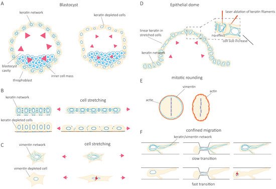
The IF network can also reorganize in response to mechanical stresses [10][11][12][13][14][18,19,20,21,22]. Rearrangement of the network is nicely illustrated in epithelial domes. In this three-dimensional in vitro epithelial sheet model, cells undergo extreme deformations while the tension across the epithelial sheet is maintained constant [15][23]. In fully expanded domes, extremely stretched cells coexist with cells that barely change their shape. The stretched cells are characterized by the formation of unusually straight bundles of keratin IFs, which extend from the nucleus to the plasma membrane. Laser ablation of these IF bundles results in a rapid increase in the cell area [15][23] ( Figure 1 D). This suggests that keratin IFs in highly stretched cells are load-bearing elements that maintain the reversibility of cell shape after large deformations. At the cellular scale, cell stretching by hypo-osmotic stress partly depolymerizes vimentin and nestin filaments and redistributes them throughout the cells [13][21]. This rearrangement is essential to cell survival after hypo-osmotic shock, confirming the contribution of IFs to cell mechanical resistance.
Accumulating evidence shows that IF networks are rearranged when cells adapt to external mechanical challenges. Cells can generate contractile or pushing forces to reshape in order to accomplish specific functions such as cell division or migration. To divide, cells must actively generate forces to accommodate their shape changes and overcome mechanical constraints by the surrounding tissue. The IF network reorganizes extensively not only to adapt to but also to promote these changes [16][17][18][19][24,25,26,27]. As cells round up to facilitate the accurate positioning of the spindle and the correct segregation of chromosomes, cortical tension generated by the actin cortex increases. In HeLa Kyoto cells, vimentin IFs contribute to this increase in cortical tension by relocalizing to the cortex, where they interact with actin to control actin organization [20][21][28,29] ( Figure 1 E). In confined environments, the loss of vimentin becomes detrimental to the segregation of chromosomes, and chromosome lagging is often observed [20][28]. However, besides vimentin IFs, HeLa cells also express keratins, which also reorganize during mitosis and affect the organization of vimentin. Indeed, when cells express nestin, the reorganization of vimentin during cell division is different. In nestin-expressing ovary (CHO) cells, C6-2 glioma, BHK-21 fibroblast, and cerebellar ST15A cells, the vimentin network disassembles at the cleavage furrow and does not localize to the cortex [22][23][30,31]. It thus seems that the reorganization of IFs that accompanies the changes in cell mechanics depends on IF proteins that are expressed. The cell-type-specific composition of the IF network needs to be taken into account in future investigations.
Migration in confined environments requires resilient mechanical support to allow cell deformation but prevent damage as they pass through complex environments. IFs appear to provide the essential mechanical support. Keratin knock-out (KO) keratinocytes migrate faster when squeezing through small pores in a Boyden chamber assay. However, they also frequently rupture and die [5][13] ( Figure 1 F). Similarly, vimentin depletion facilitates the migration of MEFs (mouse embryonic fibroblasts) in confined environments such as microchannels, collagen gels, and small pores [24][25][32,33] at the cost of nuclear alterations, nuclear envelop ruptures, and blebs [24][32] ( Figure 1 F). These observations were also confirmed in studies investigating the amoeboid migration of melanoma cancer cells [9][17]. Both keratin and vimentin networks appear to provide mechanical support to protect the nucleus against excessive deformations and maintain nuclear homeostasis during confined cell migration [24][25][26][32,33,34]. However, the switch from keratin to vimentin expression observed during the epithelial-to-mesenchymal transition (EMT) suggests that the two IF networks differentially contribute to the cell’s mechanical properties [27][28][29][30][35,36,37,38]. The microinjection of purified vimentin into MCF-7 epithelial cells changes the cell shape to a mesenchymal cell morphology [28][36]. Whether this effect is solely due to the mechanical functions of IFs or also reflects their role in intracellular signaling and cell motility remains unclear. While IFs in general, and the organization of the cytoplasmic IF network in particular, are essential to provide cell mechanical resilience, it is tempting to speculate that the mechanical specificity of each type of IF participates in cell-type-specific mechanics. Therefore, the control of IF protein expression may be central to the acquisition of cell-type-specific mechanical behavior adapted to the properties of their microenvironment and the modifications of cell behavior observed in pathological situations.
3. Mechanical Properties of IF Networks and Single Filaments In Vitro
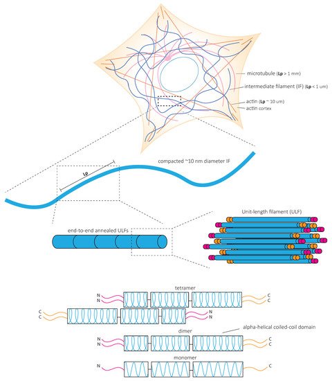
In contrast to actin microfilaments and microtubules, which interact with motors such as myosin, dynein, and kinesin, IFs do not bear molecular motors to provide mechanical forces. Instead, it is the fundamental structure of IFs that is at the heart of their mechanical properties ( Figure 23 ). In vitro studies of reconstituted IF networks and single filaments have recently shed light on the structural bases of IF mechanical properties. In this chapter, we recapitulate the results obtained with oscillatory shear rheology experiments on in vitro assembled IF networks which demonstrate their high elastic properties. We discuss how the mechanical properties of IF networks rely on inter-filament interactions ( Figure 34 ) and single filament mechanics ( Figure 45 ) based on biophysical in vitro measurements.
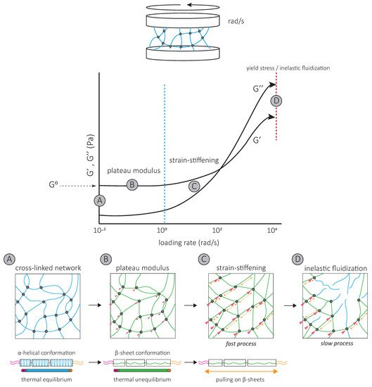
The strain-stiffening behavior of the IF network, observed at high strains, results from further filament stretching between crosslinks ( Figure 34 ) and is completely suppressed by the addition of a non-ionic surfactant [39][62]. Strain-stiffening is lost in networks formed by tailless filaments [39][40][41][42][62,63,74,75], showing that the filament C-terminal tails are responsible for the strong, attractive interactions necessary to withstand high stresses [39][62]. For instance, the characteristic side arms of neurofilament proteins are thought to be involved in the crosslinking of the network and provide resistance at large deformations [43][76].
The strain-stiffening of the network is limited by the rupture of the network [44][39][40][45][60,62,63,65] ( Figure 34 ). The stress at which the network ruptures, called the yield stress, is much higher for IF than for actin networks. When the yield stress is reached, a softening of the network is observed, which depends on the nature and degree of inter-filament interactions. The softening of vimentin IF networks is transient, which also distinguishes them from actin networks [46][47][77,78], and indicates that filaments are not permanently fractured [48][72]. Strengthening attractive interactions by the addition of divalent cations [49][40][50][45][51][61,63,64,65,68] or permanently crosslinking the network [48][72] increases the yield stress, suggesting that the softening of IF networks is due to the loss of interactions between IF proteins [48][72] ( Figure 34 ). The softening of vimentin IF networks is also loading-rate-dependent [48][72] ( Figure 34 ). At slow loading rates, the disruption of transient C-terminal inter-filament interactions counteracts the strain-stiffening response [48][72]. Here, the mechanical response is dominated by the disruption of crosslinks which occurs before or at the same time as the stiffening of the network and results in a low yield stress ( Figure 34 ) [48][72]. At fast loading rates, the strain-stiffening precedes the disruption of crosslinks, and the yield stress is much higher ( Figure 34 ) [48][72]. Similarly, transient interactions between neurofilament side arms are thought to be responsible for the reorganization of the network following disruption by large prolonged strains [43][76]. At fast deformations, these interactions provide the IF network with mechanical resilience [43][76].
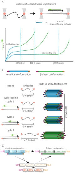
A second specific characteristic of IFs is their high stretchability and strong resistance to breakage. In vitro, single IFs can be stretched about 250% before breakage and can even reach 350% for desmin filaments [52][53][84,85]. For low strains up to 100%, a steep increase in force is observed with increasing strain [53][54][55][56][85,86,87,88]. The force–strain curve plateaus for intermediate strains, and at higher strains, the force further increases, indicating strain-stiffening of the filament [56][88] ( Figure 45 A). X-ray experiments [57][89] and mathematical modeling approaches suggest that the steep linear increase in force at low strains results from the elastic stretching of the coiled-coil α-helical domains [56][58][31][88,90,91] ( Figure 45 A). Further stretching of the α-helical domains leads to their transition to β-sheets, reflected by the force plateau at intermediate strains. The stiffening of the filaments at higher strains corresponds to the necessity to exert higher forces to extend β-sheets further ( Figure 45 A). This conformational change and the stretching of single filaments in a β-sheet conformation between filaments in IF networks are essential to the high G 0 and strain-stiffening of the network, respectively ( Figure 34 ).
4. Conclusions and Future Directions
In multicellular organisms, cells are continuously submitted to physical challenges whose nature and extent depend on microenvironment mechanics, cell motility within this environment, and the external mechanical stresses applied to the tissue. To resist these challenges, maintain tissue integrity, and ensure the survival of the organism, cell mechanical properties change in time and space [114]. Accumulating evidence points towards the IF network as a key player in cell mechanics and as an adaptable system because of its regulated composition and structure. This review advocates the idea of the IF network as a tunable intracellular scaffold integrating molecular, cellular, and tissue mechanics and providing cell-type-specific mechanical properties.
The characteristic mechanical properties of single filaments and filament networks are based on the amino acid sequence and the assembly of IF proteins. Disease-causing mutations of IFs illustrate the critical role of IFs as modulators of cell mechanics. Changes in IF-type expression, PTMs, pH, ionic concentrations, and direct and indirect interactions with other cytoskeletal components control IF composition, structure, and mechanical functions and can be modified in response to extracellular and intracellular mechanical signals. For instance, in the brain IF expression increases in response to inflammation as a consequence of injury, infections, or neurodegenerative diseases [115]. The resulting IF network rearrangements might change the mechanical properties of the IF network. Future studies should focus on uncovering the link between IF network rearrangements induced by mechanical signals and how such rearrangements impact the mechanical properties of the IF network. This will increase our knowledge on how IFs are tuned to optimize cell mechanics.
Cells not only have to resist physical stresses, but they must also sense mechanical information to direct a wide range of functions such as cell migration, proliferation, differentiation, or apoptosis. The cytoskeleton plays an essential role in sensing and transmitting the mechanical properties of the microenvironment. Although this review focused on IFs as the core of a mechanically resilient intracellular scaffold, one must also consider that IFs actively participate in the mechanotransduction process [24]. While their participation in the mechanotransduction process is becoming increasingly clear, their direct mechanosensing capacity is still debated. Since IFs can resist very large stresses, the force-sensing range of IFs has the potency to be much larger than that of actin. Recently, Cten/tensin 4—a member of the tensin family known to play a role in linking integrins to the actin cytoskeleton—was shown to interact specifically with stretched keratin filaments [116]. The protein detaches from the filaments when the stretching force decreases, making keratins a force-sensing element. Moreover, increasing the stress duration reduces the dissociation rate, which indicates that the protein binding contributes to the tensile memory of the filament. How the binding of Cten to keratin further participates in the transduction of mechanical forces has to be determined. Characterizing the IF-interacting domain of Cten to keratin will open up the door to discovering new force-sensing capacities of IFs. It is tempting to speculate that the conformational changes of IF proteins upon stretching expose numerous binding sides, which may differ depending on the composition of IFs. IF protein expression may thus not only control the mechanical properties of the cells but also modify their ability to transduce different ranges of mechanical cues as well as the cellular responses induced by these cues.
In conclusion, the unique mechanical properties of IFs, in combination with their high degree of tunability and adjustability to the mechanical needs of tissues and cells, make them key players in integrating mechanical information to support and direct cell and tissue function.