Functional Categories of S. pyogenes Immunomodulating Enzymes: Comparison
Please note this is a comparison between Version 2 by Jason Zhu and Version 1 by Mattias Collin.

Streptococcus pyogenes, or Group A Streptococcus, is an exclusively human pathogen that causes a wide variety of diseases ranging from mild throat and skin infections to severe invasive disease. The pathogenesis of S. pyogenes infection has been extensively studied, but the pathophysiology, especially of the more severe infections, is still somewhat elusive. One key feature of S. pyogenes is the expression of secreted, surface-associated, and intracellular enzymes that directly or indirectly affect both the innate and adaptive host immune systems. Undoubtedly, S. pyogenes is one of the major bacterial sources for immunomodulating enzymes. Major targets for these enzymes are immunoglobulins that are destroyed or modified through proteolysis or glycan hydrolysis.

  • Streptococcus pyogenes
  • protease
  • immunoglobulin (IgG)
  • IgG protease
  • glycan hydrolase
  • nuclease

1. Immunoglobulin Degrading and Modifying Enzymes

Without adaptive immunity, bacterial infections would have eradicated humanity a long time ago. It is therefore not surprising that antibodies are major targets for bacterial attack. Bacteria showcase a multitude of factors interfering with antibody-mediated effector functions, including enzymatic activities [11][1]. S. pyogenes expresses a remarkable number of proteins targeting and modulating human antibodies and their effector functions. These include immunoglobulin-binding surface proteins such as the M- and M-like proteins (not covered here, but for review, please see [4,11][1][2]) and unique enzymes targeting human antibodies, which will be the focus here.

21.1. Immunoglobulin Cysteine Proteinases

The Broad-Spectrum Cysteine Proteinase SpeB

The broad-range cysteine proteinase SpeB is one of the major secreted proteins in S. pyogenes (for the history of its discovery and naming, see [12][3]). It is secreted as a 40 kDa zymogen that is autocatalytically cleaved into the 28 kDa active proteinase (Figure 2A). SpeB has been implicated to have a role in the pathogenesis of S. pyogenes infections in animal models and in clinical studies (for reviews and references therein, see [12,13][3][4]) (Figure 1). SpeB has in vitro activity on a number of human proteins in the extracellular matrix and plasma (for a review see [12][3]). However, the physiological relevance of some of these activities can be debated.
SpeB is capable of breaking down immunoglobulins (Ig) A, M, D, and E into smaller fragments, and that it can also sever IgG in the hinge region to create fragment crystallizable (Fc) and fragment antigen-binding (Fab) domains [14,15][5][6]. Additionally, studies have revealed that SpeB specifically digests IgG that is antigen-bound on bacterial surfaces via its Fab-domain, while leaving IgG that is non-immunologically attached via Fc-binding surface proteins intact [16][7]. The separation of the Fc and Fab fragments in IgG predictably results in a reduction in the antibody-mediated opsonophagocytosis of S. pyogenes in vitro [17][8]. Whether the SpeB hydrolysis of antibodies is physiologically relevant has been challenged [18][9], but in vitro activity is of biotechnological interest in any instance (see below).

The Immunoglobulin G-Degrading Cysteine Proteinases IdeS/Mac-1/Mac-2
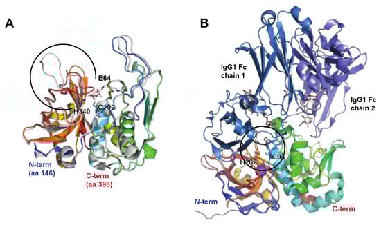
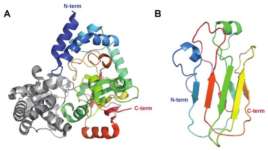
The secreted 35 kDa enzyme IdeS/Mac-1 was almost simultaneously identified by two independent research groups. One group focused on the similarities with the human protein Mac-1 (CD11b) and denoted it Mac-1, and described its antiphagocytic properties [19][10]. The other group showed that the protein is a cysteine proteinase with a remarkable specificity for human IgG, and denoted it immunoglobulin G-degrading enzyme of S. pyogenes, or IdeS [20][11]. An allelic variant of IdeS/Mac-1 denoted Mac-2 is also present in certain serotypes, as well as an enzymatically impaired variant in some serotype M28 strains [21][12]. However, the IgG protease activity seems to be the dominating feature overall [22][13]. IdeS has been extensively characterized, and it hydrolyses IgG in the lower hinge under the disulphide bridges generating a F(ab’)2 and two ½ Fc fragments (Figure 1) [23,24][14][15]. Determination of the crystal structure of IdeS both alone and in complex with IgG-Fc has shed light on its strict specificity for IgG [25,26][16][17] (Figure 2B). The strict specificity for IgG has previously been suggested to be mediated by exosite binding and interaction with another region outside of the active site [23][14]. This has been verified by the structural model of IdeS in complex with IgG, where IdeS has been demonstrated to bind across both IgG chains in the Fc region. With IdeS binding, the Fc domain peptide backbone becomes distorted, promoting cleavage between two glycine residues (Figure 2B) [26][17].
Figure 21. The streptococcal cysteine proteases SpeB and IdeS. SpeB and IdeS are both immunoglobulin cysteine proteinases with a papain fold. Common to such proteases is a catalytic site composed of a Cys and a His residue that exist as a zwitterionic pair [27][18]. (A) Structure of streptopain SpeB, PDB IDs: 4D8B (apo; shown in rainbow) and 4D8E (in complex with the small molecule inhibitor E64, shown in grey [28][19]). SpeB is composed of an N-terminal signal sequence (aa 1–27), a prodomain (aa 28–145), and a catalytic C-terminal region (aa 146–398), the structure of which is depicted here. SpeB has a unique, highly flexible glycine-rich C-terminal loop (aa 368–390, indicated by a black open circle), which, in the absence of a ligand, is positioned over the active sites Cys192 and His340 [27][18], and the side chains of which are shown as sticks. For a detailed view of the active site and the conformational changes associated with SpeB activation and substrate active site binding, please see the detailed description in [28][19]. (B) Structure of IdeS (Mac-1 and Mac-2), containing a Cys94 mutation abolishing catalytic activity, alone and in complex with the IgG1 Fc fragment, PDB IDs: 1Y08 (apo [25][16], shown in rainbow) and 8A47 (with IgG [26][17], shown in rainbow (IdeS), shades of blue (IgG heavy chains), and grey (heavy chain-attached glycans)). IdeS cleaves IgG molecules at a defined sequence between two Gly residues (shown as black sticks) in the lower hinge region in the heavy chain. This cleavage takes place in two distinct steps, with the cleavage of the first chain being faster. In the crystal structure, IdeS can be seen to clamp down over the lower hinge region of one of these Fc chains, creating a cavity in which the catalytic residues (Cys94Ser [PDB ID: 1Y08] or Cys94Ala [PDB ID: 8A47] and His262, sidechains shown as sticks) are brought into close proximity to the Gly-Gly cleavage site (black circle).

21.2. Immunoglobulin Glycan Hydrolases

The Endo-β-N-acetylglucosaminidase EndoS

When EndoS was discovered, it represented the first described enzyme from S. pyogenes with specificity for human IgG and also the first described bacterial antibody glycan hydrolase [14][5]. The ndoS gene is found in the vast majority of genome-sequenced S. pyogenes strains and is highly conserved. EndoS is a 108 kDa secreted endoglycosidase belonging to family 18 of glycosyl hydrolases and hydrolyzes the chitobiose core of the conserved N-linked glycan on Asn297 in the heavy chain on human IgG. EndoS displays similarity to other endo-β-N-acetylglucosaminidases from bacteria, most notably EndoF2 from Elisabethkingia meningoseptica (formerly Flavobacterium meningosepticum), which has been extensively used as a glycan mapping tool [29][20]. Interestingly, the other members belonging to this family of enzymes are not dependent on the IgG protein backbone for activity, while EndoS only hydrolyzes the N-linked glycan if it is attached to Asn297 of the IgG heavy chain. It does not, for instance, hydrolyze N-linked glycans if those are present in the variable regions of the heavy and light chains of IgG. EndoS has activity on most naturally occurring IgG heavy chains’ glycans (complex-type, biantennary), but has limited activity on bulkier glycans, such as high-mannose and hybrid-type glycans, including glycans with bisecting GlcNAc [30][21]. Structural determination of EndoS both alone and in complex with IgG-Fc has uncovered that its extreme specificity for IgG is dependent on both interactions between the catalytic domain and the IgG glycan as well as additional protein–protein interactions between EndoS and the constant domain of human IgG, in analogy with the exosite binding described for IdeS above [26,31,32,33][17][22][23][24] (Figure 3A,B).

The Endo-β-N-acetylglucosaminidase EndoS2
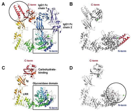
EndoS2 is an allelic variant of EndoS that has been identified in serotype M49 of S. pyogenes that displays only approximately 40% amino acid identity to EndoS. The ndoS2 gene replaces the ndoS gene in M49 strains and is in the same genetic context as ndoS in other serotypes. EndoS2 is also somewhat smaller, with an approximate size of 95 kDa, with the key structural difference to EndoS being the lack of two α-helices located towards the C-terminal end of the protein (Figure 3A,C). EndoS2 has activity on all subclasses of human (and several other subclasses of mammalian) IgG, but has, in addition, activity on the serum glycoprotein α1-acid glycoprotein [34][25]. Interestingly, EndoS2 has a much broader specificity for IgG-Fc glycans than EndoS by hydrolyzing bulkier glycans, such as high-mannose and bisecting structures [30][21]. Structural determination of EndoS2 in complex with glycan substrates showed a different type of interaction between the glycan and the enzymatic domain compared to EndoS [32][23], partly explaining the difference in glycan specificity (Figure 3D). Furthermore, a recent preprint presenting the structure of EndoS2 in complex with IgG-Fc further stresses the importance of the non-enzymatic domains for specific activity [35][26].
Figure 32. The streptococcal immunoglobulin glycan hydrolases EndoS and EndoS2. (A) Structure of the catalytically inactive EndoS Asp233Ala/Glu235Leu mutant in complex with the IgG1 Fc fragment, PDB ID: 8A49 [26][17], shown in rainbow (EndoS), shades of blue (IgG heavy chains), and blue (heavy chain-attached glycans). EndoS is composed of a proline-rich loop (aa 98–112), a glycosidase domain (aa 113–445), a leucine-rich repeat domain (LRR) (aa 446–631), an Ig domain (aa 632–764), a carbohydrate-binding domain (aa 765–923), and a C-terminal three-helix bundle domain (aa 924–995); the helix bundle is indicated (black circle). The catalytic center resides within the N-terminal glycosidase domain (black square). (B) Structure of EndoS in complex with a G2 oligosaccharide, PDB ID: 6EN3 [36][27] show in grey. In contrast to the EndoS crystallized together with the IgG Fc, which only contained EndoS amino acids 98–995, the structure in complex with the G2 oligosaccharide (blue sticks) also contains additional N-terminal residues that fold into an additional helical bundle, indicated here in red. Green spheres are Ni2+ and Ca2+ ions. (C) Structure of EndoS2, PDB ID: 6E58 [36][27] shown in rainbow colors. Like EndoS, EndoS2 has a glycosidase domain (aa 43–386) followed by a LRR domain (aa 387–547), an Ig domain (aa 548–680), and a carbohydrate-binding domain (aa 681–843) (all indicated in the figure). The main structural difference compared to EndoS is the lack of the C-terminal three-helix bundle domain, shown in panel (A). (D) Structure of EndoS2 (shown in grey) in complex with a high-mannose glycan (blue sticks), PDB ID: 6MDV [36][27]. The high-mannose glycan binds in a cleft (black circle) in the glycosidase domain. For a detailed description of the interaction interfaces between EndoS2 and its substrate glycan(s), please see [32][23].

2. Enzymes Interfering with Innate Immunity

Innate immunity is the first line of defense against invading pathogens in the body. It consists of a preformed defense mechanism that is present from birth and provides immediate protection. Innate immunity is mediated by various components, including physical barriers like the skin and mucous membranes as well as cellular and molecular mechanisms.
To combat the effects of the innate immune system, S. pyogenes expresses several enzymes degrading both complement components as well as chemokines and cytokines (Table 1). In addition to expressing complement-degrading enzymes, S. pyogenes expresses other virulence factors inhibiting complement functions, such as the recently described complement evasion factor (CEF) and streptococcal inhibitor of complement (SIC) [37,38][28][29]. Furthermore, proteolysis and modification of extracellular matrix (ECM) components that can alter barrier functions will not be discussed here (for a summary of SpeB’s activities on ECM, see [12][3]). S. pyogenes has also developed means of handling reactive oxygen species that that are generated as metabolic end products and as distinct antibacterial effectors.

2.1. Complement and Antimicrobial Peptide Degrading Enzymes

The Broad-Spectrum Cysteine Proteinase SpeB

The cysteine proteinase SpeB has been found to act on key components in both the classical and alternative pathways of complement activation. C3b, an effective opsonin that attracts phagocytic cells to infection sites, has been notably absent around soft tissue infections caused by S. pyogenes. It has also been observed that C3b levels are reduced in the blood serum of patients with streptococcal toxic shock syndrome (STSS), and that SpeB is capable of breaking down C3b in vitro [40][30]. It has further been demonstrated that SpeB degradation of C3b hampers both opsonization and the subsequent phagocytic destruction of bacteria [41][31]. Moreover, SpeB can disrupt the alternative pathway by breaking down properdin, an enhancer of complement activation [42][32].

The Serine Endopeptidase C5a, ScpA

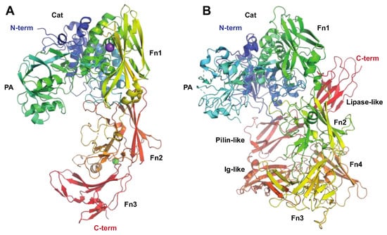
The C5a peptidase, ScpA, from S. pyogenes is one of the most extensively researched enzymes that modulate immune response (Figure 1). This enzyme specifically targets a key factor of human immunity: the chemotactic complement factor C5a. ScpA is a 130 kDa serine endopeptidase (Figure 4A) anchored to the cell wall that specifically cleaves C5a [43][33]. By doing so, it hampers the ability to attract phagocytic cells to the site of infection [44][34]. C5a is also instrumental in activating neutrophils that ingest the bacteria, highlighting the significance of ScpA’s activity [45][35]. An intriguing observation is that SpeB can liberate functional ScpA fragments, which then go on to neutralize C5a remotely from the bacterium itself [46][36].

2.2. Chemokine, Cytokine, and Kinin Active Enzymes

SpeB Activity on Immunologically Active Peptides

Chemokines are small proteins secreted from a variety of immune and non-immune cells. Not only are they powerful molecules for leukocyte signaling and differentiation, some of them also possess inherent antibacterial properties [47][37]. While most of the activities of SpeB discussed so far have anti-inflammatory effects, it has additional pro-inflammatory properties. SpeB has the ability to convert pro-interleukin-1β (IL-1β) into its biologically active form, triggering a state of inflammation that could worsen the effects of S. pyogenes infection (Figure 1) [49][38]. Another target of SpeB is H-kininogen, a component of the contact activation system that acts to inhibit proteinases. SpeB’s enzymatic action on H-kininogen, both in vitro and in vivo, leads to the release of bradykinin, a powerful vasodilator and pain inducer, which is likely to play a role in the pathogenesis of invasive S. pyogenes infections (Figure 1) [50][39]. In addition to targeting chemokines, IL-1β, and H-kininogen, SpeB has additionally been observed to induce the release of histamine and cause degranulation in mast cells [51][40]. While the exact mechanism behind its impact on mast cells remains unclear, it appears to be related to SpeB’s proteinase activity and may contribute to conditions such as STSS.

The Serine Proteinase SpyCEP

SpyCEP was originally identified as an enzyme with IL-8-degrading protelytic activity, regulated by a pheromone peptide and found in necrotizing soft-tissue isolates [52][41]. Subsequently, it was proven to be a cell-wall-anchored serine proteinase, produced as a 170 kDa zymogen, that is autocatalytically cleaved into 150 and 30 kDa forms that together form the active proteinase [53,54][42][43] (Figure 4B). SpyCEP is anchored to the cell wall by sortase, but is also released from the surface in the later growth phase [55,56][44][45]. Much like SpeB, SpyCEP also targets chemokines. SpyCEP hydrolyzes a group of ELR (Glu-Leu-Arg)-motif-containing CXC chemokines, including the strong neutrophil chemoattractant CXCL8 (IL-8) [57,58,59][46][47][48]. This contributes to S. pyogenes suppression of neutrophil migration into the site of infection (Figure 1) and consequently avoidance of opsonophagocytosis [60][49]. Intriguingly, in addition to hydrolyzing chemokines, SpyCEP also hydrolyzes the antimicrobial and signaling peptide LL-37, leading to reduced signaling activity through host receptors such as the nucleotide receptor P2X7R and the epidermal growth factor receptor (EGFR), while not affecting antibacterial activity [61,62][50][51].
Figure 43. The streptococcal subtilisin-like proteases ScpA and SpyCEP targeting the innate immune system. The C5 peptidase (ScpA) and SpyCEP are two pseudo-paralogous cell-surface-associated subtilisin-like proteases, which both are involved in evasion of the innate immune system. Common to these proteins is that they both target chemokines. (A) Structure of C5a peptidase, PDB ID: 3EIF [63][52] shown in rainbow colors. ScpA has a signal peptide, followed by a subtilisin-like catalytic domain (Cat, aa 32–583, shown in shades of blue and green) with an inserted protease-associated (PA) domain in the middle, and C-terminal A domains with fibronectin (Fn) type III domains (aa 584–1032, shown in shades of yellow to red) and W (aa 1033–1126) domains, the latter excluded in the structure. Even though ScpA is capable of degrading chemokines, its primary target is the complement component C5a. Structural modeling of the complex between ScpA and C5a suggests that the core of C5a is in close proximity to one of the Fn domains, with the C5a C-terminal tail being predicted to extend through the ScpA active site [63][52]. As for IdeS and IgG, the specificity of ScpA to C5a is likewise suggested to be mediated both by active site specificity and exosite interactions with the Fn domain, with the exosite interactions mediated by electrostatic forces. (B) Structure of SpyCEP, PDB ID: 5XYR [62][51] shown in rainbow colors. Like ScpA, SpyCEP has a signal peptide, followed by a subtilisin-like catalytic domain (Cat, aa 116–420, shown in dark blue) with an inserted protease-associated (PA) domain in the middle (aa 420–580, turquoise and green), followed by four fibronectin (Fn)-like domains (aa 580–1285, green to orange). Unlike ScpA, SpyCEP harbors three additional C-terminal domains (orange to red): a reverse-Ig-like domain (aa 1285–1398), a pilin-like domain (aa 1398–1492), and a lipase-like domain (aa 1492–1574).

3. Enzymes Acting on Chromatin and Cellular Processes

3.1. Enzymes Acting on DNA and Nucleotides

The Streptococcal Nuclease Sda1
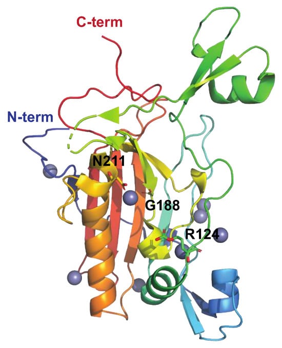
The prophage-encoded streptococcal nuclease Sda1 (Figure 54) was originally described as a secreted DNAse expressed by the M1T1 clone and is homologous to streptodornase D (SdaD) [68,69][53][54]. Like other streptococcal DNAses, Sda1 has been shown to promote streptococcal neutrophil resistance via the degradation of host DNA in NETs (Figure 1) in a murine model of necrotizing fasciitis [66][55]. In addition to degrading host DNA, Sda1 can dampen the host’s immune response by degrading streptococcal CpG-rich DNA. This suppresses Toll-like receptor 9 (TLR9)-mediated IFN-α and TNF-α production, hence decreasing macrophage bactericidal activity [70][56].
Figure 54. Structure of the streptococcal DNAase Sad1 His188Gly mutant, PDB ID: 5FGW [71][57], shown in rainbow colors. Sda1-mediated DNA degradation occurs in a sequence-nonspecific manner and is dependent on divalent cations (Zn2+ in grey). Asn211, chelating the active site divalent metal cation, is indicated, as is the general base, His188 (here replaced by Gly). Mutating Asn211 generates an inactive enzyme. The putative DNA-binding loop extends from aa 123–137, and Arg124 (side chains shown as sticks) is suggested to stabilize the transition state intermediate during cleavage of the DNA phosphodiester bond.

The Streptococcus pyogenes Nuclease A, SpnA

SpnA has been described as a cell-wall-associated DNAse with functions in the degradation of NETs (Figure 1) [67,72][58][59]. SpnA is homologous to the cell-wall-associated DNAse SsnA, the secreted nuclease A of Streptococcus suis [72][59]. Whereas no experimental structure for SpnA exists to date, the N-terminal domain is known to harbor at least three oligonucleotide-binding (OB) domains required for DNA binding during NET degradation, and, based on in vitro assays using recombinant proteins, at least two of these domains (OB2 and 3) are required for SpnA activity [67][58]. The C-terminal part of SpnA contains the DNA-degrading endo/exonuclease domain [67][58]. The DNase activity (and structural integrity) of SpnA is dependent on Ca2+ and Mg2+ ions [72][59].

The Streptococcal 5′-Nucleotidase A, S5nA

S5nA is a cell-wall-anchored ecto-5′-nucleotidase [74][60] that efficiently converts adenosine monophosphate (AMP) and, to a lesser extent, adenosine diphosphate (ADP), but not adenosine triphosphate (ATP), into the immunomodulator adenosine, facilitating the evasion of bacteria from the host immune response (Figure 1) [74][60]. Similar nucleotide-degrading enzymes have been described in other Gram-positive bacteria as well, such as the closely related Streptococcus suis, Group B Streptococcus (NudP), and Staphylococcus aureus (AdsA) [75,76][61][62]. As has been described for NudP and AdsA, S5nA can additionally convert deoxyadenosine monophosphate (dAMP) into deoxyadenosine (dAdo). dAdo is capable of inducing caspase-3-mediated apoptosis in macrophages and monocytes. Intriguingly, S5nA and SpnA have been demonstrated to generate inorganic phosphate from DNA, suggesting that these two enzymes cause a so-called “double-hit” to the host’s innate immune response by destroying NETs and killing macrophages via generation of dAdo [74][60], as has been described in S. sanguinis [77][63]. It has, however, further been demonstrated that S5nA does not significantly affect streptococcal growth in human blood, evasion of phagocytosis, or the formation of biofilm [78][64], suggesting that the main role of S5nA in streptococcal pathogenesis is indeed the destruction of macrophages.

The Streptococcal NAD(+)-Glycohydrolase, NADase
The S. pyogenes-encoded nicotinamide adenine dinucleotide (NAD)–glycohydrolase, also known as NADase, NGA, or SPN, is introduced into the host cell’s cytoplasm via cytolysin-mediated translocation (CMT) using another streptococcal virulence factor, streptolysin O (SLO), as a portal (Figure 1) [79][65]. NADase has several different activities contributing to streptococcal pathogenesis, including its ability to cleave β-NAD+ at the ribose–nicotinamide bond to generate ADP-ribose and the potent vasoactive compound nicotinamide as well as its ability to convert β-NAD+ into cyclic ADP-ribose, a potent second messenger. In order to protect itself against the detrimental effects of NADase, S. pyogenes expresses an endogenous inhibitor of NADase, IFS [80][66] (Figure 65). IFS inhibits glycohydrolase activity by acting as a competitive inhibitor of the β-NAD+ substrate.
Figure 65. Structure of streptococcal NADase. (A) The structure of NADase (shown in rainbow) with the endogenous inhibitor for NADase (IFS, grey) as determined by X-ray crystallography, PDB ID: 4KT6 [81][67]. Streptococcal NADase consists of two domains, of which the N-terminal domain (aa 1–190) is required for translocation into the host cell, and the C-terminal domain (aa 191–145) harbors the β-NAD+ glycohydrolase activity. The crystallized structure only contains the C-terminal catalytic domain. (B) The structure for the N-terminal domain as determined by nuclear magnetic resonance (NMR) spectroscopy, PDB ID: 7JI1 [82][68] shown in rainbow colors.

The C-di-AMP Synthase DacA

C-di-AMP synthases like DacA, found in many bacteria, are responsible for synthesizing cyclic-di-adenosine monophosphate (c-di-AMP), which is a second messenger molecule involved in various cellular processes. In S. pyogenes, it has been shown to have indirect immunomodulatory effects through the regulation of several pathways, including SpeB expression [83][69]. However, it has recently been shown to be involved in the activation of a type I interferon response mediated through the intracellular sensor stimulator of interferon genes (STING) in macrophages, contributing to disease severity. This is pronounced in individuals with a STING genotype, leading to reduced c-di-AMP binding in combination with S. pyogenes strains with high NADase activity [84][70].

3.2. Enzymatic Effects on Pyroptosis and Autophagy

SpeB Activity on Gasdermins

Gasdermins are host proteins involved in a process called pyroptosis, which is a type of cell death associated with inflammation and the host’s response to bacterial infections [85][71]. Gasdermins are cleaved by various proteases, including caspases, to release their N-terminal fragments, which form pores in the cell membrane, leading to cell lysis and inflammation. It has recently been shown that SpeB can activate gasdermins in keratinocytes that undergo pyroptosis, leading to killing of the bacteria, and that mice lacking gasdermins are more susceptible to infection (Figure 1) [86][72]. This most likely represents an ancient sensing mechanism for dangerous proteases, and represents an immunomodulating activity of SpeB that is beneficial for the host rather than for S. pyogenes.

SpeB Activity on Ubiquitin-Binding Proteins

Ubiquitin-binding proteins are involved in various cellular processes, including protein degradation, DNA repair, and signal transduction. They recognize and interact with ubiquitin moieties attached to target proteins leading to the controlled destruction, autophagy, of labeled proteins. Autophagy is recognized as a vital component of the innate immune response against intracellular bacteria. It has recently been shown that S. pyogenes has the capacity to evade ubiquitylation and evade recognition by the host autophagy marker LC3, as well as the ubiquitin-LC3 adaptor proteins NDP52, p62, and NBR1 [87][73]. This phenomenon depends upon the expression of SpeB, as an isogenic mutant of M1T1 lacking SpeB was more susceptible to autophagic targeting and demonstrated reduced intracellular replication [87][73]. In the same study, SpeB was shown to directly degrade p62, NDP52, and NBR1.

2.3.3. Enzymes Acting on Other Cellular Processes

The Streptococcal Arginine Deiminase SAGP
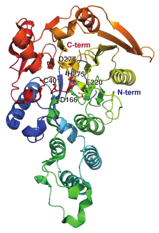
Like many other bacteria, S. pyogenes generates a diverse array of enzymes from different categories, including metabolic and housekeeping enzymes. Most of these enzymes have no proven direct link to immune evasion. However, among these enzymes, the streptococcal acid glycoprotein (SAGP) stands out (Figure 76). Initially, SAGP was characterized as an antitumor protein [88][74]. It exhibits arginine deiminase activity (breakdown of arginine to citrulline and NH3) and suppresses the proliferation and differentiation of T-lymphocytes in vitro (Figure 1) [89,90][75][76]. Furthermore, SAGP has been suggested to play a role in the survival of S. pyogenes under acidic conditions though the buffering activity of NH3, potentially aiding in intracellular/lysosomal survival [91][77].
Figure 76. Structure of the streptococcal acid glycoprotein SAGP or arginine deiminase ADI, PDB ID: 4BOF ([92][78], shown in rainbow colors). The active site residues Asp166, Glu220, His275, Asp77, and Cys401 are indicated. Individual mutation of these residues impaired the catalytic activity of ADI as determined by arginine conversion to citrulline. Intriguingly, the Asp211Ala and Asp277Ala mutants were deemed the most suitable as vaccine candidates due to their preserved structural integrity compared to wt ADI, as well as the production of high-titer antisera against these. Moreover, neither mutant disrupted the B-cell epitopes of ADI. Importantly, there is no human ortholog of ADI, further strengthening its possible use as a future vaccine target.

References

  1. Collin, M.; Kilian, M. Bacterial modulation of Fc effector functions. In Antibody Fc; Elsevier: Amsterdam, The Netherlands, 2014; pp. 317–332.
  2. Walker, M.J.; Barnett, T.C.; McArthur, J.D.; Cole, J.N.; Gillen, C.M.; Henningham, A.; Sriprakash, K.S.; Sanderson-Smith, M.L.; Nizet, V. Disease manifestations and pathogenic mechanisms of Group A Streptococcus. Clin. Microbiol. Rev. 2014, 27, 264–301.
  3. Nelson, D.C.; Garbe, J.; Collin, M. Cysteine proteinase SpeB from Streptococcus pyogenes—A potent modifier of immunologically important host and bacterial proteins. Biol. Chem. 2011, 392, 1077–1088.
  4. Carroll, R.K.; Musser, J.M. From transcription to activation: How group A streptococcus, the flesh-eating pathogen, regulates SpeB cysteine protease production. Mol. Microbiol. 2011, 81, 588–601.
  5. Collin, M.; Olsén, A. EndoS, a novel secreted protein from Streptococcus pyogenes with endoglycosidase activity on human IgG. EMBO J. 2001, 20, 3046–3055.
  6. Collin, M.; Olsén, A. Effect of SpeB and EndoS from Streptococcus pyogenes on human immunoglobulins. Infect. Immun. 2001, 69, 7187–7189.
  7. Eriksson, A.; Norgren, M. Cleavage of antigen-bound immunoglobulin G by SpeB contributes to streptococcal persistence in opsonizing blood. Infect. Immun. 2003, 71, 211–217.
  8. Collin, M.; Svensson, M.D.; Sjöholm, A.G.; Jensenius, J.C.; Sjöbring, U.; Olsén, A. EndoS and SpeB from Streptococcus pyogenes inhibit immunoglobulin-mediated opsonophagocytosis. Infect. Immun. 2002, 70, 6646–6651.
  9. Persson, H.; Vindebro, R.; von Pawel-Rammingen, U. The streptococcal cysteine protease SpeB is not a natural immunoglobulin-cleaving enzyme. Infect. Immun. 2013, 81, 2236–2241.
  10. Lei, B.; DeLeo, F.R.; Hoe, N.P.; Graham, M.R.; Mackie, S.M.; Cole, R.L.; Liu, M.; Hill, H.R.; Low, D.E.; Federle, M.J.; et al. Evasion of human innate and acquired immunity by a bacterial homolog of CD11b that inhibits opsonophagocytosis. Nat. Med. 2001, 7, 1298–1305.
  11. von Pawel-Rammingen, U.; Johansson, B.P.; Björck, L. IdeS, a novel streptococcal cysteine proteinase with unique specificity for immunoglobulin G. EMBO J. 2002, 21, 1607–1615.
  12. Agniswamy, J.; Lei, B.; Musser, J.M.; Sun, P.D. Insight of host immune evasion mediated by two variants of group A Streptococcus Mac protein. J. Biol. Chem. 2004, 279, 52789–52796.
  13. Söderberg, J.J.; Engström, P.; von Pawel-Rammingen, U. The intrinsic immunoglobulin g endopeptidase activity of streptococcal Mac-2 proteins implies a unique role for the enzymatically impaired Mac-2 protein of M28 serotype strains. Infect. Immun. 2008, 76, 2183–2188.
  14. Vincents, B.; von Pawel-Rammingen, U.; Björck, L.; Abrahamson, M. Enzymatic characterization of the streptococcal endopeptidase, IdeS, reveals that it is a cysteine protease with strict specificity for IgG cleavage due to exosite binding. Biochemistry 2004, 43, 15540–15549.
  15. Söderberg, J.J.; von Pawel-Rammingen, U. The streptococcal protease IdeS modulates bacterial IgGFc binding and generates 1/2Fc fragments with the ability to prime polymorphonuclear leucocytes. Mol. Immunol. 2008, 45, 3347–3353.
  16. Wenig, K.; Chatwell, L.; von Pawel-Rammingen, U.; Björck, L.; Huber, R.; Sondermann, P. Structure of the streptococcal endopeptidase IdeS, a cysteine proteinase with strict specificity for IgG. Proc. Natl. Acad. Sci. USA 2004, 101, 17371–17376.
  17. Sudol, A.S.L.; Butler, J.; Ivory, D.P.; Tews, I.; Crispin, M. Extensive substrate recognition by the streptococcal antibody-degrading enzymes IdeS and EndoS. Nat. Commun. 2022, 13, 7801.
  18. Dardenne, L.E.; Werneck, A.S.; de Oliveira Neto, M.; Bisch, P.M. Electrostatic properties in the catalytic site of papain: A possible regulatory mechanism for the reactivity of the ion pair. Proteins 2003, 52, 236–253.
  19. González-Páez, G.E.; Wolan, D.W. Ultrahigh and high resolution structures and mutational analysis of monomeric Streptococcus pyogenes SpeB reveal a functional role for the glycine-rich C-terminal loop. J. Biol. Chem. 2012, 287, 24412–24426.
  20. Trimble, R.B.; Tarentino, A.L. Identification of distinct endoglycosidase (endo) activities in Flavobacterium meningosepticum: Endo F1, Endo F2, and Endo F3. Endo F1 and Endo H hydrolyze only high mannose and hybrid glycans. J. Biol. Chem. 1991, 266, 1646–1651.
  21. Sjögren, J.; Cosgrave, E.F.J.; Allhorn, M.; Nordgren, M.; Björk, S.; Olsson, F.; Fredriksson, S.; Collin, M. EndoS and EndoS2 hydrolyze Fc-glycans on therapeutic antibodies with different glycoform selectivity and can be used for rapid quantification of high-mannose glycans. Glycobiology 2015, 25, 1053–1063.
  22. Du, J.J.; Klontz, E.H.; Guerin, M.E.; Trastoy, B.; Sundberg, E.J. Structural insights into the mechanisms and specificities of IgG-active endoglycosidases. Glycobiology 2019, 30, 268–279.
  23. Klontz, E.H.; Trastoy, B.; Deredge, D.; Fields, J.K.; Li, C.; Orwenyo, J.; Marina, A.; Beadenkopf, R.; Günther, S.; Flores, J.; et al. Molecular basis of broad spectrum N-Glycan specificity and processing of therapeutic IgG monoclonal antibodies by Endoglycosidase S2. ACS Cent. Sci. 2019, 5, 524–538.
  24. Trastoy, B.; Lomino, J.V.; Pierce, B.G.; Carter, L.G.; Günther, S.; Giddens, J.P.; Snyder, G.A.; Weiss, T.M.; Weng, Z.; Wang, L.-X.; et al. Crystal structure of Streptococcus pyogenes EndoS, an immunomodulatory endoglycosidase specific for human IgG antibodies. Proc. Natl. Acad. Sci. USA 2014, 111, 6714–6719.
  25. Sjögren, J.; Struwe, W.B.; Cosgrave, E.F.J.; Rudd, P.M.; Stervander, M.; Allhorn, M.; Hollands, A.; Nizet, V.; Collin, M. EndoS2 is a unique and conserved enzyme of serotype M49 group A Streptococcus that hydrolyses N-linked glycans on IgG and α1-acid glycoprotein. Biochem. J. 2013, 455, 107–118.
  26. Sudol, A.S.L.; Tews, I.; Crispin, M. Bespoke conformation and antibody recognition distinguishes the streptococcal immune evasion factors EndoS and EndoS2. bioRxiv 2023.
  27. Trastoy, B.; Klontz, E.; Orwenyo, J.; Marina, A.; Wang, L.-X.; Sundberg, E.J.; Guerin, M.E. Structural basis for the recognition of complex-type N-glycans by Endoglycosidase S. Nat. Commun. 2018, 9, 1874.
  28. Aghababa, H.; Ting, Y.T.; Pilapitiya, D.; Loh, J.M.S.; Young, P.G.; Proft, T. Complement evasion factor (CEF), a novel immune evasion factor of Streptococcus pyogenes. Virulence 2022, 13, 225–240.
  29. Åkesson, P.; Sjöholm, A.G.; Björck, L. Protein SIC, a novel extracellular protein of Streptococcus pyogenes interfering with complement function. J. Biol. Chem. 1996, 271, 1081–1088.
  30. Terao, Y.; Mori, Y.; Yamaguchi, M.; Shimizu, Y.; Ooe, K.; Hamada, S.; Kawabata, S. Group A streptococcal cysteine protease degrades C3 (C3b) and contributes to evasion of innate immunity. J. Biol. Chem. 2008, 283, 6253–6260.
  31. Kuo, C.-F.; Lin, Y.-S.; Chuang, W.-J.; Wu, J.-J.; Tsao, N. Degradation of complement 3 by streptococcal pyrogenic exotoxin B inhibits complement activation and neutrophil opsonophagocytosis. Infect. Immun. 2008, 76, 1163–1169.
  32. Tsao, N.; Tsai, W.-H.; Lin, Y.-S.; Chuang, W.-J.; Wang, C.-H.; Kuo, C.-F. Streptococcal pyrogenic exotoxin B cleaves properdin and inhibits complement-mediated opsonophagocytosis. Biochem. Biophys. Res. Commun. 2006, 339, 779–784.
  33. Cleary, P.P.; Prahbu, U.; Dale, J.B.; Wexler, D.E.; Handley, J. Streptococcal C5a peptidase is a highly specific endopeptidase. Infect. Immun. 1992, 60, 5219–5223.
  34. Wexler, D.E.; Cleary, P.P. Purification and characteristics of the streptococcal chemotactic factor inactivator. Infect. Immun. 1985, 50, 757–764.
  35. DeMaster, E.; Schnitzler, N.; Cheng, Q.; Cleary, P. M+ group a streptococci are phagocytized and killed in whole blood by C5a-activated polymorphonuclear leukocytes. Infect. Immun. 2002, 70, 350–359.
  36. Berge, A.; Björck, L. Streptococcal cysteine proteinase releases biologically active fragments of streptococcal surface proteins. J. Biol. Chem. 1995, 270, 9862–9867.
  37. Yang, D.; Chen, Q.; Hoover, D.; Staley, P.; Tucker, K.; Lubkowski, J.; Oppenheim, J. Many chemokines including CCL20/MIP-3α display antimicrobial activity. J. Leukoc. Biol. 2003, 74, 448–455.
  38. Kapur, V.; Majesky, M.W.; Li, L.L.; Black, R.A.; Musser, J.M. Cleavage of interleukin 1β (IL-1β) precursor to produce active IL-1β by a conserved extracellular cysteine protease from Streptococcus pyogenes. Proc. Natl. Acad. Sci. USA 1993, 90, 7676–7680.
  39. Herwald, H.; Collin, M.; Müller-Esterl, W.; Björck, L. Streptococcal cysteine proteinase releases kinins: A novel virulence mechanism. J. Exp. Med. 1996, 184, 665–673.
  40. Watanabe, Y.; Todome, Y.; Ohkuni, H.; Sakurada, S.; Ishikawa, T.; Yutsudo, T.; Fischetti, V.A.; Zabriskie, J.B. Cysteine protease activity and histamine release from the human mast cell line HMC-1 stimulated by recombinant streptococcal pyrogenic exotoxin B/streptococcal cysteine protease. Infect. Immun. 2002, 70, 3944–3947.
  41. Hidalgo-Grass, C.; Dan-Goor, M.; Maly, A.; Eran, Y.; Kwinn, L.A.; Nizet, V.; Ravins, M.; Jaffe, J.; Peyser, A.; Moses, A.E.; et al. Effect of a bacterial pheromone peptide on host chemokine degradation in group A streptococcal necrotising soft-tissue infections. Lancet 2004, 363, 696–703.
  42. Turner, C.E.; Kurupati, P.; Jones, M.D.; Edwards, R.J.; Sriskandan, S. Emerging role of the interleukin-8 cleaving enzyme SpyCEP in clinical Streptococcus pyogenes infection. J. Infect. Dis. 2009, 200, 555–563.
  43. Zingaretti, C.; Falugi, F.; Nardi-Dei, V.; Pietrocola, G.; Mariani, M.; Liberatori, S.; Gallotta, M.; Tontini, M.; Tani, C.; Speziale, P.; et al. Streptococcus pyogenes SpyCEP: A chemokine-inactivating protease with unique structural and biochemical features. FASEB J. 2010, 24, 2839–2848.
  44. Chiappini, N.; Seubert, A.; Telford, J.L.; Grandi, G.; Serruto, D.; Margarit, I.; Janulczyk, R. Streptococcus pyogenes SpyCEP influences host-pathogen interactions during infection in a murine air pouch model. PLoS ONE 2012, 7, e40411.
  45. Lawrenson, R.A.; Sriskandan, S. Cell Envelope Proteinase A (Streptococcus). Handbook of Proteolytic Enzymes; Elsevier: Amsterdam, The Netherlands, 2013; pp. 3195–3202.
  46. Edwards, R.J.; Taylor, G.W.; Ferguson, M.; Murray, S.; Rendell, N.; Wrigley, A.; Bai, Z.; Boyle, J.; Finney, S.J.; Jones, A.; et al. Specific C-terminal cleavage and inactivation of interleukin-8 by invasive disease isolates of Streptococcus pyogenes. J. Infect. Dis. 2005, 192, 783–790.
  47. Sumby, P.; Zhang, S.; Whitney, A.R.; Falugi, F.; Grandi, G.; Graviss, E.A.; Deleo, F.R.; Musser, J.M. A chemokine-degrading extracellular protease made by group A Streptococcus alters pathogenesis by enhancing evasion of the innate immune response. Infect. Immun. 2008, 76, 978–985.
  48. Zinkernagel, A.S.; Timmer, A.M.; Pence, M.A.; Locke, J.B.; Buchanan, J.T.; Turner, C.E.; Mishalian, I.; Sriskandan, S.; Hanski, E.; Nizet, V. The IL-8 protease SpyCEP/ScpC of group A Streptococcus promotes resistance to neutrophil killing. Cell Host Microbe 2008, 4, 170–178.
  49. Soderholm, A.T.; Barnett, T.C.; Korn, O.; Rivera-Hernandez, T.; Seymour, L.M.; Schulz, B.L.; Nizet, V.; Wells, C.A.; Sweet, M.J.; Walker, M.J. Group A Streptococcus M1T1 intracellular infection of primary tonsil epithelial cells dampens levels of secreted IL-8 through the action of SpyCEP. Front. Cell. Infect. Microbiol. 2018, 8, 160.
  50. Biswas, D.; Ambalavanan, P.; Ravins, M.; Anand, A.; Sharma, A.; Lim, K.X.Z.; Tan, R.Y.M.; Lim, H.Y.; Sol, A.; Bachrach, G.; et al. LL-37-mediated activation of host receptors is critical for defense against group A streptococcal infection. Cell Rep. 2021, 34, 108766.
  51. Jobichen, C.; Tan, Y.C.; Prabhakar, M.T.; Nayak, D.; Biswas, D.; Pannu, N.S.; Hanski, E.; Sivaraman, J. Structure of ScpC, a virulence protease from Streptococcus pyogenes, reveals the functional domains and maturation mechanism. Biochem. J. 2018, 475, 2847–2860.
  52. Kagawa, T.F.; O’Connell, M.R.; Mouat, P.; Paoli, M.; O’Toole, P.W.; Cooney, J.C. Model for substrate interactions in C5a peptidase from Streptococcus pyogenes: A 1.9 Å crystal structure of the active form of ScpA. J. Mol. Biol. 2009, 386, 754–772.
  53. Aziz, R.K.; Ismail, S.A.; Park, H.-W.; Kotb, M. Post-proteomic identification of a novel phage-encoded streptodornase, Sda1, in invasive M1T1 Streptococcus pyogenes. Mol. Microbiol. 2004, 54, 184–197.
  54. Aziz, R.K.; Edwards, R.A.; Taylor, W.W.; Low, D.E.; McGeer, A.; Kotb, M. Mosaic prophages with horizontally acquired genes account for the emergence and diversification of the globally disseminated M1T1 clone of Streptococcus pyogenes. J. Bacteriol. 2005, 187, 3311–3318.
  55. Buchanan, J.T.; Simpson, A.J.; Aziz, R.K.; Liu, G.Y.; Kristian, S.A.; Kotb, M.; Feramisco, J.; Nizet, V. DNase expression allows the pathogen group A Streptococcus to escape killing in neutrophil extracellular traps. Curr. Biol. 2006, 16, 396–400.
  56. Uchiyama, S.; Andreoni, F.; Schuepbach, R.A.; Nizet, V.; Zinkernagel, A.S. DNase Sda1 allows invasive M1T1 Group A Streptococcus to prevent TLR9-dependent recognition. PLoS Pathog. 2012, 8, e1002736.
  57. Moon, A.F.; Krahn, J.M.; Lu, X.; Cuneo, M.J.; Pedersen, L.C. Structural characterization of the virulence factor Sda1 nuclease from Streptococcus pyogenes. Nucleic Acids Res. 2016, 44, 3946–3957.
  58. Chang, A.; Khemlani, A.; Kang, H.; Proft, T. Functional analysis of Streptococcus pyogenes nuclease A (SpnA), a novel group A streptococcal virulence factor. Mol. Microbiol. 2011, 79, 1629–1642.
  59. Hasegawa, T.; Minami, M.; Okamoto, A.; Tatsuno, I.; Isaka, M.; Ohta, M. Characterization of a virulence-associated and cell-wall-located DNase of Streptococcus pyogenes. Microbiology 2010, 156 Pt 1, 184–190.
  60. Zheng, L.; Khemlani, A.; Lorenz, N.; Loh, J.M.S.; Langley, R.J.; Proft, T. Streptococcal 5′-Nucleotidase A (S5nA), a novel Streptococcus pyogenes virulence factor that facilitates immune evasion. J. Biol. Chem. 2015, 290, 31126–31137.
  61. Firon, A.; Dinis, M.; Raynal, B.; Poyart, C.; Trieu-Cuot, P.; Kaminski, P.A. Extracellular nucleotide catabolism by the Group B Streptococcus ectonucleotidase NudP increases bacterial survival in blood. J. Biol. Chem. 2014, 289, 5479–5489.
  62. Thammavongsa, V.; Schneewind, O.; Missiakas, D.M. Enzymatic properties of Staphylococcus aureus adenosine synthase (AdsA). BMC Biochem. 2011, 12, 56.
  63. Fan, J.; Zhang, Y.; Chuang-Smith, O.N.; Frank, K.L.; Guenther, B.D.; Kern, M.; Schlievert, P.M.; Herzberg, M.C. Ecto-5′-nucleotidase: A candidate virulence factor in Streptococcus sanguinis experimental endocarditis. PLoS ONE 2012, 7, e38059.
  64. Dangel, M.-L.; Dettmann, J.-C.; Haßelbarth, S.; Krogull, M.; Schakat, M.; Kreikemeyer, B.; Fiedler, T. The 5′-nucleotidase S5nA is dispensable for evasion of phagocytosis and biofilm formation in Streptococcus pyogenes. PLoS ONE 2019, 14, e0211074.
  65. Madden, J.C.; Ruiz, N.; Caparon, M. Cytolysin-mediated translocation (CMT): A functional equivalent of type III secretion in gram-positive bacteria. Cell 2001, 104, 143–152.
  66. Meehl, M.A.; Pinkner, J.S.; Anderson, P.J.; Hultgren, S.J.; Caparon, M.G. A novel endogenous inhibitor of the secreted streptococcal NAD-glycohydrolase. PLoS Pathog. 2005, 1, e35.
  67. Yoon, J.Y.; An, D.R.; Yoon, H.J.; Kim, H.S.; Lee, S.J.; Im, H.N.; Jang, J.Y.; Suh, S.W. High-resolution crystal structure of Streptococcus pyogenes β-NAD+ glycohydrolase in complex with its endogenous inhibitor IFS reveals a highly water-rich interface. J. Synchrotron. Radiat. 2013, 20 Pt 6, 962–967.
  68. Velarde, J.J.; Piai, A.; Lichtenstein, I.J.; Lynskey, N.N.; Chou, J.J.; Wessels, M.R. Structure of the Streptococcus pyogenes NAD+ glycohydrolase translocation domain and its essential role in toxin binding to oropharyngeal keratinocytes. J. Bacteriol. 2022, 204, e0036621.
  69. Fahmi, T.; Faozia, S.; Port, G.; Cho, K.H. The second messenger c-di-AMP regulates diverse cellular pathways involved in stress response, biofilm formation, cell wall Homeostasis, SpeB expression and virulence in Streptococcus pyogenes. Infect. Immun. 2019, 87, e00147-19.
  70. Movert, E.; Bolarin, J.S.; Valfridsson, C.; Velarde, J.; Skrede, S.; Nekludov, M.; Hyldegaard, O.; Arnell, P.; Svensson, M.; Norrby-Teglund, A.; et al. Interplay between human STING genotype and bacterial NADase activity regulates inter-individual disease variability. Nat. Commun. 2023, 14, 4008.
  71. Broz, P.; Pelegrín, P.; Shao, F. The gasdermins, a protein family executing cell death and inflammation. Nat. Rev. Immunol. 2020, 20, 143–157.
  72. LaRock, D.L.; Johnson, A.F.; Wilde, S.; Sands, J.S.; Monteiro, M.P.; LaRock, C.N. Group A Streptococcus induces GSDMA-dependent pyroptosis in keratinocytes. Nature 2022, 605, 527–531.
  73. Barnett, T.C.; Liebl, D.; Seymour, L.M.; Gillen, C.M.; Lim, J.Y.; Larock, C.N.; Davies, M.R.; Schulz, B.L.; Nizet, V.; Teasdale, R.D.; et al. The globally disseminated M1T1 clone of group A Streptococcus evades autophagy for intracellular replication. Cell Host Microbe 2013, 14, 675–682.
  74. Kanaoka, M.; Fukita, Y.; Taya, K.; Kawanaka, C.; Negoro, T.; Agui, H. Antitumor activity of streptococcal acid glycoprotein produced by Streptococcus pyogenes Su. Jpn. J. Cancer Res. 1987, 78, 1409–1414.
  75. Degnan, B.A.; Palmer, J.M.; Robson, T.; Jones, C.E.; Fischer, M.; Glanville, M.; Mellor, G.D.; Diamond, A.G.; Kehoe, M.A.; Goodacre, J.A. Inhibition of human peripheral blood mononuclear cell proliferation by Streptococcus pyogenes cell extract is associated with arginine deiminase activity. Infect. Immun. 1998, 66, 3050–3058.
  76. Starikova, E.A.; Mammedova, J.T.; Ozhiganova, A.; Leveshko, T.A.; Lebedeva, A.M.; Sokolov, A.V.; Isakov, D.V.; Karaseva, A.B.; Burova, L.A.; Kudryavtsev, I.V. Streptococcal arginine deiminase inhibits T lymphocyte differentiation in vitro. Microorganisms 2023, 11, 2585.
  77. Degnan, B.A.; Fontaine, M.C.; Doebereiner, A.H.; Lee, J.J.; Mastroeni, P.; Dougan, G.; Goodacre, J.A.; Kehoe, M.A. Characterization of an isogenic mutant of Streptococcus pyogenes Manfredo lacking the ability to make streptococcal acid glycoprotein. Infect. Immun. 2000, 68, 2441–2448.
  78. Henningham, A.; Ericsson, D.J.; Langer, K.; Casey, L.W.; Jovcevski, B.; Chhatwal, G.S.; Aquilina, J.A.; Batzloff, M.R.; Kobe, B.; Walker, M.J. Structure-informed design of an enzymatically inactive vaccine component for group A Streptococcus. MBio 2013, 4, e00509-13.
More
Video Production Service