Your browser does not fully support modern features. Please upgrade for a smoother experience.
Please note this is a comparison between Version 1 by Ali Athafah Tomah and Version 2 by Camila Xu.
Trichoderma is the asexual stage of the filamentous Hypocrea genus belonging to the Ascomycota fungi division. The species of this genus are free-living saprophytic fungi found in all soils, with an average presence in temperate and tropical soils of nearly 101-103 culturable propagules per gram.
- Trichoderma spp.
- cell-free culture filtrate
- mycosynthesis
- nanoparticles
1. Introduction (General Properties of Trichoderma)
Although the genus Trichoderma was first discovered by Persoon in 1794, it was not until Weindling published his first full paper on Trichoderma lignorum in 1934 that its role as a biological control agent was realized. Weindling’s paper showed that this species can control plant diseases, and this discovery led to a renewed interest in Trichoderma as a potential biocontrol agent [1]. Trichoderma is the asexual stage of the filamentous Hypocrea genus belonging to the Ascomycota fungi division. The species of this genus are free-living saprophytic fungi found in all soils, with an average presence in temperate and tropical soils of nearly 101-103 culturable propagules per gram [2][3][2,3]. These fungi reproduce asexually by the production of conidia and chlamydospores and in wild habitats by ascospores [4]. The genus Trichoderma is one of the most frequently isolated soil microorganisms. It has several useful properties, including being non-pathogenic to humans and plants, environmentally friendly, easy to isolate and culture, capable of rapid growth on a variety of inexpensive organic substrates, and able to produce a wide range of proteins, enzymes, and secondary metabolites [5][6][7][5,6,7]. The species of this genus are genetically quite diverse, with differences in capabilities among strains. These fungi have been widely used as biocontrol agents. Some Trichoderma spp. are known to control plant diseases through a variety of mechanisms, either indirectly (by competing for nutrients and space, modifying the environmental conditions, or promoting plant growth and plant defense mechanisms and antibiosis) or directly (by mechanisms such as mycoparasitism or the synergistic action of several mechanisms) [8]. Trichoderma spp. produce a variety of antibiotics and secondary metabolites that play an important role in inhibiting pathogens. The antibiotic’s mechanism is via low-molecular-weight diffusible organic compounds excreted by Trichoderma that inhibit the growth of the pathogen. The bioactive compounds secreted by Trichoderma are natural compounds that are chemically different, non-polar, and low molecular mass (less than 3000 Daltons) [9][10][9,10] and include polyketides, alkaloids, terpenoids, non-ribosomally biosynthesized peptides (NRPs), and metabolites of mixed biogenesis [10][11][10,11]. The compounds, which are typically composed of 5–20 amino acid residues, have a high content of α-aminoisobutyric acid (Aib) with an acylated N-terminus and a complex C-terminus that may consist of a free or methoxy-substituted 2-amino alcohol, amine, amide, free amino acid or sugar alcohol [12][13][12,13].
The antibiotic production of over 180 secondary metabolites, representing different classes of chemical compounds exhibiting biocontrol activity, has been reported for isolates of Trichoderma [10][14][10,14]. For example, gliovirin is produced from Gliocladium virens and has antimicrobial active against of Pythium ultimum [15], while gliotoxin from T. virens [16] inhibits the mycelium of both P. ultimum and Rhizoctonia solani [17]. Some species produce types of peptaibols (linear peptides), such as trichokonin VI from T. pseudokoningii SMF2, which has a wide antimicrobial spectrum against several bacteria, yeasts, and filamentous fungi [18], and trichorzianine from T. harzianum, which exhibits antibacterial activity against S. aureus [19], while koninginin A and B (polyketides group) are secreted by T. koningii [20][21][20,21]. Pyrone 6-PP was first discovered in a culture broth of T. viride [22] and is classified as a volatile organic compound. The 6-PP compound displayed good antifungal activity against Aspergillus flavus, Penicillium expansum, and Fusarium acuminatum [23]. Other antifungal compounds isolated from Trichoderma spp., belonging to different chemical classes, have been used in plant protection as an environmentally friendly and efficient management tool against a variety of phytopathogens.
These metabolites can be either overproduced or combined with other metals to create new formulations that are more efficient for use in several fields, including in the control of plant diseases. Biomolecules can bind to metals through their proteins and amino acid residues, forming a coating on the surface of the NPs. This coating, known as capping, can increase the stability of the NPs and prevent them from aggregating [24][25][24,25]. Subsequent stabilization can also be provided by free amino groups or cysteine residues or through the electrostatic attraction resulting from negative carboxyl groups provided by mycelial cell wall enzymes present in the filtrate. Furthermore, the ability of the thiol (-SH) group to form disulfides—perhaps the most important bridging structure in nature—with cysteine subunits of endogenous proteins renders them unique among the functional groups utilized in nanotechnology to form thiolated disulfide bonds that tightly bind with noble metals, leading to the formation of stable NPs [24][26][27][24,26,27]. For example, FTIR analysis confirmed that a gliotoxin bioactive compound secreted by T. virens is responsible for capping and reducing Ag+ and the biosynthesis of silver NPs through binding of each of the negative carboxyl groups, sulfur, and oxygen from the hydroxyl groups of gliotoxin with silver [28].
2. Trichogenic Nanoparticles
The term “mycosynthesis” was first used to describe the synthesis of NPs using the fungus F. acuminatum by Ingle et al. [29]. Since then, the term “Myconanotechnology” has expanded to include literature studies that describe NPs synthesized using fungi [30]. The term “Trichogenic” has recently been used to describe the biogenic synthesis of selenium NPs using six species of Trichoderma [31]. Among the most important fungi that are used in biological control against phytopathogens, the Trichoderma species have the potential to be used to synthesize NPs on a large scale by using an environmentally friendly production process. According to the inventory list conducted by Cai and Druzhinina [32], there are around 460 species with valid names in the Trichoderma genus that have been deposited in public updated databases available on the International Subcommission on Taxonomy of Trichoderma website (www.trichoderma.info, accessed on 25 February 2022). However, and from a thorough inventory of literature studies, researchers found that only about 13 species belonging to the Trichoderma genus show the ability for NP synthesis, including T. harzianum, T. asperellum, T. viride, T. atroviride, T. virens, T. longibrachiatum, T. pseudokoningii, T. reesei, T. koningii, T. brevicompactum, T. citrinoviride, T. hamatum, and T. gamsii. These species are known to be applied globally as biological control agents against different plant pathogens. Although the number of Trichoderma species has been increasing, research on the synthesis of NPs using this genus is still in its early stages. More research is needed to explore the potential of Trichoderma species for nanoparticle synthesis. The first report on the use of Trichoderma in the synthesis of silver NPs by Mukherjee [33] dates back to 2008. This strategy has attracted much more attention in the most recent decade as the synthesis mechanism for various NPs using diverse metals such as silver (Ag), gold (Au), copper (Cu), zinc (Zn), and so on. NPs of both silver and gold are considered more secure in contrast to other metallic NPs [34]. Through a survey of the literature published from the years 2008 to 2021, more than 100 research papers published in the approved journals adopted the use of Trichoderma in the synthesis of NPs from different metals. Using the percentage equation [(M/N) × 100)], where M means the number of studies that used Trichoderma in the synthesis of NPs from a specific metal and N means the number of research studies that used Trichoderma in the synthesis of NPs from different minerals, the proportional analysis shows that silver metal occupied the leading position in the synthesis of NPs by the Trichoderma species, which was 54% of the total of other metals (Figure 1A). On the other hand, most of the Trichoderma spp. applied in biological control were suggested to have the capacity to biosynthesize NPs. Five different species of Trichoderma, viz., T. asperellum, T. harzianum, T. longibrachiatum, T. pseudokoningii, and T. virens, led in the production of silver NPs (AgNPs) [35]. Spherical AgNPs with sizes from 2 to 15 nm were formed using cell filtrates of T. inhamatum [36]. Production of silver NPs was achieved through extracellular reduction by six isolates of T. virens, and a single and aggregated form was obtained, which was uniform in shape and had a size of 8–60 nm [37]. Meanwhile, six species belonging to the Trichoderma genus, viz., T. asperellum, T. harzianum, T. atroviride, T. virens, T. longibrachiatum, and T. brevicompactum, were successful in the biogenic synthesis of selenium NPs (SeNPs) [31].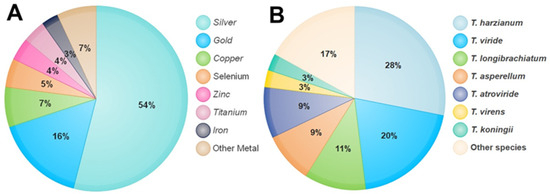
Figure 1. The percentage of metals used in the synthesis of NPs by Trichoderma species (A). Percentage of Trichoderma species used in the synthesis of NPs (B).