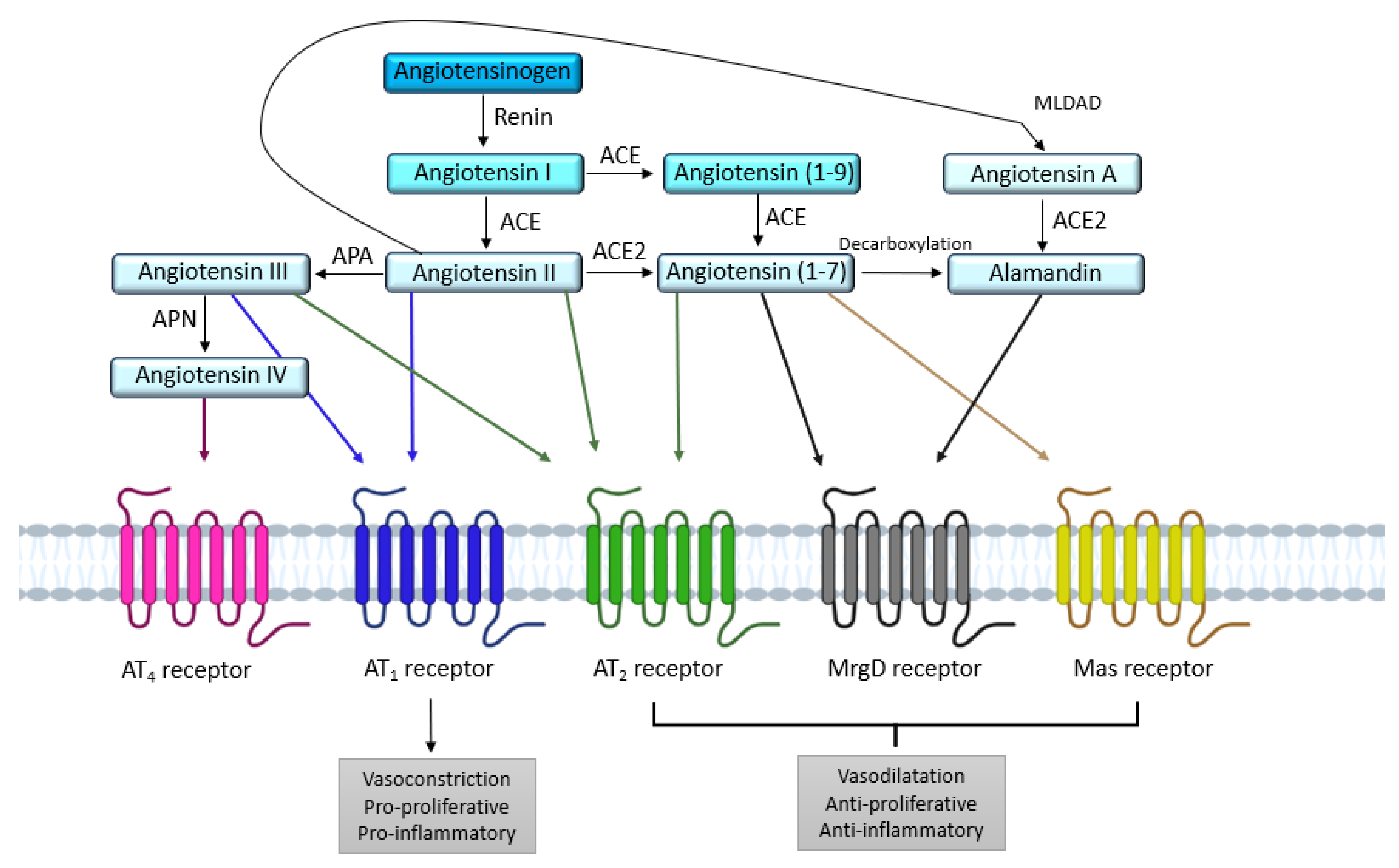
The AT1 receptor has mainly been associated with the pathological effects of the renin-angiotensin system (RAS) (e.g., hypertension, heart and kidney diseases), and constitutes a major therapeutic target. In contrast, the AT2 receptor is presented as the protective arm of this RAS, and its targeting via specific agonists is mainly used to counteract the effects of the AT1 receptor. The discovery of a local RAS has highlighted the importance of the balance between AT1/AT2 receptors at the tissue level. Disruption of this balance is suggested to be detrimental. The fine tuning of this balance is not limited to the regulation of the level of expression of these two receptors. Other mechanisms still largely unexplored, such as S-nitrosation of the AT1 receptor, homo- and heterodimerization, and the use of AT1 receptor-biased agonists, may significantly contribute to and/or interfere with the settings of this AT1/AT2 equilibrium.
- AT1 receptor
- AT2 receptor
- angiotensin II
- AT1/AT2 balance
- biased agonism
1. Introduction
2. AT1 and AT2 Angiotensin II Receptors
2.1. AT
1
Receptor
2.1.1. Structure
2.1.2. Signaling
G Protein Pathway
β-Arrestins
After stimulation by Ang II, the AT1 receptor is phosphorylated on its intracellular C-terminal serine and threonine residues by GPCR kinases (GRKs). This phosphorylation increases the affinity of β-arrestin-1 and β-arrestin-2 equally for the AT1 receptor. The β-arrestins’ recruitment is known to inhibit G protein-induced signaling by interfering with the conformation of the receptor; it is also known to initiate, with the help of clathrins and AP-2 adaptor proteins, the internalization and sequestration of the AT1 receptor coupled to its ligand, as well as the membrane recycling of the receptors [34,45][23][31]. Originally, arrestins were identified as central players in the desensitization and internalization of GPCRs. In addition to modulating GPCR signaling, in 1999, Robert Lefkowitz’s team showed that β-arrestins can also initiate a second wave of signaling [46][32].NADPH
Griendling et al. were the first to demonstrate the implication of nicotinamide adenine dinucleotide phosphate (NADPH) in oxidative stress mediated by the AT1 receptor [49][33]. Using rat aortic VSMCs, they showed that treatment of VSMCs with Ang II for 4–6 h caused a nearly threefold increase in intracellular O2- consumption. Ang II stimulates the activity of NAD(P)H oxidase (NOX), and thus generates reactive oxygen species (ROS) [49][33]. NOX comprises five subunits, and in the absence of stimulation, some of its subunits are cytosolic, while others are membrane-bound [50][34]. Ang II, via processes involving several players such as c-Src, PLD, PKC, PI3K, and transactivation of EGFR (epidermal growth factor receptor), induces phosphorylation of the p47phox subunit, which causes the formation of a complex between cytosolic subunits, followed by transfer to the membrane, wherein the complex associates with membrane subunits to give the active form of the oxidase [50][34]. This will lead to the production of reactive oxygen species (ROS) such as H2O2 or superoxide. These ROS are able to activate transcription factors such as activator protein-1 (AP-1) and nuclear factor kβ (NF-kβ), which will induce the expression of pro-inflammatory genes [44][30].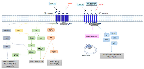
2.2. AT
2
Receptor
2.2.1. Structure
2.2.2. Signaling
G Protein Pathway
Bradykinin
Siragy et al. has suggested that AT2 receptor activity is mediated by stimulation of bradykinin (BK) production [69][51]. This hypothesis was later confirmed by showing that AT2 receptor inhibits the activity of Na+/H+ exchangers, resulting in acidification of the cell environment, which ultimately results in the release of BK [70][52]. AT2 receptor-dependent stimulation of BK receptors (B2 receptor) seems to activate protein kinase A (PKA), which phosphorylates eNOS [71][53].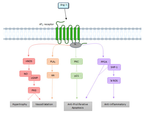
3. The Functional AT1/AT2 Receptors Balance
3.1. Systemic Cardiovascular Impact
3.2. AT
1
/AT
2
Balance in the Brain and Cerebral Circulation
3.2.1. Cerebral Circulation
The discovery of the presence of cerebral Ang II in 1971 revealed a cerebral RAS in dogs and rats [78][55]. Subsequently, AT1 and AT2 receptors were localized in cerebral blood vessels in rats [51][35], and then in the human brain [79][56]. In a rat cranial window model, Vincent et al. showed that Ang II-induced vasoconstriction of cerebral arterioles was abolished when an AT1 receptor antagonist was used, resulting in vasodilation, which was itself abolished when AT2 receptor antagonists were used [80][57]. In physiological conditions, the AT1 receptor will allow vasoconstriction of cerebral arterioles while the AT2 receptor has the opposite effect. The AT1 and AT2 receptors and changes in the AT1/AT2 equilibrium are particularly important contributors to the regulation of cerebral circulation. Indeed, the vasoconstriction of cerebral arterioles observed during Ang II stimulation is the sum of AT1 receptor-dependent vasoconstriction and AT2 receptor-dependent vasodilation in physiological conditions.3.2.2. Cardiovascular Regulation
The two receptors are expressed inside or near the medulla oblongata, the brain region which regulates cardiac rhythm and blood pressure. They are exclusively found in the neurons rather than the glia. The localization of AT1 receptors in the brain has been determined primarily by receptor autoradiography [32,85,86][58][59][60]. These studies demonstrated a wide distribution of AT1 receptors’ expression in the brain, including several regions involved in cardiovascular regulation. This distribution of AT1 receptors in the brain was confirmed by the development of transgenic mice expressing the AT1 receptor fused with eGFP [87][61]. High densities of AT1 receptors are found in the subfornical organ (SFO), the paraventricular nucleus (PVN), the area postrema (AP), the nucleus of the solitary tract (NTS), and the rostral ventrolateral medulla (RVLM) [87][61]. AT1 receptors are almost exclusively localized in neurons rather than in microglia, or astrocytes in these cardiovascular control centers [88][62]. Furthermore, the AT1 receptor appears to be predominantly expressed on glutamatergic neurons [89][63].3.2.3. Neuroinflammation
Inflammation is another example of the dysregulation of the AT1/AT2 receptors’ balance in the brain. Cells present in the brain such as astrocytes or microglial cells are the main sources of inflammation. In general, the activation of the AT1 receptor in macrophages leads to the activation of the pro-inflammatory axis of the RAS, while activation of the AT2 receptor promotes the activation of an anti-inflammatory axis [94,95][64][65]. In contrast, in pathological conditions such as inflammation, these receptors (and in particular, AT1/NOX signaling) are upregulated. NOX-derived superoxides are amplified by the activation of NF-kβ and the RhoA/ROCK pathway, leading to the production of ROS. In addition, through a feedback mechanism, activation of the RhoA/ROCK pathway enables increased expression of the AT1 receptor via NF-kβ [96][66].3.3. Cellular Cycle
The proliferation of normal cells is regulated by kinases called cyclin-dependent kinases (CDKs). The main actors in this cell cycle are cyclins, which regulate CDKs, enabling cells to progress through the cell cycle [99][67]. AT1 receptors promote cell proliferation pathways, in particular via the MAPK pathway, which allows the expression of cyclin and CDKs, itself allowing the advancement of cells in the cell cycle (Figure 6) [100][68].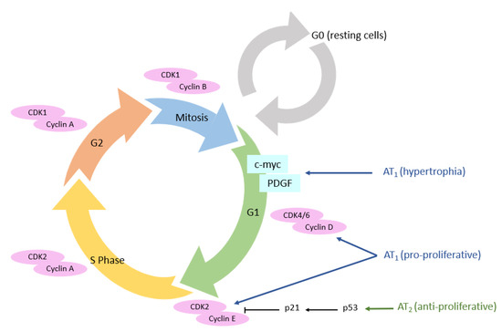
3.4. Wound Healing
4. Mechanisms Regulating the AT1/AT2 Functional Balance
4.1. Functional Opposition vs. Expression Level
4.2. Direct AT
1
/AT
2
Receptors Interactions
4.2.1. AT1 Receptor Dimerization
Abdallah et al. showed that increased levels of AT1 receptor homodimers were present on monocytes from patients with hypertension, which is an atherogenic risk factor, and that they were related to increased Ang II-dependent monocyte activity and adhesiveness [153][91]. This increase leads to the formation of atherosclerotic lesions. In this study, in addition to showing that inhibition of Ang II release prevents the formation of AT1 receptor homodimers, they observed that dimerized receptors increase Gq/11-mediated inositol phosphate signaling [153][91]. The AT1 receptor and B2 receptor can form a heterodimeric complex. This heterodimerization between the AT1 receptor and B2 receptor in HEK-293 cells increased the efficacy and potency of Ang II, but decreased the potency and the efficacy of BK. To confirm this, the authors compared the Ang II- or BK-stimulated increase in inositol phosphates in HEK-293 cells expressing the indicated receptors.4.2.2. AT2 Receptor Dimerization
AT2 receptor homodimerization was first described by Miura et al. in PC12W cells and CHO cells transfected with AT2R [159][92]. They also showed that these AT2 receptor homodimers allow constitutive signaling that leads to apoptosis. Furthermore, dimerization and pro-apoptotic signaling were not altered following AT2 receptor stimulation, suggesting that this homodimerization is ligand-independent, which has also been reported in transfected HEK-293 cells [160][93]. Like AT1 receptors, AT2 receptors can form heterodimers with B2 receptors. AT2 receptors mediate a vasodilatory cascade that includes BK, NO, and cGMP. Using a KO mouse model for B2R, they showed that when AT2 and B2 receptors are simultaneously activated in vivo, NO and cGMP production increases [162][94]. In a PC12W cell model, heterodimerization of these receptors was shown without any stimulation, suggesting the presence of constitutive heterodimers [72][95]. The AT2 receptors is also able to form a heterodimeric complex with MasR [164][96]. This dimerization influences the RAS-protective Ang II/AT2 axis, resulting in increased NO production and promoting diuretic–natriuretic response in obese Zucker rats [164][96]. Several studies suggest that these receptors may be functionally interdependent; indeed, the AT2 receptor antagonist (PD123319) reduced the vasodepressor effects of the MasR agonist Ang-(1-7) [165][97]. Similarly, Ang-(1-7) mediated endothelium-dependent vasodilation in the cerebral arteries [166][98] and aortic rings of salt-fed animals [167][99] was inhibited by PD123319 as well as the MasR antagonist (A-779). Last but not least, AT2/AT1 heterodimerization was first described by AbdAlla et al. in PC-12 cells, rat fetal fibroblasts, and human myometrial tissue samples [170][100]. They showed that AT2/AT1 dimerization was constitutive and led to inhibition of the AT1 receptor-mediated G protein pathway. This inhibition of AT1 receptor signaling does not require AT2 receptor activation, as shown by the fact that AT2/AT1 heterodimerization is not affected by AT2 receptor antagonists such as PD123319, and by the persistence of the effect in cells with dimers containing an AT2 receptor mutant that is unable to bind agonists or to initiate AT2 signaling. Attenuation of AT1 receptor signaling by the AT2 receptor (via calcium signaling, ERK1/2 MAPK activation) as well as constitutive AT2/AT1 dimerization has been confirmed in studies by other groups in HeLa cells [171][101] or HEK-293 transfected with AT2/AT1 [172][102]. AT2/AT1 dimerization also appears to impact AT2 intracellular trafficking, as AT2/AT1 dimers internalize upon Ang II stimulation (whereas AT2 alone is unable to do so) [171][101].4.3. Post-Translational Modifications
4.3.1. N-Glycosylation
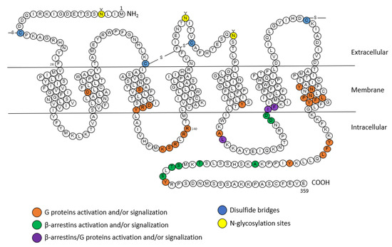
AT1 receptor function is regulated by various post-translational modifications such as N-glycosylation [173][103]. To study the effects of N-glycosylation on extracellular loops (ECL), artificial N-glycosylation sequences were incorporated into ECL1, ECL2 and ECL3 [174][104]. In ECL1, N-glycosylation causes a very significant decrease in the ligand affinity and surface expression of the receptor; in ECL2, it leads to the synthesis of a misfolded receptor, and in ECL3 N-glycosylation produces mutant receptors with normal affinity and low surface expression. These results show that N-glycosylation sites alter many properties of the AT1 receptor, such as targeting, folding, affinity, and surface expression [174][104].4.3.2. Phosphorylation
Phosphorylation is another post-translational modification that can occur on the AT1 receptor [177][105]. Phosphorylation of AT1 receptor serine/threonine residues is required for β-arrestin recruitment and receptor internalization [178][106]. In contrast, in the kidneys and arteries, GRK4 has been shown to exacerbate urinary sodium retention and vasoconstriction by increasing AT1 receptor expression [179][107]. The AT2 receptor is rapidly phosphorylated on a serine by PKC after activation by Ang II. The functional role of AT2 receptor phosphorylation is not known. When triggered by AT1 receptor activation, it appears to modulate the AT2 receptor’s effects, as opposed to AT1’s effects [180][108].4.3.3. S-Nitrosation
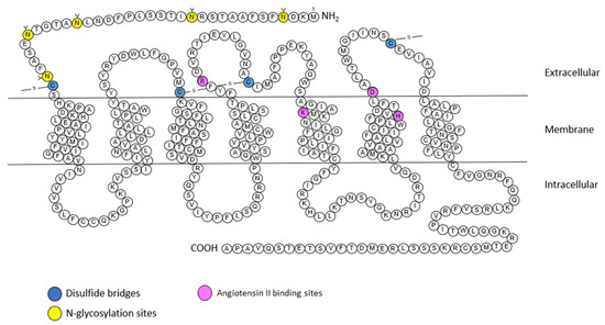
S-nitrosation is a mode of post-translational modification that allows the addition of a NO group to the sulfur atom of specific cysteine residues. The AT1 receptor contains ten cysteine residues. Four of these cysteines are involved in disulfide bridges at the extracellular loops, one is located on the cytoplasmic tail, and the other five are distributed in the transmembrane domains of the receptor. The affinity of the AT1 receptor for Ang II is decreased in the presence of sodium nitroprusside (SNP), an NO donor. An assessment of the affinity of different mutated AT1 receptors for each of five cysteines revealed the cysteine involved in sensitivity to sodium nitroprusside, cysteine residue 289 [182][109]. The AT2 receptor has 14 cysteine residues. It has seen that the four cysteines in the extracellular loops are engaged in disulfide bridges. The remaining ten cysteines are distributed on the transmembrane domains and the cytoplasmic tail.5. Possible Ways to Tune the AT1/AT2 Functional Balance
5.1. Pushing the Balance Using Agonist/Antagonist Ligands
One of the most obvious means to act on the AT1/AT2 functional balance is to use agonists or antagonists towards one or the other receptor. In order to rebalance the AT1/AT2 balance, the use of specific molecules targeting the receptors seems to be an interesting avenue. Indeed, this would make it possible to block or activate one specific receptor, which cannot be achieved with ACE inhibitors, for example, as they prevent both receptors’ activation by inhibiting Ang I cleavage. AT1 receptor antagonists were the first molecules discovered in this sense. In the case of pathologies associated with AT1 receptor, the ideal is to be able to abolish the overexpression of the receptor responsible for the imbalance in order to orient it in favor of the AT2 receptor. AT1 receptor antagonists are widely used to treat hypertension as well as cardiac diseases (heart failure or myocardial infarction). In vitro and in vivo, losartan is a reversible competitive AT1 receptor antagonist that inhibits the Ang II-induced vasoconstriction of blood [184][110]. However, complete blockade of the receptor then amounts to promoting activation of the AT2 receptor, which will tip the balance in the other direction instead of bringing it back to equilibrium. Another method would be to act on the AT2 receptor itself by using an agonist of the latter. For this, molecules capable of specifically activating the receptor have been developed, such as CGP42112A, which is a peptide, or C21, which is a synthetic compound. A study on the effects of CGP42112A in the same SHR model was performed, and the authors tested CGP42112A in the presence or absence of candesartan. The results showed that the use of candesartan alone at a high concentration lowered blood pressure in SHR rats, and that CGP42112A only provided a depressant effect in the presence of candesartan [185][111].5.2. Selective Activation of the β-Arrestin Pathway
The use of biased agonists is another strategy to regulate the AT1/AT2 receptors’ balance. A biased agonist is a receptor-specific ligand capable of selectively activating a single signaling pathway by preferentially stabilizing one of the receptor conformations. This phenomenon is also called “functional selectivity”. In the case of the AT1 receptor, several biased agonists have been developed that allow AT1 receptor to adopt an alternative active conformation [45][31]. For example SII, TRV120027, and TRV120023 selectively activate the β-arrestin pathway while inhibiting the G protein pathway [197][112]. SII is a modified Ang II peptide, which can trigger the phosphorylation of the AT1 receptor, and thus β-arrestin recruitment [203][113]. SII elicits GRK6 and β-arrestin 2-dependent ERK activation, and promotes β-arrestin-regulated Akt activity and mTOR phosphorylation to stimulate protein synthesis [204][114]. TRV120023 has been reported to only recruit β-arrestin while blocking G protein activation, enhancing myocyte contractility but without promoting hypertrophy, as seen with Ang II [205][115]. TRV120027 has been studied in several preclinical studies in a heart failure model, and has shown promising results [206][116]. A recent study just demonstrated that β-arrestin signaling, mediated by the PAR-1 receptor, produces prolonged activation of MAPK 42/44, which increases PDGF-β secretion. It has been shown that after ischemic stroke, PDGF-β secretion can provide increased protection of endothelial function and barrier integrity [207][117]. Similarly, there are biased agonists that selectively activate the G protein pathway, such as TRV120055 or TRV120056 [33][22].6. Conclusions
In summary, the AT1/AT2 balance is very important from a physiological point of view, and that the disturbance of this balance leads to the appearance of pathologies, most often as a result of the dominance of the AT1 receptor over the AT2 receptor. However, emerging studies have shown that secondary signaling of β-arrestins could have beneficial effects. This is why a finer regulation of this balance via S-nitrosation of the receptors or the use of biased agonists seems to be interesting, and could open new therapeutic perspectives.References
- Henrion, D.; Chillon, J.-M.; Capdeville-Atkinson, C.; Vinceneux-Feugier, M.; Atkinson, J. Chronic treatment with the angiotensin I converting enzyme inhibitor, perindopril, protects in vitro carbachol-induced vasorelaxation in a rat model of vascular calcium overload. Br. J. Pharmacol. 1991, 104, 966–972.
- Lartaud, I.; Bray-des-Boscs, L.; Chillon, J.M.; Atkinson, J.; Capdeville-Atkinson, C. In vivo cerebrovascular reactivity in Wistar and Fischer 344 rat strains during aging. Am. J. Physiol. Heart Circ. Physiol. 1993, 264, H851–H858.
- Régrigny, O.; Atkinson, J.; Capdeville-Atkinson, C.; Limiñana, P.; Chillon, J.-M. Effect of Lovastatin on Cerebral Circulation in Spontaneously Hypertensive Rats. Hypertension 2000, 35, 1105–1110.
- Atkinson, J. Stroke, high blood pressure and the renin–angiotensin–aldosterone system—New developments. Front. Pharm. 2011, 2, 22.
- Tigerstedt, R.; Bergman, P.Q. Niere und Kreislauf 1. Skand. Arch. Für Physiol. 1898, 8, 223–271.
- Guyton, A.C. Blood Pressure Control—Special Role of the Kidneys and Body Fluids. Science 1991, 252, 1813–1816.
- Skeggs, L.T.; Lentz, K.E.; Gould, A.B.; Hochstrasser, H.; Kahn, J.R. Biochemistry and kinetics of the renin-angiotensin system. Fed. Proc. 1967, 26, 42–47.
- Murphy, T.J.; Alexander, R.W.; Griendling, K.K.; Runge, M.S.; Bernstein, K.E. Isolation of a cDNA encoding the vascular type-1 angiotensin II receptor. Nature 1991, 351, 233–236.
- Mukoyama, M.; Nakajima, M.; Horiuchi, M.; Sasamura, H.; Pratt, R.E.; Dzau, V.J. Expression cloning of type 2 angiotensin II receptor reveals a unique class of seven-transmembrane receptors. J. Biol. Chem. 1993, 268, 24539–24542.
- Sevá Pessôa, B.; van der Lubbe, N.; Verdonk, K.; Roks, A.J.M.; Hoorn, E.J.; Danser, A.H.J. Key developments in renin–angiotensin–aldosterone system inhibition. Nat. Rev. Nephrol. 2013, 9, 26–36.
- Horiuchi, M.; Akishita, M.; Dzau, V.J. Recent Progress in Angiotensin II Type 2 Receptor Research in the Cardiovascular System. Hypertension 1999, 33, 613–621.
- Gasparo, M.D.; Catt, K.J.; Inagami, T.; Wright, J.W.; Unger, T. International Union of Pharmacology. XXIII. The Angiotensin II Receptors. Pharmacol. Rev. 2000, 52, 415–472.
- Campbell, D.J.; Habener, J.F. Angiotensinogen gene is expressed and differentially regulated in multiple tissues of the rat. J. Clin. Investig. 1986, 78, 31–39.
- Hunyady, L.; Catt, K.J. Pleiotropic AT1 Receptor Signaling Pathways Mediating Physiological and Pathogenic Actions of Angiotensin II. Mol. Endocrinol. 2006, 20, 953–970.
- Forrester, S.J.; Booz, G.W.; Sigmund, C.D.; Coffman, T.M.; Kawai, T.; Rizzo, V.; Scalia, R.; Eguchi, S. Angiotensin II Signal Transduction: An Update on Mechanisms of Physiology and Pathophysiology. Physiol. Rev. 2018, 98, 1627–1738.
- Pueyo, M.E.; N’Diaye, N.; Michel, J.-B. Angiotensin Il-elicited signal transduction via AT1 receptors in endothelial cells. Br. J. Pharmacol. 1996, 118, 79–84.
- Busche, S.; Gallinat, S.; Bohle, R.-M.; Reinecke, A.; Seebeck, J.; Franke, F.; Fink, L.; Zhu, M.; Sumners, C.; Unger, T. Expression of Angiotensin AT1 and AT2 Receptors in Adult Rat Cardiomyocytes after Myocardial Infarction. Am. J. Pathol. 2000, 157, 605–611.
- Siragy, H.M. AT1 and AT2 receptor in the kidney: Role in health and disease. Semin. Nephrol. 2004, 24, 93–100.
- Lenkei, Z.; Palkovits, M.; Corvol, P.; Llorens-Cortès, C. Expression of Angiotensin Type-1 (AT1) and Type-2 (AT2) Receptor mRNAs in the Adult Rat Brain: A Functional Neuroanatomical Review. Front. Neuroendocrinol. 1997, 18, 383–439.
- Johren, O.; Saavedra, J.M. Expression of AT1A and AT1B angiotensin II receptor messenger RNA in forebrain of 2-wk-old rats. Am. J. Physiol. Endocrinol. Metab. 1996, 271, E104–E112.
- Zhang, H.; Unal, H.; Gati, C.; Han, G.W.; Liu, W.; Zatsepin, N.A.; James, D.; Wang, D.; Nelson, G.; Weierstall, U.; et al. Structure of the Angiotensin Receptor Revealed by Serial Femtosecond Crystallography. Cell 2015, 161, 833–844.
- Wingler, L.M.; Skiba, M.A.; McMahon, C.; Staus, D.P.; Kleinhenz, A.L.W.; Suomivuori, C.-M.; Latorraca, N.R.; Dror, R.O.; Lefkowitz, R.J.; Kruse, A.C. Angiotensin and biased analogs induce structurally distinct active conformations within a GPCR. Science 2020, 367, 888–892.
- Suomivuori, C.-M.; Latorraca, N.R.; Wingler, L.M.; Eismann, S.; King, M.C.; Kleinhenz, A.L.W.; Skiba, M.A.; Staus, D.P.; Kruse, A.C.; Lefkowitz, R.J.; et al. Molecular mechanism of biased signaling in a prototypical G protein–coupled receptor. Science. 2020, 367, 881–887.
- Aplin, M.; Bonde, M.M.; Hansen, J.L. Molecular determinants of angiotensin II type 1 receptor functional selectivity. J. Mol. Cell. Cardiol. 2009, 46, 15–24.
- Balakumar, P.; Jagadeesh, G. Structural determinants for binding, activation, and functional selectivity of the angiotensin AT1 receptor. J. Mol. Endocrinol. 2014, 53, R71–R92.
- Kaschina, E.; Unger, T. Angiotensin AT1/AT2 Receptors: Regulation, Signalling and Function. Blood Press. 2003, 12, 70–88.
- Rao, G.N.; Lassegue, B.; Alexander, R.W.; Griendling, K.K. Angiotensin 11 stimulates phosphorylation of high-molecular-mass cytosolic phospholipase A2 in vascular smooth-muscle cells. Biochem. J. 1994, 299, 197–201.
- Touyz, R.M.; Schiffrin, E.L. Signal transduction mechanisms mediating the physiological and pathophysiological actions of angiotensin II in vascular smooth muscle cells. Pharmacol. Rev. 2000, 52, 639–672.
- Ohtsu, H.; Suzuki, H.; Nakashima, H.; Dhobale, S.; Frank, G.D.; Motley, E.D.; Eguchi, S. Angiotensin II Signal Transduction Through Small GTP-Binding Proteins: Mechanism and Significance in Vascular Smooth Muscle Cells. Hypertension 2006, 48, 534–540.
- Kawai, T.; Forrester, S.J.; O’Brien, S.; Baggett, A.; Rizzo, V.; Eguchi, S. AT1 receptor signaling pathways in the cardiovascular system. Pharmacol. Res. 2017, 125, 4–13.
- Violin, J.D.; Lefkowitz, R.J. β-Arrestin-biased ligands at seven-transmembrane receptors. Trends Pharmacol. Sci. 2007, 28, 416–422.
- Luttrell, L.M.; Lefkowitz, R.J. The role of β-arrestins in the termination and transduction of G-protein-coupled receptor signals. J. Cell Sci. 2002, 115, 455–465.
- Griendling, K.K.; Minieri, C.A.; Ollerenshaw, J.D.; Alexander, R.W. Angiotensin II stimulates NADH and NADPH oxidase activity in cultured vascular smooth muscle cells. Circ. Res. 1994, 74, 1141–1148.
- Touyz, R.M.; Briones, A.M. Reactive oxygen species and vascular biology: Implications in human hypertension. Hypertens. Res. 2011, 34, 5–14.
- Tsutsumi, K.; Saavedra, J.M. Characterization and development of angiotensin II receptor subtypes (AT1 and AT2) in rat brain. Am. J. Physiol. 1991, 261, R209–R216.
- Ichiki, T.; Herold, C.L.; Kambayashi, Y.; Bardhan, S.; Inagami, T. Cloning of the cDNA and the genomic DNA of the mouse angiotensin II type 2 receptor. Biochim. Biophys. Acta (BBA) Biomembr. 1994, 1189, 247–250.
- Rompe, F.; Artuc, M.; Hallberg, A.; Alterman, M.; Ströder, K.; Thöne-Reineke, C.; Reichenbach, A.; Schacherl, J.; Dahlöf, B.; Bader, M.; et al. Direct Angiotensin II Type 2 Receptor Stimulation Acts Anti-Inflammatory Through Epoxyeicosatrienoic Acid and Inhibition of Nuclear Factor κB. Hypertension 2010, 55, 924–931.
- Zhang, H.; Han, G.W.; Batyuk, A.; Ishchenko, A.; White, K.L.; Patel, N.; Sadybekov, A.; Zamlynny, B.; Rudd, M.T.; Hollenstein, K.; et al. Structural basis for selectivity and diversity in angiotensin II receptors. Nature 2017, 544, 327–332.
- Feng, Y.-H.; Saad, Y.; Karnik, S.S. Reversible inactivation of AT2 angiotensin II receptor from cysteine-disulfide bond exchange. FEBS Lett. 2000, 484, 133–138.
- Heerding, J.N.; Yee, D.K.; Jacobs, S.L.; Fluharty, S.J. Mutational analysis of the angiotensin II type 2 receptor: Contribution of conserved extracellular amino acids. Regul. Pept. 1997, 72, 97–103.
- Yee, D.K.; Kisley, L.R.; Heerding, J.N.; Fluharty, S.J. Mutation of a conserved fifth transmembrane domain lysine residue (Lys215) attenuates ligand binding in the angiotensin II type 2 receptor. Brain Res. Mol. Brain Res. 1997, 51, 238–241.
- Turner, C.A.; Cooper, S.; Pulakat, L. Role of the His273 located in the sixth transmembrane domain of the Angiotensin II receptor subtype AT2 in ligand–receptor interaction. Biochem. Biophys. Res. Commun. 1999, 257, 704–707.
- Steckelings, U.M.; Kaschina, E.; Unger, T. The AT2 receptor—A matter of love and hate. Peptides 2005, 26, 1401–1409.
- Turu, G.; Szidonya, L.; Gáborik, Z.; Buday, L.; Spät, A.; Clark, A.J.L.; Hunyady, L. Differential beta-arrestin binding of AT1 and AT2 angiotensin receptors. FEBS Lett. 2006, 580, 41–45.
- Padia, S.H.; Carey, R.M. AT2 receptors: Beneficial counter-regulatory role in cardiovascular and renal function. Pflug. Arch. Eur. J. Physiol. 2013, 465, 99–110.
- Bottari, S.P.; Taylor, V.; King, I.N.; Bogdal, Y.; Whitebread, S.; de Gasparo, M. Angiotensin II AT2 receptors do not interact with guanine nucleotide binding proteins. Eur. J. Pharmacol. Mol. Pharmacol. 1991, 207, 157–163.
- Zhang, J.; Pratt, R.E. The AT2 Receptor Selectively Associates with Giα2 and Giα3 in the Rat Fetus. J. Biol. Chem. 1996, 271, 15026–15033.
- Stennett, A.K.; Qiao, X.; Falone, A.E.; Koledova, V.V.; Khalil, R.A. Increased vascular angiotensin type 2 receptor expression and NOS-mediated mechanisms of vascular relaxation in pregnant rats. Am. J. Physiol. Heart Circ. Physiol. 2009, 296, H745–H755.
- Vinturache, A.E.; Smith, F.G. Angiotensin type 1 and type 2 receptors during ontogeny: Cardiovascular and renal effects. Vasc. Pharmacol. 2014, 63, 145–154.
- Alexander, L.D.; Ding, Y.; Alagarsamy, S.; Cui, X. Angiotensin II stimulates fibronectin protein synthesis via a Gβγ/arachidonic acid-dependent pathway. Am. J. Physiol. Ren. Physiol. 2014, 307, F287–F302.
- Siragy, H.M.; Carey, R.M. The subtype 2 (AT2) angiotensin receptor mediates renal production of nitric oxide in conscious rats. J. Clin. Investig. 1997, 100, 264–269.
- Tsutsumi, Y.; Matsubara, H.; Masaki, H.; Kurihara, H.; Murasawa, S.; Takai, S.; Miyazaki, M.; Nozawa, Y.; Ozono, R.; Nakagawa, K.; et al. Angiotensin II type 2 receptor overexpression activates the vascular kinin system and causes vasodilation. J. Clin. Investig. 1999, 104, 925–935.
- Yayama, K.; Hiyoshi, H.; Imazu, D.; Okamoto, H. Angiotensin II Stimulates Endothelial NO Synthase Phosphorylation in Thoracic Aorta of Mice With Abdominal Aortic Banding Via Type 2 Receptor. Hypertension 2006, 48, 958–964.
- Anderson, W.P.; Selig, S.E.; Korner, P.I. Role of Angiotensin II in the Hypertension Induced by Renal Artery Stenosis. Clin. Exp. Hypertens. Part A Theory Pract. 1984, 6, 299–314.
- Fischer-Ferraro, C.; Nahmod, V.E.; Goldstein, D.J.; Finkielman, S. Angiotensin and renin in rat and dog brain. J. Exp. Med. 1971, 133, 353–361.
- Barnes, J.M.; Steward, L.J.; Barber, P.C.; Barnes, N.M. Identification and characterisation of angiotensin II receptor subtypes in human brain. Eur. J. Pharmacol. 1993, 230, 251–258.
- Vincent, J.-M.; Kwan, Y.W.; Lung Chan, S.; Perrin-Sarrado, C.; Atkinson, J.; Chillon, J.-M. Constrictor and Dilator Effects of Angiotensin II on Cerebral Arterioles. Stroke 2005, 36, 2691–2695.
- Ohyama, K.; Yamano, Y.; Sano, T.; Nakagomi, Y.; Hamakubo, T.; Morishima, I.; Inagami, T. Disulfide bridges in extracellular domains of angiotensin II receptor type IA. Regul. Pept. 1995, 57, 141–147.
- Chen, D.; Jancovski, N.; Bassi, J.K.; Nguyen-Huu, T.-P.; Choong, Y.-T.; Palma-Rigo, K.; Davern, P.J.; Gurley, S.B.; Thomas, W.G.; Head, G.A.; et al. Angiotensin Type 1A Receptors in C1 Neurons of the Rostral Ventrolateral Medulla Modulate the Pressor Response to Aversive Stress. J. Neurosci. 2012, 32, 2051–2061.
- De Kloet, A.D.; Wang, L.; Ludin, J.A.; Smith, J.A.; Pioquinto, D.J.; Hiller, H.; Steckelings, U.M.; Scheuer, D.A.; Sumners, C.; Krause, E.G. Reporter mouse strain provides a novel look at angiotensin type-2 receptor distribution in the central nervous system. Brain Struct. Funct. 2016, 221, 891–912.
- Gonzalez, A.D.; Wang, G.; Waters, E.M.; Gonzales, K.L.; Speth, R.C.; Van Kempen, T.A.; Marques-Lopes, J.; Young, C.N.; Butler, S.D.; Davisson, R.L.; et al. Distribution of angiotensin type 1a receptor-containing cells in the brains of bacterial artificial chromosome transgenic mice. Neuroscience 2012, 226, 489–509.
- Elsaafien, K.; de Kloet, A.D.; Krause, E.G.; Sumners, C. Brain Angiotensin Type-1 and Type-2 Receptors in Physiological and Hypertensive Conditions: Focus on Neuroinflammation. Curr. Hypertens. Rep. 2020, 22, 48.
- De Kloet, A.D.; Wang, L.; Pitra, S.; Hiller, H.; Smith, J.A.; Tan, Y.; Nguyen, D.; Cahill, K.M.; Sumners, C.; Stern, J.E.; et al. A Unique “Angiotensin-Sensitive” Neuronal Population Coordinates Neuroendocrine, Cardiovascular, and Behavioral Responses to Stress. J. Neurosci. 2017, 37, 3478–3490.
- Valero-Esquitino, V.; Lucht, K.; Namsolleck, P.; Monnet-Tschudi, F.; Stubbe, T.; Lucht, F.; Liu, M.; Ebner, F.; Brandt, C.; Danyel, L.A.; et al. Direct angiotensin type 2 receptor (AT2R) stimulation attenuates T-cell and microglia activation and prevents demyelination in experimental autoimmune encephalomyelitis in mice. Clin. Sci. 2015, 128, 95–109.
- Kim, J.-H.; Afridi, R.; Cho, E.; Yoon, J.H.; Lim, Y.-H.; Lee, H.-W.; Ryu, H.; Suk, K. Soluble ANPEP Released From Human Astrocytes as a Positive Regulator of Microglial Activation and Neuroinflammation: Brain Renin–Angiotensin System in Astrocyte–Microglia Crosstalk. Mol. Cell. Proteom. 2022, 21, 100424.
- Rodriguez-Perez, A.I.; Borrajo, A.; Rodriguez-Pallares, J.; Guerra, M.J.; Labandeira-Garcia, J.L. Interaction between NADPH-oxidase and Rho-kinase in angiotensin II-induced microglial activation: NADPH-Oxidase and Rho-Kinase Interaction. Glia 2015, 63, 466–482.
- Campbell, G.J.; Hands, E.L.; Van de Pette, M. The Role of CDKs and CDKIs in Murine Development. Int. J. Mol. Sci. 2020, 21, 5343.
- Han, H.J.; Han, J.Y.; Heo, J.S.; Lee, S.H.; Lee, M.Y.; Kim, Y.H. ANG II-stimulated DNA synthesis is mediated by ANG II receptor-dependent Ca2+/PKC as well as EGF receptor-dependent PI3K/Akt/mTOR/p70S6K1 signal pathways in mouse embryonic stem cells. J. Cell. Physiol. 2007, 211, 618–629.
- Yu, L.; Meng, W.; Ding, J.; Cheng, M. Klotho inhibits angiotensin II-induced cardiomyocyte hypertrophy through suppression of the AT1R/beta catenin pathway. Biochem. Biophys. Res. Commun. 2016, 473, 455–461.
- Zhou, L.; Liu, Y. Wnt/β-catenin signaling and renin–angiotensin system in chronic kidney disease. Curr. Opin. Nephrol. Hypertens. 2016, 25, 100–106.
- Fujihara, S.; Morishita, A.; Ogawa, K.; Tadokoro, T.; Chiyo, T.; Kato, K.; Kobara, H.; Mori, H.; Iwama, H.; Masaki, T. The angiotensin II type 1 receptor antagonist telmisartan inhibits cell proliferation and tumor growth of esophageal adenocarcinoma via the AMPKα/mTOR pathway in vitro and in vivo. Oncotarget 2017, 8, 8536–8549.
- Samukawa, E.; Fujihara, S.; Oura, K.; Iwama, H.; Yamana, Y.; Tadokoro, T.; Chiyo, T.; Kobayashi, K.; Morishita, A.; Nakahara, M.; et al. Angiotensin receptor blocker telmisartan inhibits cell proliferation and tumor growth of cholangiocarcinoma through cell cycle arrest. Int. J. Oncol. 2017, 51, 1674–1684.
- Aleksiejczuk, M.; Gromotowicz-Poplawska, A.; Marcinczyk, N.; Przylipiak, A.; Chabielska, E. The expression of the renin-angiotensin-aldosterone system in the skin and its effects on skin physiology and pathophysiology. J. Physiol. Pharmacol. 2019, 70.
- Silva, I.M.S.; Assersen, K.B.; Willadsen, N.N.; Jepsen, J.; Artuc, M.; Steckelings, U.M. The role of the renin-angiotensin system in skin physiology and pathophysiology. Exp. Dermatol. 2020, 29, 891–901.
- Viswanathan, M.; Saavedra, J.M. Expression of angiotensin II AT2 receptors in the rat skin during experimental wound healing. Peptides 1992, 13, 783–786.
- Steckelings, U.M.; Henz, B.M.; Wiehstutz, S.; Unger, T.; Artuc, M. Differential expression of angiotensin receptors in human cutaneous wound healing. Br. J. Dermatol. 2005, 153, 887–893.
- Takeda, H.; Katagata, Y.; Kondo, S. Immunohistochemical study of angiotensin receptors in human anagen hair follicles and basal cell carcinoma. Br. J. Dermatol. 2002, 147, 276–280.
- Jiang, X.; Wu, F.; Xu, Y.; Yan, J.-X.; Wu, Y.-D.; Li, S.-H.; Liao, X.; Liang, J.-X.; Li, Z.-H.; Liu, H.-W. A novel role of angiotensin II in epidermal cell lineage determination: Angiotensin II promotes the differentiation of mesenchymal stem cells into keratinocytes through the p38 MAPK, JNK and JAK2 signalling pathways. Exp. Dermatol. 2019, 28, 59–65.
- Jadhav, S.S.; Sharma, N.; Meeks, C.J.; Mordwinkin, N.M.; Espinoza, T.B.; Roda, N.R.; DiZerega, G.S.; Hill, C.K.; Louie, S.G.; Rodgers, K.E. Effects of combined radiation and burn injury on the renin-angiotensin system: CRBI and renin-angiotensin system. Wound Repair. Regen. 2013, 21, 131–140.
- Kamiñska, M.; Mogielnicki, A.; Stankiewicz, A.; Kramkowski, K.; Domaniewski, T.; Buczko, W.; Chabielska, E. Angiotensin Ii Via At1 Receptor Accelerates Arterial Thrombosis In Renovascular Hypertensive Rats. J. Physiol. Pharmacol. 2005, 56, 571–585.
- Bernasconi, R.; Nyström, A. Balance and circumstance: The renin angiotensin system in wound healing and fibrosis. Cell. Signal. 2018, 51, 34–46.
- Yahata, Y.; Shirakata, Y.; Tokumaru, S.; Yang, L.; Dai, X.; Tohyama, M.; Tsuda, T.; Sayama, K.; Iwai, M.; Horiuchi, M.; et al. A Novel Function of Angiotensin II in Skin Wound Healing. J. Biol. Chem. 2006, 281, 13209–13216.
- Faghih, M.; Hosseini, S.M.; Smith, B.; Ansari, A.M.; Lay, F.; Ahmed, A.K.; Inagami, T.; Marti, G.P.; Harmon, J.W.; Walston, J.D.; et al. Knockout of Angiotensin AT2 receptors accelerates healing but impairs quality. Aging 2015, 7, 1185–1197.
- Karppinen, S.-M.; Heljasvaara, R.; Gullberg, D.; Tasanen, K.; Pihlajaniemi, T. Toward understanding scarless skin wound healing and pathological scarring. F1000Res 2019, 8, 787.
- Murphy, A.M.; Wong, A.L.; Bezuhly, M. Modulation of angiotensin II signaling in the prevention of fibrosis. Fibrogenes. Tissue Repair. 2015, 8, 7.
- Ito, M.; Oliverio, M.I.; Mannon, P.J.; Best, C.F.; Maeda, N.; Smithies, O.; Coffman, T.M. Regulation of blood pressure by the type 1A angiotensin II receptor gene. Proc. Natl. Acad. Sci. USA 1995, 92, 3521–3525.
- Daviet, L.; Lehtonen, J.Y.; Tamura, K.; Griese, D.P.; Horiuchi, M.; Dzau, V.J. Cloning and characterization of ATRAP, a novel protein that interacts with the angiotensin II type 1 receptor. J. Biol. Chem. 1999, 274, 17058–17062.
- Lopez-Ilasaca, M.; Liu, X.; Tamura, K.; Dzau, V.J. The angiotensin II type I receptor-associated protein, ATRAP, is a transmembrane protein and a modulator of angiotensin II signaling. Mol. Biol. Cell. 2003, 14, 5038–5050.
- Mogi, M.; Iwai, M.; Horiuchi, M. Emerging Concepts of Regulation of Angiotensin II Receptors: New Players and Targets for Traditional Receptors. Arterioscler. Thromb. Vasc. Biol. 2007, 27, 2532–2539.
- Wruck, C.J.; Funke-Kaiser, H.; Pufe, T.; Kusserow, H.; Menk, M.; Schefe, J.H.; Kruse, M.L.; Stoll, M.; Unger, T. Regulation of transport of the angiotensin AT2 receptor by a novel membrane-associated Golgi protein. Arterioscl. Thromb. Vasc. Biol. 2005, 25, 57–64.
- AbdAlla, S.; Lother, H.; Langer, A.; el Faramawy, Y.; Quitterer, U. Factor XIIIA Transglutaminase Crosslinks AT1 Receptor Dimers of Monocytes at the Onset of Atherosclerosis. Cell 2004, 119, 343–354.
- Miura, S.; Karnik, S.S.; Saku, K. Constitutively Active Homo-oligomeric Angiotensin II Type 2 Receptor Induces Cell Signaling Independent of Receptor Conformation and Ligand Stimulation. J. Biol. Chem. 2005, 280, 18237–18244.
- Porrello, E.R.; Pfleger, K.D.G.; Seeber, R.M.; Qian, H.; Oro, C.; Abogadie, F.; Delbridge, L.M.D.; Thomas, W.G. Heteromerization of angiotensin receptors changes trafficking and arrestin recruitment profiles. Cell. Signal. 2011, 23, 1767–1776.
- Abadir, P.M.; Carey, R.M.; Siragy, H.M. Angiotensin AT2 Receptors Directly Stimulate Renal Nitric Oxide in Bradykinin B2-Receptor–Null Mice. Hypertension 2003, 42, 600–604.
- Abadir, P.M.; Periasamy, A.; Carey, R.M.; Siragy, H.M. Angiotensin II Type 2 Receptor–Bradykinin B2 Receptor Functional Heterodimerization. Hypertension 2006, 48, 316–322.
- Patel, S.N.; Ali, Q.; Samuel, P.; Steckelings, U.M.; Hussain, T. Angiotensin II Type 2 Receptor and Receptor Mas Are Colocalized and Functionally Interdependent in Obese Zucker Rat Kidney. Hypertension 2017, 70, 831–838.
- Walters, P.E.; Gaspari, T.A.; Widdop, R.E. Angiotensin-(1–7) Acts as a Vasodepressor Agent Via Angiotensin II Type 2 Receptors in Conscious Rats. Hypertension 2005, 45, 960–966.
- Durand, M.J.; Raffai, G.; Weinberg, B.D.; Lombard, J.H. Angiotensin-(1-7) and low-dose angiotensin II infusion reverse salt-induced endothelial dysfunction via different mechanisms in rat middle cerebral arteries. Am. J. Physiol. Heart Circ. Physiol. 2010, 299, H1024–H1033.
- Roks, A.J.; Nijholt, J.; van Buiten, A.; van Gilst, W.H.; de Zeeuw, D.; Henning, R.H. Low sodium diet inhibits the local counter-regulator effect of angiotensin-(1-7) on angiotensin II. J. Hypertens. 2004, 22, 2355–2361.
- AbdAlla, S.; Lother, H.; Abdel-tawab, A.M.; Quitterer, U. The Angiotensin II AT2 Receptor Is an AT1Receptor Antagonist. J. Biol. Chem. 2001, 276, 39721–39726.
- Inuzuka, T.; Fujioka, Y.; Tsuda, M.; Fujioka, M.; Satoh, A.O.; Horiuchi, K.; Nishide, S.; Nanbo, A.; Tanaka, S.; Ohba, Y. Attenuation of ligand-induced activation of angiotensin II type 1 receptor signaling by the type 2 receptor via protein kinase C. Sci Rep 2016, 6, 21613.
- Rivas-Santisteban, R.; Rodriguez-Perez, A.I.; Muñoz, A.; Reyes-Resina, I.; Labandeira-García, J.L.; Navarro, G.; Franco, R. Angiotensin AT1 and AT2 receptor heteromer expression in the hemilesioned rat model of Parkinson’s disease that increases with levodopa-induced dyskinesia. J. Neuroinflamm. 2020, 17, 243.
- Lanctôt, P.M.; Leclerc, P.C.; Escher, E.; Leduc, R.; Guillemette, G. Role of N-glycosylation in the expression and functional properties of human AT1 receptor. Biochemistry 1999, 38, 8621–8627.
- Lanctot, P.M.; Leclerc, P.C.; Clément, M.; Auger-Messier, M.; Escher, E.; Leduc, R.; Guillemette, G. Importance of N-glycosylation positioning for cell-surface expression, targeting, affinity and quality control of the human AT1 receptor. Biochem. J. 2005, 390, 367–376.
- Qian, H. Association of -Arrestin 1 with the Type 1A Angiotensin II Receptor Involves Phosphorylation of the Receptor Carboxyl Terminus and Correlates with Receptor Internalization. Mol. Endocrinol. 2001, 15, 1706–1719.
- Kule, C.E.; Karoor, V.; Day, J.N.E.; Thomas, W.G.; Baker, K.M.; Dinh, D.; Acker, K.A.; Booz, G.W. Agonist-dependent internalization of the angiotensin II type one receptor (AT1): Role of C-terminus phosphorylation in recruitment of β-arrestins. Regul. Pept. 2004, 120, 141–148.
- Chen, K.; Fu, C.; Chen, C.; Jose, P.A.; Zeng, C. Role of GRK4 in the Regulation of Arterial AT1 Receptor in Hypertension. J. Am. Soc. Hypertens. 2016, 10, e3.
- Olivares-Reyes, J.A.; Smith, R.D.; Hunyady, L.; Shah, B.H.; Catt, K.J. Agonist-induced Signaling, Desensitization, and Internalization of a Phosphorylation-deficient AT1A Angiotensin Receptor. J. Biol. Chem. 2001, 276, 37761–37768.
- Leclerc, P.C.; Lanctot, P.M.; Auger-Messier, M.; Escher, E.; Leduc, R.; Guillemette, G. S-nitrosylation of cysteine 289 of the AT1 receptor decreases its binding affinity for angiotensin II: S -nitrosylation of the AT1 receptor. Br. J. Pharmacol. 2006, 148, 306–313.
- Sica, D.A.; Gehr, T.W.B.; Ghosh, S. Clinical Pharmacokinetics of Losartan. Clin. Pharmacokinet. 2005, 44, 797–814.
- Barber, M.N.; Sampey, D.B.; Widdop, R.E. AT2 Receptor Stimulation Enhances Antihypertensive Effect of AT1 Receptor Antagonist in Hypertensive Rats. Hypertension 1999, 34, 1112–1116.
- Violin, J.D.; DeWire, S.M.; Yamashita, D.; Rominger, D.H.; Nguyen, L.; Schiller, K.; Whalen, E.J.; Gowen, M.; Lark, M.W. Selectively engaging β-arrestins at the angiotensin II type 1 receptor reduces blood pressure and increases cardiac performance. J. Pharmacol. Exp. Ther. 2010, 335, 572–579.
- Cui, Y.; Kassmann, M.; Nickel, S.; Zhang, C.; Alenina, N.; Anistan, Y.M.; Schleifenbaum, J.; Bader, M.; Welsh, D.G.; Huang, Y.; et al. Myogenic Vasoconstriction Requires Canonical Gq/11 Signaling of the Angiotensin II Type 1 Receptor. J. Am. Heart Assoc. 2022, 11, e022070.
- Jean-Charles, P.-Y.; Kaur, S.; Shenoy, S.K. G Protein–Coupled Receptor Signaling Through β-Arrestin–Dependent Mechanisms. J. Cardiovasc. Pharmacol. 2017, 70, 142–158.
- Ma, Z.; Viswanathan, G.; Sellig, M.; Jassal, C.; Choi, I.; Garikipati, A.; Xiong, X.; Nazo, N.; Rajagopal, S. β-Arrestin–Mediated Angiotensin II Type 1 Receptor Activation Promotes Pulmonary Vascular Remodeling in Pulmonary Hypertension. JACC Basic Transl. Sci. 2021, 6, 854–869.
- Boerrigter, G.; Soergel, D.G.; Lark, M.W.; Burnett, J.C. TRV120027, a Novel Beta-Arrestin Biased Ligand at the Angiotensin II Type I Receptor, Unloads the Heart and Maintains Renal Function When Added to Furosemide in Experimental Heart Failure. J. Card. Fail. 2011, 17, S63–S64.
- Kanki, H.; Sasaki, T.; Matsumura, S.; Yokawa, S.; Yukami, T.; Shimamura, M.; Sakaguchi, M.; Furuno, T.; Suzuki, T.; Mochizuki, H. β-arrestin-2 in PAR-1-biased signaling has a crucial role in endothelial function via PDGF-β in stroke. Cell Death Dis. 2019, 10, 100.
